HDAC8 / Histone deacetylase 8 · IHC design guide

Design Immunohistochemistry for HDAC8

Plan HDAC8 paraffin IHC around its general nuclear staining pattern (HPA tissue IHC). Use pancreatic exocrine glandular cells, which show high staining, as a positive tissue reference (HPA tissue IHC).

Evidence assembled Oct 2026 · For research use; verify linked source records and product datasheet before use
Immunohistochemistry protocol sheet for HDAC8 (IHC for HDAC8): expected localisation General nuclear staining in tissue IHC (HPA tissue IHC), antibody PA1591, validated IHC image, and IHC protocol steps
Printable HDAC8 IHC protocol sheet — expected localisation General nuclear staining in tissue IHC (HPA tissue IHC), antibody PA1591, controls and protocol steps. Open the full HDAC8 IHC guide →

HDAC8 Immunohistochemistry Experimental Design Guide

Expected localisation, validated protocols, controls and antibodies — the at-a-glance facts below, then the full design guide.

Must know before staining
Expected localisation General nuclear staining in tissue IHC (HPA tissue IHC)
Staining pattern Nuclear staining across tissue cell types (HPA tissue IHC)
Antigen retrieval Tris-EDTA pH 9.0 HIER, 95–98 °C, 20 min (rule: nuclear antigen)
Positive control ⓘ Adrenal gland+4 more · see all
Negative control ⓘ Adipose tissue+4 more · see all
Important caveats
Reasons your staining may differ from the expected pattern.
Fixation Keep fixation consistent across sections (standard IHC practice; not target-specific). Selected-image fixative and duration unreported (datasheet PA1591); verify before use.
Caveat The HPA antibody may detect products of multiple genes (HPA tissue IHC)
Regulation Higher expression in pancreas (UniProt)
Isoform / epitope 6 isoforms; epitope coverage is unknown (UniProt)
Section 1

Recommended HDAC8 IHC & IF Protocols

The catalog antibody has its own IHC-P protocol (datasheet); the options below summarize three published HDAC8 staining protocols (PMC8798431; PMC13214310; PMC11297794).

Recommended immunohistochemistry (IHC-P) protocol parameters
SampleTissue sections; selected-image fixative not specified (standard IHC workflow)
FixationImage fixative and duration unreported (datasheet PA1591); verify before use.
Sectioning4–5 µm sections on charged slides (standard)
DeparaffinisationXylene, graded ethanol series to water (standard)
Antigen retrievalHeat-induced epitope retrieval in Tris-EDTA buffer, pH 9.0, 20 min at 95–98 °C (standard rule: nuclear antigen)
Peroxidase block3% H2O2, 10 min, room temperature (standard)
Blocking10% normal serum of the secondary host, 30 min, room temperature (standard)
Primary antibodyRabbit anti-HDAC8, 0.5-1μg/ml (datasheet PA1591)
Primary incubationOvernight at 4 °C (standard)
DetectionHRP-polymer secondary, DAB chromogen 5–10 min (standard)
CounterstainHematoxylin, blue, dehydrate and mount (standard)
Expected resultHDAC8-positive staining in glandular cells of adrenal gland (HPA tissue IHC: High). HPA tissue profile: General nuclear expression. No signal in the no-primary control.
💡Decision noteStart the catalog antibody with Tris-EDTA pH 9.0 HIER at 95–98 °C for 20 min (page antigen-retrieval rule).
Section 2

What Is the Expected HDAC8 Staining Pattern?

In paraffin-section IHC, expect mainly nuclear HDAC8 staining, with nucleoli spared; cytoplasmic staining can occur in cells showing smooth muscle differentiation (UniProt Q9BY41 subcellular location). Adrenal, salivary and thyroid glandular cells, pancreatic exocrine cells, bone marrow hematopoietic cells, and tonsillar non-germinal center cells show high staining (HPA: tissue IHC). HPA rates its tissue pattern Supported, with medium staining–RNA consistency and a warning that the antibody targets protein from more than one gene (HPA: tissue IHC reliability).

What am I looking at on my slide?
Distinct nuclear staining in pancreatic exocrine glandular cells, with little nucleolar staining.This fits the expected compartment and a high-staining cell population (UniProt Q9BY41: nuclear, excluded from nucleoli; HPA: high in pancreatic exocrine glandular cells). Judge the pattern in the named cells, since an organ-wide intensity label would hide cell-type differences.
Predominantly cytoplasmic staining in glandular cells, or staining concentrated in nucleoli.Treat this as a compartment mismatch and investigate artefact or off-target signal (UniProt Q9BY41: nuclear, excluded from nucleoli). Cytoplasmic signal is biologically plausible in cells showing smooth muscle differentiation, so assess that cell identity before rejecting the pattern (UniProt Q9BY41: subcellular location).
Strong color in adipocytes or soft-tissue fibroblasts while the expected nuclear pattern is absent.These cell populations were not detected in HPA tissue IHC (HPA: adipocytes; soft-tissue fibroblasts). Consider cross-reactivity or endogenous detection activity; HPA also cautions that its tissue antibody targets protein from more than one gene (HPA: reliability note).
Diffuse color covers nuclei, cytoplasm and extracellular areas without clear cell boundaries.This does not resolve the reported general nuclear pattern (HPA: tissue IHC profile). Review background controls, blocking, detection chemistry and antibody dilution as general IHC practice. Diffuse color alone cannot establish HDAC8 localization or identify which assay component caused it.
No staining in pancreatic exocrine cells or adrenal glandular cells on an otherwise interpretable section.Both populations had high staining in HPA tissue IHC (HPA: pancreas; adrenal gland). Check a known-positive section and the detection workflow before calling the sample negative (general IHC practice); HPA's Supported rating is not a guarantee for every specimen (HPA: reliability).
💡Expected HDAC8 appearanceCall a result consistent when identifiable glandular or hematopoietic cells show predominantly nuclear staining, potentially high in the HPA-listed populations; prominent nucleolar color, indiscriminate diffuse color or strong staining in HPA-undetected cell types calls for control review (HPA: tissue IHC; UniProt Q9BY41: subcellular location).
How each factor affects the staining
Cell identity and tissue contextHPA reports high staining in selected glandular and hematopoietic populations, but not detected in adipocytes and soft-tissue fibroblasts (HPA: tissue IHC). Compare the named cell types within the section rather than assigning one expected intensity to every cell.
Compartment and smooth muscle differentiationHDAC8 is annotated in nucleus, chromosome and cytoplasm, with nucleolar exclusion; cytoplasmic localization is reported in cells showing smooth muscle differentiation (UniProt Q9BY41: subcellular location). Interpret cytoplasmic color against cell morphology and nuclear staining.
Antibody evidence and specificityHPA048560 is rated Supported for IHC, while the HPA tissue profile reports medium staining–RNA consistency and cautions that the antibody targets protein from more than one gene (HPA: antibody validation; tissue IHC reliability). Morphology and controls remain necessary for attribution.
Isoforms and antigen locationUniProt lists 6 HDAC8 isoforms and a 1–377 chain with no signal peptide, propeptide or transmembrane segment (UniProt Q9BY41: isoforms, processing, topology). The supplied sources give no antibody epitope, so they cannot predict isoform-specific staining or retrieval needs.
What can IF/ICC show?Use nuclear localization, nucleolar exclusion and the smooth muscle cytoplasmic exception as interpretation context (UniProt Q9BY41: subcellular location). HPA supplies no main ICC/IF location or cell-line images here, so it provides no observed IF pattern to confirm (HPA: subcellular record).
Why is my staining missing, weak or wrong?
SituationLikely causeNext action
Expected positive cells appear blank.Weak or failed detection is possible; HPA reports high staining in pancreatic exocrine and adrenal glandular cells (HPA: tissue IHC). A blank section alone does not identify a target-specific fixation effect.Run a known-positive tissue section alongside the sample; verify retrieval, primary-antibody application and detection reagents using the antibody's IHC instructions (general IHC practice).
Nucleoli stain more strongly than surrounding nuclei.This conflicts with reported nucleolar exclusion (UniProt Q9BY41: subcellular location). Background or off-target binding is possible, but the image alone cannot distinguish them.Inspect a primary-omission control and a positive tissue section; compare nuclear and nucleolar color at the same detection settings (general IHC practice).
Cytoplasm dominates the signal.Cytoplasmic HDAC8 is reported in cells showing smooth muscle differentiation (UniProt Q9BY41: subcellular location); the same appearance in unrelated cells needs scrutiny.Identify the stained cells morphologically, check for the expected nuclear pattern in nearby positive cells, and compare with detection controls (general IHC practice; HPA: general nuclear expression).
Unexpected cell types stain strongly.HPA found adipocytes and soft-tissue fibroblasts not detected and cautions that its tissue antibody targets protein from more than one gene (HPA: tissue IHC; reliability note). Endogenous detection activity is another general IHC possibility.Compare an appropriate negative cell population, a positive tissue section and a primary-omission control; review blocking and detection chemistry (general IHC practice).
Color is diffuse across most of the section.The reported tissue pattern is generally nuclear (HPA: tissue IHC profile). Widespread diffuse color can reflect nonspecific staining or detection background (general IHC practice).Check the primary-omission control, blocking, washes and antibody dilution; interpret HDAC8 only where cellular boundaries and nuclear localization remain clear (general IHC practice; HPA: general nuclear expression).
A low-signal tissue is read as a failed assay.HPA reports low staining in cerebellar granular-layer cells and splenic red-pulp cells, alongside higher staining in selected other cell types (HPA: tissue IHC). Expected intensity depends on the cells examined.Compare the named cell population with a HPA-listed high-staining positive control, such as pancreatic exocrine cells, before judging assay performance (HPA: tissue IHC; general IHC practice).

Sample controls for HDAC8 IHC & IF

🧪Run pancreas first: exocrine glandular cells should stain (HPA: High in pancreatic exocrine glandular cells). Use adipose tissue as a negative comparison, focusing on adipocytes (HPA: Not detected in adipocytes); on the pancreas slide, treat counterstain-only cells as a background reference without assuming a specific non-exocrine cell type is HDAC8-negative.
Positive control tissue: Adrenal gland (Glandular cells, HPA High)
Negative control tissue: Adipose tissue (HPA Not detected)
ICC-IF cell lines (HPA subcellular resource): HPA carries no ICC-IF cell line for HDAC8; derive a cell-line control from the positive tissue's cell type (Glandular cells) and confirm it by RNA or western blot first.
Technical controls: Include a no-primary (secondary-only) section, an irrelevant immunoglobulin matched to the primary antibody’s host species and isotype or clonality, and HDAC8-knockout tissue if available; peptide competition is an alternative when the immunizing peptide is available. For pancreatic sections, check background with the no-primary control and block endogenous peroxidase before chromogenic detection; assess endogenous biotin if using a biotin-based detection system.
⚠️Feasibility: The supplied evidence reports no HDAC8-specific fixation window or antigen-retrieval dependence, so optimize retrieval on the paraffin sections. The selected PA1591 tissue-IHC caption does not state a fixative (caption: fixative unreported), and the supplied evidence does not establish whether frozen sections or IF are easier. No HDAC8-specific pancreatic staining artefact is reported in the supplied evidence.

HPA tissue IHC evidence for HDAC8

Comprehensive Human Protein Atlas IHC scoring per tissue (reliability: Supported — Medium consistency between antibody staining and RNA expression data. Caution, targets protein from more than one gene.). Rows are taken directly from the HPA tissue chart — click any row's HPA link to view the source.

Positive expression · recommended positive controls

TissueCell typeLevelEvidenceSource
Adrenal gland Glandular cells High Protein (IHC) HPA →
Bone marrow Hematopoietic cells High Protein (IHC) HPA →
Pancreas Exocrine glandular cells High Protein (IHC) HPA →
Salivary gland Glandular cells High Protein (IHC) HPA →
Thyroid gland Glandular cells High Protein (IHC) HPA →

Undetected expression · recommended negative controls

TissueCell typeLevelEvidenceSource
Adipose tissue Adipocytes Not detected Protein (IHC) HPA →
Bronchus Basal cells Not detected Protein (IHC) HPA →
Caudate Glial cells Not detected Protein (IHC) HPA →
Cervix Glandular cells Not detected Protein (IHC) HPA →
Nasopharynx Basal cells Not detected Protein (IHC) HPA →
Section 3

Advanced HDAC8 IHC Tips

Troubleshoot HDAC8 staining in paraffin sections by checking retrieval, compartment, cell identity and controls before comparing staining intensity.

What should I change when HDAC8 nuclear staining is weak after retrieval?
Start with Tris-EDTA pH 9.0 heat-induced retrieval at 95–98 °C for 20 min (page retrieval setting: nuclear-antigen rule). Process a control section alongside the specimen, then compare nuclear signal and tissue integrity before changing the primary antibody conditions (standard IHC practice). If staining remains weak, test a shorter or longer retrieval time on matched sections; excessive heating can damage morphology and make nuclear scoring unreliable (standard IHC practice). Record the buffer, measured temperature and time for each section, because HDAC8 is generally nuclear but can also occur in the cytoplasm of cells showing smooth muscle differentiation (HPA: general nuclear expression; UniProt Q9BY41: subcellular location).
Could fixation explain weak HDAC8 staining in paraffin sections?
Target-specific fixation sensitivity is unknown from the supplied evidence; the PA1591 tissue-IHC caption identifies a paraffin section but does not state its fixative (PA1591 tissue-IHC caption). For a comparison, prepare matched sections from material fixed under documented conditions, such as 10% neutral buffered formalin, while keeping retrieval and detection identical (standard IHC practice). Examine nuclear detail and background as well as signal strength, since poor preservation can defeat a technically strong chromogenic stain (standard IHC practice). Do not attribute a difference to HDAC8 phosphorylation at serine 39 or its lack of a transmembrane segment without direct fixation evidence (UniProt Q9BY41: modified residue and topology).
How should I assess cytoplasmic HDAC8 staining alongside nuclear staining?
Score nuclear and cytoplasmic staining separately: the tissue atlas describes general nuclear expression, while UniProt also places HDAC8 in the cytoplasm and on chromosomes (HPA: tissue profile; UniProt Q9BY41: subcellular location). Cytoplasmic staining is particularly plausible in cells showing smooth muscle differentiation (UniProt Q9BY41: subcellular location). Review serial sections with preserved architecture and a cell-type marker before assigning the signal to those cells (standard IHC practice). Exclude nucleoli from the expected nuclear pattern, and compare compartment-specific staining against a no-primary control at the same chromogen development time (UniProt Q9BY41: nucleolar exclusion; standard IHC practice).
Can HDAC8 isoforms or phosphorylation alter how I interpret IHC staining?
HDAC8 has 6 listed isoforms, and serine 39 is an annotated phosphorylation site (UniProt Q9BY41: isoforms and modified residues). The supplied evidence does not map the catalog antibody’s epitope, so its recognition of individual isoforms or phosphorylation states is unknown (supplied antibody evidence). Check the antibody’s documented immunogen or epitope information before interpreting a weak stain as loss of all HDAC8 protein (standard IHC practice). Compare matched sections using identical retrieval, antibody conditions and chromogen development, and report staining by compartment; an apparent shift from nucleus to cytoplasm cannot, by itself, identify an isoform (standard IHC practice; UniProt Q9BY41: subcellular location).
How can IF help check a disputed HDAC8 IHC pattern?
Use IF on a separate matched specimen to check whether an IHC signal occupies nuclei or cells showing smooth muscle differentiation (HPA: general nuclear expression; UniProt Q9BY41: cytoplasmic location). Multiplex HDAC8 with a smooth muscle alpha-actin marker and a nuclear counterstain, then assess overlap at the cell level rather than treating adjacent signals as colocalisation (UniProt Q9BY41: alpha-SMA association; standard IF practice). Choose a far-red fluorophore when green tissue autofluorescence obscures signal, and include single-label controls (standard IF practice). Because HDAC8 has no transmembrane segment and includes nuclear pools, test gentle permeabilisation, such as 0.1% Triton X-100 for 5 min, against an unpermeabilised control (UniProt Q9BY41: topology and location; standard IF practice).
How do I distinguish HDAC8 staining from chromogenic background?
Compare the stained section with a no-primary control processed through the same peroxidase block, secondary reagent and DAB development (standard chromogenic IHC practice). Apply a hydrogen peroxide block according to the detection system’s instructions, then check whether residual colour tracks endogenous peroxidase-rich areas rather than nuclei (standard chromogenic IHC practice; HPA: general nuclear expression). If diffuse colour rises with stronger antibody conditions, titrate the IHC-validated antibody and keep wash conditions and development time fixed across comparisons (standard IHC practice). Interpret faint staining cautiously because the tissue atlas reports only medium agreement with RNA and warns that its antibodies may detect protein from more than one gene (HPA: reliability description).
What should I measure when comparing HDAC8 IHC across sections? ⚠ ANSWER MARKED FOR VERIFICATION
Define the cell population and compartment before scoring, since HDAC8 is generally nuclear but can be cytoplasmic in cells showing smooth muscle differentiation (HPA: tissue profile; UniProt Q9BY41: subcellular location). For nuclear staining, record the percentage of positive cells and an H-score from 0–300, using intensity grades 0–3 applied consistently across sections (standard IHC scoring practice). For sparse populations, report positive cells per mm² of viable, annotated tissue and normalise counts to the corresponding cell population or tissue area (standard IHC quantification practice). Keep section thickness, imaging, counterstain and DAB development consistent, and report cytoplasmic scores separately (standard IHC practice).
When is an apparent HDAC8-positive cell convincing?
A convincing result follows an interpretable cell pattern and survives comparison with a no-primary control and a positive-control section processed in the same run (standard IHC practice). Nuclear staining fits the tissue atlas profile; cytoplasmic staining warrants cell-identity checks, particularly where smooth muscle differentiation is suspected (HPA: general nuclear expression; UniProt Q9BY41: cytoplasmic location). Discount colour concentrated at section edges, in necrotic tissue, or where endogenous enzyme activity persists after blocking (standard chromogenic IHC practice). Do not assign HDAC8 identity from staining alone: the atlas rates agreement with RNA as medium and cautions that its antibody staining can include protein from more than one gene (HPA: reliability description).
Boster reagents

Best HDAC8 / Histone deacetylase 8 IHC Antibodies

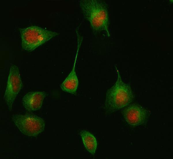
Anti-HDAC8 IHC data show human lung cancer paraffin sections (PA1591 image caption); IF data show HeLa cells (M01843-1 image caption). Both antibodies list Human, Mouse, Rat reactivity (catalog).

Real IHC data Anti-HDAC8 antibody, PA1591, IHC(P) IHC(P): Human Lung Cancer Tissue
Anti-Histone deacetylase 8 HDAC8 Antibody ®
Cat # PA1591
Real IF data IF analysis of HDAC8 using anti-HDAC8 antibody (M01843-1) and anti-Beta Tubulin antibody (M01857-3). HDAC8 was detected in immunocytochemical section of HELA cell. Enzyme antigen retrieval was performed using IHC enzyme antigen retrieval reagent (AR0022) for 15 mins. The cells were blocked with 10% goat serum. And then incubated at 1:50 with rabbit anti-HDAC8 Antibody (M01843-1) and mouse anti-Beta Tubulin antibody (M01857-3) overnight at 4°C. Cy3 Conjugated Goat Anti-Rabbit IgG (BA1032) and DyLight®488 Conjugated Goat Anti-Mouse IgG (BA1126) were used as secondary antibody at 1:500 dilution and incubated for 30 minutes at 37°C. Visualize using a fluorescence microscope and filter sets appropriate for the label used.
Anti-HDAC8/Histone Deacetylase 8 Rabbit Monoclonal Antibody
Cat # M01843-1

PA1591 has an IHC image of human lung cancer tissue in paraffin sections and lists Human, Mouse, Rat reactivity (PA1591 image caption; catalog). M01843-1 has an IF/ICC image of HeLa cells and lists Human, Mouse, Rat reactivity (M01843-1 image caption; catalog).

Which to pick: Choose PA1591 for tissue IHC: its own image documents paraffin sections of human lung cancer tissue, with an IHC dilution of 0.5–1 μg/ml; the fixative is unreported (PA1591 image caption; catalog). Choose rabbit monoclonal M01843-1 for IF/ICC in cells: its own image shows HeLa cells, and its listed IF dilution is 1:50 (M01843-1 image caption; catalog). For mouse or rat samples, both list reactivity; choose by application, since PA1591 lists IHC and M01843-1 lists IF/ICC (catalog: PA1591; catalog: M01843-1).

Each figure is that product's own IHC / IF validation image from its datasheet.

References

  1. UniProt Consortium. UniProt entry Q9BY41 (HDAC8_HUMAN, Histone deacetylase 8).
  2. Human Protein Atlas. HDAC8 tissue IHC expression (reliability: Supported).
  3. Human Protein Atlas. HDAC8 subcellular location (ICC-IF): Highest expression in NB4: 42.0 nTPM.
  4. Human Protein Atlas. HDAC8 antibody validation summary (1 antibodies).
  5. Over-expression of HDAC8 down-regulate CDKN2A is associated with worse prognosis of esophageal squamous cell carcinoma. Translational cancer research 2020 — PMC8798431.
  6. Chronic high-fat diet decreases global histone H4 acetylation and increases HDAC8 expression in mouse testes. Biochemistry and biophysics reports 2026 — PMC13214310.
  7. Anti-tumour activity of Panobinostat in oesophageal adenocarcinoma and squamous cell carcinoma cell lines. Clinical epigenetics 2024 — PMC11297794.
  8. HDAC8, A Potential Therapeutic Target, Regulates Proliferation and Differentiation of Bone Marrow Stromal Cells in Fibrous Dysplasia. Stem cells translational medicine 2019 — PMC6344909.
  9. PubMed PMID:10748112 — UniProt-cited evidence.
  10. PubMed PMID:10926844 — UniProt-cited evidence.
  11. PubMed PMID:10922473 — UniProt-cited evidence.