This website uses cookies to ensure you get the best experience on our website.


- Table of Contents
Plan HDGF paraffin IHC around distinct nuclear staining across tissues (HPA tissue IHC). This guide covers fixation, the catalog antibody’s 0.5–1 μg/mL IHC range (datasheet A01057), chromogenic detection, and nuclear scoring.

Expected localisation, validated protocols, controls and antibodies — the at-a-glance facts below, then the full design guide.
| Expected localisation | Nuclear staining in tissues (HPA tissue IHC) | |
| Staining pattern | Distinct nuclear staining across tissues (HPA tissue IHC) | |
| Antigen retrieval | Citrate pH 6 HIER, heat-mediated (datasheet A01057) | |
| Positive control | Adrenal gland+4 more · see all | |
| Negative control | None in HPA (detected in all 45 tissues); use no-primary + isotype controls |
| Fixation | Keep fixation consistent across sections (standard IHC practice; not target-specific) | |
| Caveat | Secretion may separate protein staining from tissue RNA (HPA tissue IHC) | |
| Regulation | No staining regulator specified (UniProt) | |
| Isoform / epitope | 3 isoforms; epitope differences unspecified (UniProt) |
The catalog antibody’s IHC protocol is followed by published HDGF staining protocols for synovial tissue, cervical adenocarcinoma, and endometrial carcinoma (PMC9169164; PMC4264238; PMC3947826).
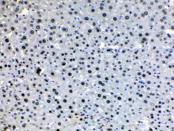
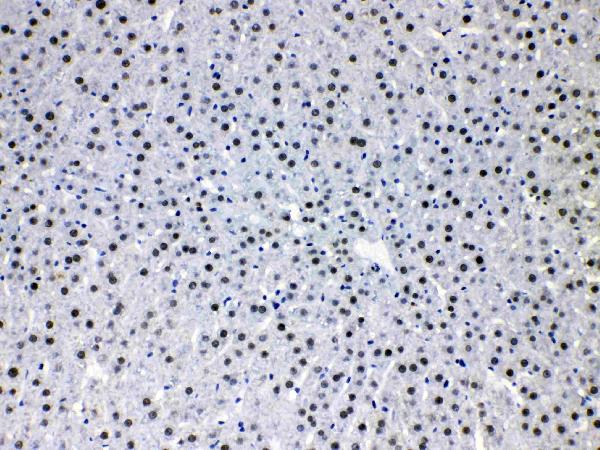
| Sample | Paraffin-embedded mouse liver tissues; fixative not specified (datasheet A01057) |
| Fixation | Image fixative and duration unreported (datasheet A01057); verify before use. |
| Sectioning | 4–5 µm sections on charged slides (standard) |
| Deparaffinisation | Xylene, graded ethanol series to water (standard) |
| Antigen retrieval | Heat retrieval: Citrate pH 6, 20 min (datasheet A01057) |
| Peroxidase block | 3% H2O2, 10 min, room temperature (standard) |
| Blocking | 10% goat serum (datasheet A01057) |
| Primary antibody | Rabbit anti-HDGF, 0.5-1μg/ml (datasheet A01057) |
| Primary incubation | Overnight at 4 °C (datasheet A01057) |
| Detection | Streptavidin-biotin complex (SABC), DAB chromogen (datasheet A01057) |
| Counterstain | Hematoxylin, blue, dehydrate and mount (standard) |
| Expected result | HDGF-positive staining in glandular cells of adrenal gland (HPA tissue IHC: High). HPA tissue profile: Distinct nuclear expression in all tissues. No signal in the no-primary control. |
HDGF should show distinct nuclear staining across tissues in paraffin-section IHC (HPA tissue IHC: Enhanced; distinct nuclear expression in all tissues). High staining is reported in specified glandular, hematopoietic, respiratory epithelial, granular-layer, and endothelial cells (HPA tissue IHC: High in listed cells). Nucleoplasm is the main ICC-IF location (HPA subcellular: enhanced). HDGF also has cytoplasmic and secreted locations and no transmembrane segment (UniProt P51858).
| Distinct nuclear signal in the expected cells, with interpretable counterstained nuclei. | This matches the reported IHC pattern (HPA tissue IHC: distinct nuclear expression in all tissues). High signal is documented in breast glandular cells and bone marrow hematopoietic cells, among other listed examples (HPA tissue IHC: High). Compare cells within the same section before assigning an intensity score. |
| Predominantly diffuse cytoplasmic signal, with little or no nuclear definition. | Question an IHC result that lacks the reported distinct nuclear pattern (HPA tissue IHC: nuclear expression). Cytoplasmic HDGF is biologically possible (UniProt P51858: cytoplasm), so compartment alone cannot prove artefact; assess whether the pattern persists with an independently validated antibody (HPA antibodies: 2 IHC Enhanced antibodies). |
| Strong staining in an unexpected cell population while the documented positive population is weak. | Consider cross-reactivity or endogenous chromogen-generating activity before calling that population HDGF positive (general IHC practice). For example, colon endothelial cells are documented as High (HPA tissue IHC: colon endothelial cells); staining elsewhere in colon needs its own validation. Broad tissue expression does not establish every cell type as High (UniProt P51858: ubiquitous; HPA tissue IHC: selected High cells). |
| Uniform haze, precipitate, or staining across tissue structures without clear nuclear contrast. | Treat this as background until controls and morphology support a cell-specific result (general IHC practice). Check blocking, washes, chromogen development, and a no-primary control (general IHC practice); the reported HDGF IHC pattern has distinct nuclei (HPA tissue IHC: nuclear expression). |
| No nuclear signal in a section expected to contain a documented High cell population. | This is an assay warning, not evidence that HDGF is absent (HPA tissue IHC: High in listed cells). First confirm that the relevant cells are present, then review antibody dilution, retrieval, detection, and control performance (general IHC practice). The supplied sources do not report HDGF-specific fixation sensitivity. |
| Antibody evidence | HPA048728 and HPA053422 are IHC Enhanced; CAB026035 is IHC Supported (HPA antibodies). An independently validated antibody can help resolve a disputed pattern (general IHC practice). |
| Antigen retrieval | Record and optimize the retrieval condition for the chosen IHC-validated antibody (general IHC practice). Neither HPA tissue IHC nor UniProt P51858 supplies an HDGF-specific retrieval or fixation effect. |
| Isoforms and epitope | UniProt lists 3 HDGF isoforms and a PWWP domain at residues 12–69 (UniProt P51858). The supplied evidence does not identify antibody epitopes or establish isoform-specific staining. |
| Intracellular versus secreted protein | Nuclear IHC is the documented tissue pattern (HPA tissue IHC). HDGF can also occur in cytoplasm and inside secreted exosomes, and may be released as free protein (UniProt P51858); extracellular staining alone needs corroboration. |
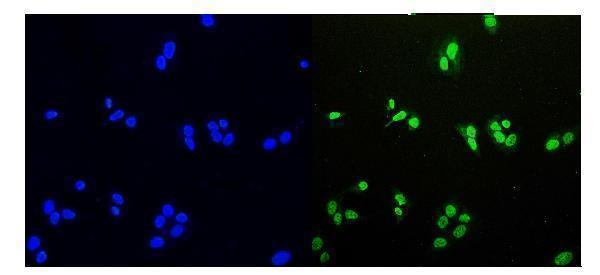
| IF/ICC: where should signal appear? | Primarily in the nucleoplasm (HPA subcellular: enhanced); HPA reports ICC Enhanced for HPA048728 and HPA053422 (HPA antibodies). This localisation cross-check does not define an IF/ICC protocol. |
| Situation | Likely cause | Next action |
|---|---|---|
| Known-positive cells have no nuclear signal. | The expected cells may be absent, or the IHC run may have failed (HPA tissue IHC: listed High cells; general IHC practice). | Confirm the cell population on the section; check a positive control, antibody dilution, retrieval, and detection in that order (general IHC practice). Do not infer HDGF-specific fixation loss from this result. |
| Nuclei stain weakly or unevenly across a positive section. | Section handling, retrieval, reagent coverage, or development may vary across the slide (general IHC practice). | Compare staining with tissue morphology and a control section; repeat with consistent section coverage and the antibody's validated IHC conditions (general IHC practice). |
| Diffuse chromogen obscures nuclear boundaries. | Background from insufficient washing, blocking, or excess antibody or chromogen is possible (general IHC practice). | Run a no-primary control, improve washing or blocking, and titrate antibody or development as needed (general IHC practice). Score only distinguishable cellular staining. |
| Signal is chiefly cytoplasmic or extracellular. | These locations are biologically plausible (UniProt P51858: cytoplasm; exosomal secretion), but differ from the distinct nuclear tissue-IHC profile (HPA tissue IHC). | Check nuclear counterstain and controls, then compare with an independent IHC Enhanced antibody if available (HPA antibodies: HPA048728, HPA053422; general IHC practice). |
| A different cell population stains more strongly than the documented High cells. | Cross-reactivity or endogenous detection activity is possible (general IHC practice); HPA's listed High cells do not validate every stained population (HPA tissue IHC). | Identify cells by morphology, inspect a no-primary control, and seek agreement with another validated antibody before assigning HDGF expression (general IHC practice). |
| IHC appears nuclear, but a companion IF/ICC image appears diffuse. | HPA reports enhanced nucleoplasmic ICC-IF localisation (HPA subcellular); diffuse fluorescence can arise from background or image settings (general IF practice). | Review IF controls, nuclear counterstain, exposure, and cell boundaries (general IF practice). Interpret each assay with its own controls; no IF/ICC protocol is specified here. |
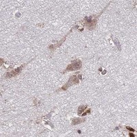
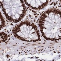
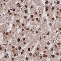
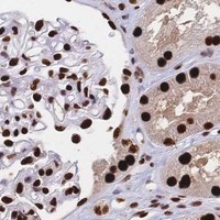


Comprehensive Human Protein Atlas IHC scoring per tissue (reliability: Enhanced — High consistency between antibody staining and RNA expression data. At least one protein variant secreted, tissue location of RNA and protein might differ and correlation is complex.). Rows are taken directly from the HPA tissue chart — click any row's HPA link to view the source.
| Tissue | Cell type | Level | Evidence | Source |
|---|---|---|---|---|
| Adrenal gland | Glandular cells | High | Protein (IHC) | HPA → |
| Appendix | Glandular cells | High | Protein (IHC) | HPA → |
| Bone marrow | Hematopoietic cells | High | Protein (IHC) | HPA → |
| Breast | Glandular cells | High | Protein (IHC) | HPA → |
| Bronchus | Respiratory epithelial cells | High | Protein (IHC) | HPA → |
| Tissue | Cell type | Level | Evidence | Source |
|---|---|---|---|---|
| None in HPA: HDGF is detected in all 45 scored tissues. Use a no-primary (secondary-only) and an isotype control instead. | ||||
Use nuclear staining as the main reference for HDGF in chromogenic paraffin-section IHC, while evaluating cytoplasmic or extracellular signal separately (HPA tissue IHC; UniProt P51858).
A01057 has IHC images from human, mouse and rat tissues and an IF image from U20S cells; M01057-1 is listed for human and mouse IHC/IF (catalog: image captions, applications, reactivity).
A01057 has paraffin-section IHC images from mouse and rat liver and human intestinal and lung cancer tissues, plus an IF image from U20S cells (catalog: image captions). M01057-1 is listed for human and mouse IHC and IF/ICC, but has no supplied IHC or IF image (catalog: applications, reactivity, image captions).
Which to pick: For tissue IHC, choose A01057 when a matching paraffin-section example matters; its caption documents citrate retrieval at pH 6 for 20 minutes and primary antibody at 1 μg/ml overnight at 4°C (A01057: IHC captions). For IF/ICC, A01057 has a U20S cell IF example at 2 μg/ml, while M01057-1 offers a rabbit monoclonal option, clone 26H94, listed for IF/ICC without a supplied image (A01057: IF caption; M01057-1: catalog applications and clone). For cross-species tissue work, A01057 covers human, mouse and rat; M01057-1 lists human and mouse, with no supplied IHC processing example, and the fixative is unreported in A01057’s paraffin-section captions (catalog: reactivity and IHC captions).