HERPUD1 / Homocysteine-responsive endoplasmic reticulum-resident ubiquitin-like domain member 1 protein · IHC design guide

Design Immunohistochemistry for HERPUD1

Plan HERPUD1 chromogenic IHC in paraffin sections using parathyroid glandular cells as a positive reference (HPA tissue IHC). Assess cytoplasmic staining (HPA tissue IHC) against the protein’s ER membrane location (UniProt), and compare with unstained lymph-node germinal center cells (HPA tissue IHC).

Evidence assembled Oct 2026 · For research use; verify linked source records and product datasheet before use
Immunohistochemistry protocol sheet for HERPUD1 (IHC for HERPUD1): expected localisation Cytoplasmic staining (HPA tissue IHC); ER membrane (UniProt), antibody A04908-2, validated IHC image, and IHC protocol steps
Printable HERPUD1 IHC protocol sheet — expected localisation Cytoplasmic staining (HPA tissue IHC); ER membrane (UniProt), antibody A04908-2, controls and protocol steps. Open the full HERPUD1 IHC guide →

HERPUD1 Immunohistochemistry Experimental Design Guide

Expected localisation, validated protocols, controls and antibodies — the at-a-glance facts below, then the full design guide.

Must know before staining
Expected localisation Cytoplasmic staining (HPA tissue IHC); ER membrane (UniProt)
Staining pattern Widespread cytoplasmic staining across cell types (HPA tissue IHC)
Antigen retrieval EDTA pH 8.0 HIER, heat-mediated (datasheet A04908-2)
Positive control ⓘ Parathyroid gland+4 more · see all
Negative control ⓘ Lymph node+2 more · see all
Important caveats
Reasons your staining may differ from the expected pattern.
Fixation Keep paraffin-section fixation consistent across samples. (standard IHC practice; not target-specific)
Caveat Staining has only medium consistency with RNA (HPA tissue IHC)
Regulation Homocysteine-responsive (UniProt)
Isoform / epitope 4 isoforms; map epitopes to cytoplasmic or ER-lumenal regions (UniProt)
Section 1

Recommended HERPUD1 IHC & IF Protocols

The catalog antibody uses EDTA pH 8.0 heat retrieval (datasheet A04908-2). Four published HERPUD1 IHC methods provide tissue-specific examples (PMC12589607; PMC5645377; PMC9769045; PMC12079719).

Recommended immunohistochemistry (IHC-P) protocol parameters
SampleParaffin-embedded human appendiceal carcinoid tissue; fixative not specified (datasheet A04908-2)
FixationImage fixative and duration unreported (datasheet A04908-2); verify before use.
Sectioning4–5 µm sections on charged slides (standard)
DeparaffinisationXylene, graded ethanol series to water (standard)
Antigen retrievalHeat retrieval: EDTA pH 8.0 (datasheet A04908-2); 20 min, 95–100 °C (standard)
Peroxidase block3% H2O2, 10 min, room temperature (standard)
Blocking10% goat serum (datasheet A04908-2)
Primary antibodyRabbit anti-HERPUD1, 2-5 μg/ml (datasheet A04908-2)
Primary incubationOvernight at 4 °C (datasheet A04908-2)
DetectionHRP-conjugated secondary, DAB chromogen (datasheet A04908-2)
CounterstainHematoxylin, blue, dehydrate and mount (standard)
Expected resultHERPUD1-positive staining in glandular cells of parathyroid gland (HPA tissue IHC: High). HPA tissue profile: Ubiquitous cytoplasmic expression. No signal in the no-primary control.
💡Decision noteStart with EDTA pH 8.0 heat retrieval for the catalog antibody (datasheet A04908-2). Use an article’s reported retrieval conditions when reproducing its method (PMC12589607; PMC9769045; PMC12079719).
Section 2

What Is the Expected HERPUD1 Staining Pattern?

HERPUD1 is an endoplasmic reticulum (ER) membrane protein with 2 transmembrane segments (UniProt Q15011 topology). In tissue IHC, expect predominantly cytoplasmic staining, with High signal in parathyroid and thyroid glandular cells (HPA tissue IHC). HPA describes the overall pattern as ubiquitous cytoplasmic expression, but rates its tissue IHC profile Approved with medium consistency against RNA data (HPA tissue IHC).

What am I looking at on my slide?
Cytoplasmic signal is strong in parathyroid or thyroid glandular cells.This matches the strongest listed tissue IHC observations (HPA: High in parathyroid and thyroid glandular cells). A cytoplasmic pattern fits HERPUD1's ER membrane location (UniProt Q15011 subcellular location); chromogenic IHC alone does not prove that every stained structure is ER.
Signal is exclusively nuclear, with little or no cytoplasmic staining.Treat a nuclear-only pattern as suspect: UniProt places HERPUD1 in the ER membrane, and HPA reports cytoplasmic tissue staining (UniProt Q15011 subcellular location; HPA tissue IHC). Review morphology, counterstain, and detection controls before scoring it as HERPUD1 (general IHC practice).
Strong staining appears in germinal center cells or oral squamous epithelial cells.These listed cell populations were Not detected by HPA; unexpected strong signal could reflect cross-reactivity or endogenous detection activity (HPA tissue IHC; general IHC practice). Assess the particular cells stained rather than labeling an entire lymph node or oral mucosa section negative.
Chromogen covers many structures without a discernible cellular pattern.Diffuse background cannot support a HERPUD1 localisation call (general IHC practice). Check whether a no-primary control shows similar color, then review blocking, washes, detection reagent, and counterstain intensity (general IHC practice). HPA's ubiquitous cytoplasmic profile does not justify featureless staining (HPA tissue IHC).
Parathyroid or thyroid glandular cells have no detectable cytoplasmic signal.That result conflicts with the listed High observations (HPA tissue IHC). First check tissue preservation and assay controls; then review the antibody's IHC validation and whether the antigen retrieval and dilution used produce interpretable staining (general IHC practice). Do not infer HERPUD1-specific fixation sensitivity from this result.
💡Expected HERPUD1 appearanceCall a section positive when glandular cells show clear cytoplasmic staining, expected to be High in parathyroid or thyroid (HPA tissue IHC), consistent with ER membrane localisation (UniProt Q15011); nuclear-only color or strong staining in HPA Not detected cell populations warrants control review before interpretation (HPA tissue IHC; general IHC practice).
How each factor affects the staining
Cell population and tissue choiceUse parathyroid or thyroid glandular cells as strong observed reference populations (HPA: High). Adipocytes and several other listed populations are Medium, whereas hippocampal neurons are Low (HPA tissue IHC). A weak result in a Low population has less diagnostic value than one in a High population.
Subcellular topology and epitopeHERPUD1 has cytoplasmic regions at residues 1–263 and 311–391, a short lumenal region at 285–289, and 2 membrane spans (UniProt Q15011 topology). The supplied record gives no epitope for the assay antibody, so topology cannot establish its staining access or retrieval needs.
Isoforms and antibody validationUniProt lists 4 isoforms (UniProt Q15011 isoforms). The supplied HPA antibodies have Approved IHC status, while the tissue profile has medium consistency against RNA data (HPA antibodies; HPA tissue IHC). Neither record establishes which isoforms a particular antibody detects or makes every cellular signal specific.
Antigen retrieval and chromogenic detectionRetrieval conditions, blocking, and a no-primary control can help distinguish usable tissue staining from assay background (general IHC practice). The supplied UniProt and HPA records give no HERPUD1-specific retrieval requirement or fixation effect; assess these conditions with assay controls rather than predicting a target-specific response.
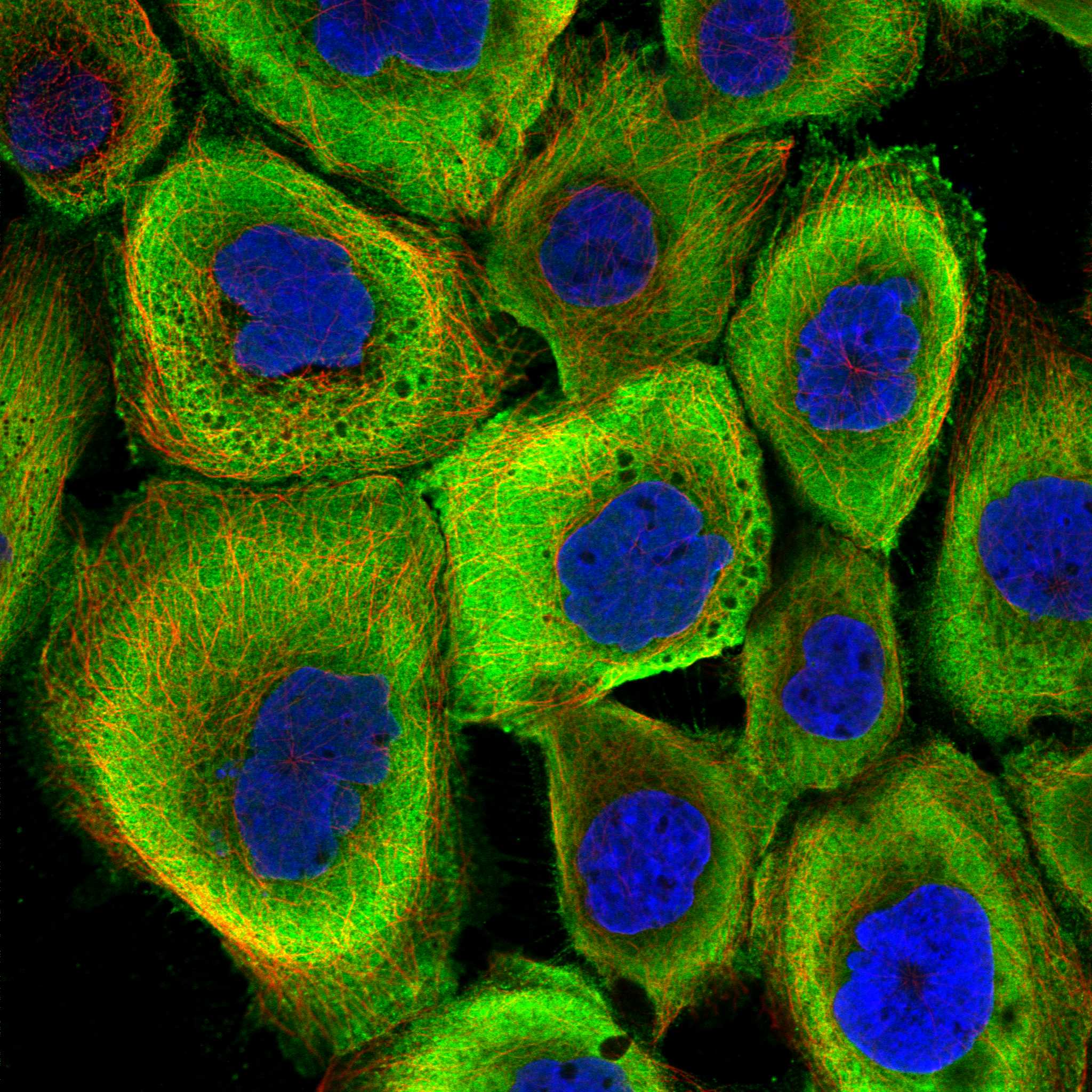
IF/ICC localisation Q&AWhat should IF/ICC show? HPA reports plasma membrane and cytosol localisation, both marked uncertain, in its ICC-IF summary (HPA subcellular). UniProt places HERPUD1 at the ER membrane (UniProt Q15011 subcellular location). Treat the uncertain ICC-IF annotation as context, not a substitute for interpreting the tissue IHC pattern.
Why is my staining missing, weak or wrong?
SituationLikely causeNext action
No signal in parathyroid or thyroid glandular cells.The result misses populations listed as High (HPA tissue IHC); the cause cannot be assigned from the supplied target records.Verify tissue and detection controls, then review retrieval, primary dilution, and chromogen development for the IHC assay (general IHC practice).
Only nuclei are strongly colored.A nuclear-only pattern conflicts with ER membrane localisation and HPA's cytoplasmic tissue profile (UniProt Q15011; HPA tissue IHC).Inspect counterstain and a no-primary control; confirm that cytoplasmic color is distinguishable before scoring cells (general IHC practice).
Germinal center cells stain strongly in lymph node.HPA lists these cells as Not detected; cross-reactivity or endogenous detection activity is possible (HPA tissue IHC; general IHC practice).Compare adjacent cell populations and a no-primary control, then reassess antibody specificity if the unexpected cellular pattern persists (general IHC practice).
Color is diffuse across the section.Nonspecific background or detection activity can obscure cellular localisation (general IHC practice).Check the no-primary control, blocking, washes, detection reagent, and chromogen development; score only a resolved cellular pattern (general IHC practice).
A Low population appears negative.Hippocampal neurons, skeletal muscle myocytes, and tonsil germinal center cells are listed as Low (HPA tissue IHC); their signal may be harder to judge.Compare with a listed High glandular population and assay controls before calling the stain unsuccessful (HPA tissue IHC; general IHC practice).
IF/ICC shows plasma membrane or cytosolic signal.HPA labels both ICC-IF locations uncertain, while UniProt assigns HERPUD1 to the ER membrane (HPA subcellular; UniProt Q15011).Interpret that image within its IF/ICC assay and controls; use the cytoplasmic tissue IHC observations when scoring paraffin sections (HPA tissue IHC; general IHC practice).

Sample controls for HERPUD1 IHC & IF

🧪Run parathyroid gland first; its glandular cells should show strong staining (HPA: parathyroid gland, glandular cells High). Use lymph node germinal center cells as the negative tissue (HPA: lymph node, germinal center cells Not detected); on the parathyroid slide, adjacent nonglandular cells can serve as an internal background reference only if they are visibly unstained, because HPA does not designate an internal-negative cell type (HPA: parathyroid gland, glandular cells High).
Positive control tissue: Parathyroid gland (Glandular cells, HPA High)
Negative control tissue: Lymph node (HPA Not detected)
ICC-IF cell lines (HPA subcellular resource): HPA ICC-IF images show HERPUD1 in A-431, U-251MG, U2OS, with annotated localisation: Plasma membrane (uncertain), Cytosol (uncertain) (HPA subcellular).
Technical controls: Include no-primary and secondary-only controls, plus nonimmune rabbit IgG matched to the primary antibody’s verified clonality (selected-SKU tissue-IHC caption: rabbit primary antibody; standard IHC practice). Confirm specificity with HERPUD1-knockout material or an immunizing-peptide block if available, and quench endogenous peroxidase before HRP/DAB detection in parathyroid sections (selected-SKU tissue-IHC caption: HRP/DAB; standard IHC practice).
⚠️Feasibility: A target-specific fixation window or fixation effect is unreported, and the selected paraffin-section caption does not state the fixative (selected-SKU tissue-IHC caption: fixative not stated). Heat retrieval in EDTA at pH 8.0 was used, but whether retrieval is required is unreported (selected-SKU tissue-IHC caption: EDTA retrieval). The evidence does not establish whether frozen sections or IF are easier; IF assessment of the ER membrane signal would require permeabilization, and no parathyroid-specific artefact is reported (UniProt Q15011: ER membrane; standard IF practice; supplied tissue-IHC caption).

HPA tissue IHC evidence for HERPUD1

Comprehensive Human Protein Atlas IHC scoring per tissue (reliability: Approved — Medium consistency between antibody staining and RNA expression data.). Rows are taken directly from the HPA tissue chart — click any row's HPA link to view the source.

Positive expression · recommended positive controls

TissueCell typeLevelEvidenceSource
Parathyroid gland Glandular cells High Protein (IHC) HPA →
Thyroid gland Glandular cells High Protein (IHC) HPA →
Adipose tissue Adipocytes Medium Protein (IHC) HPA →
Adrenal gland Glandular cells Medium Protein (IHC) HPA →
Appendix Glandular cells Medium Protein (IHC) HPA →

Undetected expression · recommended negative controls

TissueCell typeLevelEvidenceSource
Lymph node Germinal center cells Not detected Protein (IHC) HPA →
Oral mucosa Squamous epithelial cells Not detected Protein (IHC) HPA →
Spleen Cells in red pulp Not detected Protein (IHC) HPA →
Section 3

Advanced HERPUD1 IHC Tips

Troubleshoot HERPUD1 staining in paraffin sections by checking retrieval, compartment, controls and scoring before interpreting differences between samples.

Which retrieval condition should I try first for HERPUD1 IHC?
Start with heat-mediated antigen retrieval in EDTA at pH 8.0 for paraffin sections (datasheet A04908-2). The selected tissue image used this retrieval before 2 μg/ml primary antibody overnight at 4°C (datasheet A04908-2). If staining is weak, vary heating or cooling time one variable at a time while holding antibody concentration and detection chemistry constant (standard IHC practice). Compare signal and morphology in a matched positive section, such as appendix glandular cells, because stronger staining after prolonged heating is useful only if tissue structure remains interpretable (HPA: medium in appendix glandular cells; standard IHC practice).
Could fixation explain weak or uneven HERPUD1 staining?
The selected paraffin-section caption does not state a fixative, so HERPUD1-specific fixation sensitivity is unknown (datasheet A04908-2: fixative unreported). Record fixative, tissue thickness and time in fixative for each specimen, and compare matched sections processed through the same pH 8.0 EDTA retrieval (standard IHC practice; datasheet A04908-2). A pilot using 10% neutral buffered formalin and controlled fixation intervals can reveal handling-related differences without assuming a HERPUD1-specific effect (standard IHC practice). Interpret weak staining alongside tissue preservation and a concurrently processed positive control; neither staining patterns nor protein topology establishes which fixation condition preserves this antibody's epitope (standard IHC practice).
What staining pattern fits HERPUD1, and how should membrane staining be judged?
Expect predominantly cytoplasmic staining in tissue sections, consistent with ubiquitous cytoplasmic expression reported by HPA and the protein's endoplasmic reticulum membrane assignment (HPA: tissue profile; UniProt Q15011: subcellular location). Its transmembrane segments span residues 264–284 and 290–310, with most of the sequence facing the cytoplasm (UniProt Q15011: topology). A fine perinuclear or reticular pattern can therefore be assessed against an ER marker when resolution permits (UniProt Q15011: subcellular location; standard IHC practice). HPA also reports uncertain plasma membrane and cytosol locations in cell imaging, so treat an isolated sharp membrane rim as a finding requiring controls, not definitive HERPUD1 localisation (HPA: subcellular location).
Can an unknown epitope make a negative section misleading?
Yes: HERPUD1 has 4 reported isoforms, while the supplied tissue-IHC caption does not identify this antibody's epitope (UniProt Q15011: isoforms; datasheet A04908-2: caption). Before treating absence of stain as absence of protein, obtain the immunogen or mapped epitope and check whether it is shared by the isoforms under study (standard IHC practice; UniProt Q15011: isoforms). The protein has a ubiquitin-like region at residues 10–72, phosphorylation at residue 135, and two transmembrane segments, but these features do not locate this antibody's binding site (UniProt Q15011: domains, modified residues and topology). Compare retrieval conditions with matched controls rather than assigning weak staining to a particular isoform or modification without epitope evidence (standard IHC practice).
How should I assess HERPUD1 in a separate multiplex IF experiment?
For a separate IF/ICC experiment, pair HERPUD1 with an ER marker and a marker for the expected cell population, such as glandular cells in an appropriate tissue, then inspect both cellular identity and compartment (UniProt Q15011: ER membrane; HPA: glandular-cell staining; standard IF practice). Choose spectrally separated fluorophores and consider a far-red channel for the weaker signal when tissue autofluorescence overlaps shorter wavelengths (standard IF practice). Permeabilisation depends on the mapped epitope: residues 1–263 and 311–391 are cytoplasmic, whereas residues 285–289 face the ER lumen (UniProt Q15011: topology). Because no antibody epitope or IF fixation condition is supplied, optimise access and fixation with controls rather than importing conditions from the paraffin IHC image (datasheet A04908-2: caption; standard IF practice).
How can I separate HERPUD1 signal from chromogenic background?
The selected image used 10% goat serum blocking, a rabbit primary at 2 μg/ml, and peroxidase-based DAB detection (datasheet A04908-2). Include a no-primary section to assess secondary reagent, endogenous enzyme and chromogen background, and apply an appropriate peroxidase block as part of the chromogenic workflow (standard IHC practice). If diffuse DAB persists, compare matched sections while adjusting antibody concentration or wash stringency one variable at a time (standard IHC practice). Judge improvement in viable cell interiors rather than at folds, section edges or damaged regions, where deposits can obscure the cytoplasmic pattern expected for HERPUD1 (HPA: tissue profile; standard IHC practice).
How should HERPUD1 staining be scored across tissue samples? ⚠ ANSWER MARKED FOR VERIFICATION
Define the cell population and compartment before scoring; for example, assess cytoplasmic signal in glandular cells rather than pooling every stained structure (HPA: ubiquitous cytoplasmic expression; HPA: glandular-cell staining; standard IHC practice). An H-score combines the percentage of cells at each intensity and spans 0–300; alternatively, report percent positive cells or positive-cell density per mm² (standard IHC practice). Normalise counts to viable cells or evaluable tissue area within the same annotated compartment, and keep retrieval, imaging and threshold settings consistent across batches (standard IHC practice). Report the scoring rule and controls, because HPA rates its tissue staining as Approved with medium consistency against RNA data (HPA: reliability).
When is an apparent HERPUD1-positive cell likely to be artefactual?
Give greatest weight to reproducible cytoplasmic staining in viable cells, supported by the ER membrane assignment and the tissue-IHC profile (UniProt Q15011: subcellular location; HPA: ubiquitous cytoplasmic expression; standard IHC practice). Parathyroid and thyroid glandular cells are reported high, while lymph-node germinal center cells are reported undetected, providing context for matched control sections rather than absolute specificity tests (HPA: tissue staining). Be cautious with an isolated nuclear deposit or sharp membrane rim, especially when it tracks a section edge or necrotic area (UniProt Q15011: ER membrane; HPA: uncertain plasma membrane location; standard IHC practice). Check no-primary and peroxidase-block controls before attributing brown precipitate to HERPUD1, since endogenous enzyme activity can generate chromogenic signal (standard IHC practice).
Boster reagents

Best HERPUD1 / Homocysteine-responsive endoplasmic reticulum-resident ubiquitin-like domain member 1 protein IHC Antibodies

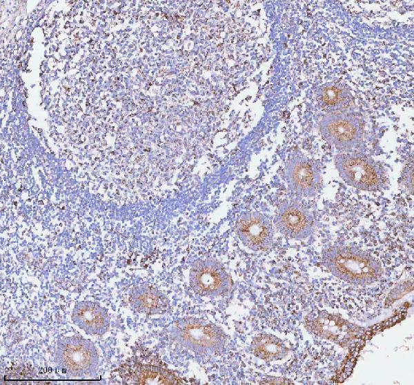
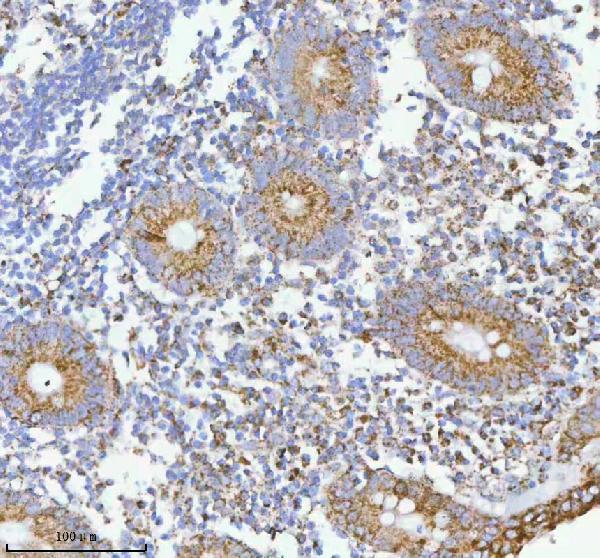
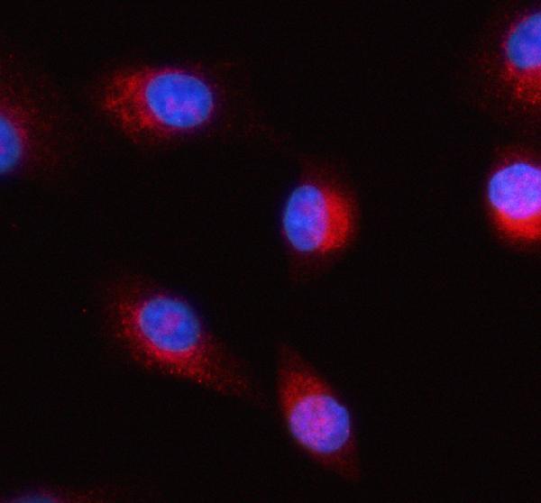
A04908-2 has IHC images from human paraffin sections and IF images from A549 cells and human paraffin sections (catalog image captions).

Real IHC data IHC analysis of HERP/HERPUD1 using anti-HERP/HERPUD1 antibody (A04908-2). HERP/HERPUD1 was detected in a paraffin-embedded section of human appendiceal carcinoid tissue. Heat mediated antigen retrieval was performed in EDTA buffer (pH 8.0, epitope retrieval solution). The tissue section was blocked with 10% goat serum. The tissue section was then incubated with 2 μg/ml rabbit anti-HERP/HERPUD1 Antibody (A04908-2) overnight at 4°C. Peroxidase Conjugated Goat Anti-rabbit IgG was used as secondary antibody and incubated for 30 minutes at 37°C. The tissue section was developed using HRP Conjugated Rabbit IgG Super Vision Assay Kit (Catalog # SV0002) with DAB as the chromogen.
Anti-HERP/HERPUD1 Antibody ®
Cat # A04908-2

A04908-2 has IHC data from human appendiceal carcinoid and breast cancer paraffin sections (catalog IHC image captions). A04908-2 also has IF data from A549 cells and human appendix and ovarian paraffin sections (catalog IF image captions).

Which to pick: For tissue IHC, choose A04908-2; its paraffin-section captions document EDTA retrieval at pH 8.0 and 2 μg/ml primary antibody, while the fixative is unreported (catalog IHC image captions). For IF/ICC, A04908-2 lists both applications and has cell and tissue IF images (catalog applications and IF image captions). For cross-species work, A04908-2 lists human, mouse and rat reactivity, although the supplied IHC and IF images show human samples only (catalog reactivity and image captions).

Each figure is that product's own IHC / IF validation image from its datasheet.

References

  1. UniProt Consortium. UniProt entry Q15011 (HERP1_HUMAN, Homocysteine-responsive endoplasmic reticulum-resident ubiquitin-like domain member 1 protein).
  2. Human Protein Atlas. HERPUD1 tissue IHC expression (reliability: Approved).
  3. Human Protein Atlas. HERPUD1 subcellular location (ICC-IF): Localized to the plasma membrane and cytosol..
  4. Human Protein Atlas. HERPUD1 antibody validation summary (4 antibodies).
  5. HERPUD1 mediates palmitic acid-induced UPR sustaining TNBC aggressiveness and is destabilized by CK2 pharmacological inhibition. Cell death & disease 2025 — PMC12589607.
  6. Herpud1 negatively regulates pathological cardiac hypertrophy by inducing IP3 receptor degradation. Scientific reports 2017 — PMC5645377.
  7. HERPUD1 promotes ovarian cancer cell survival by sustaining autophagy and inhibit apoptosis via PI3K/AKT/mTOR and p38 MAPK signaling pathways. BMC cancer 2022 — PMC9769045.
  8. Deciphering the Molecular Crosstalk of Endoplasmic Reticulum Stress and Immune Infiltration in Endometriosis. American journal of reproductive immunology (New York, N.Y. : 1989) 2025 — PMC12079719.
  9. PubMed PMID:10922362 — UniProt-cited evidence.
  10. PubMed PMID:7584026 — UniProt-cited evidence.
  11. PubMed PMID:11112790 — UniProt-cited evidence.