HEXA / Beta-hexosaminidase subunit alpha · IHC design guide

Design Immunohistochemistry for HEXA

Plan HEXA IHC in paraffin sections using lung macrophages and gallbladder glandular cells as positive staining references (HPA tissue IHC). The guide covers fixation, localisation and the catalog antibody’s 0.5–1 μg/mL IHC dilution (datasheet: PA1787).

Evidence assembled Oct 2026 · For research use; verify linked source records and product datasheet before use
Immunohistochemistry protocol sheet for HEXA (IHC for HEXA): expected localisation Lysosomal tissue staining is reported (HPA tissue IHC)., antibody PA1787, validated IHC image, and IHC protocol steps
Printable HEXA IHC protocol sheet — expected localisation Lysosomal tissue staining is reported (HPA tissue IHC)., antibody PA1787, controls and protocol steps. Open the full HEXA IHC guide →

HEXA Immunohistochemistry Experimental Design Guide

Expected localisation, validated protocols, controls and antibodies — the at-a-glance facts below, then the full design guide.

Must know before staining
Expected localisation Lysosomal tissue staining is reported (HPA tissue IHC).
Staining pattern Lysosomal staining in lung macrophages and gallbladder glandular cells (HPA tissue IHC).
Antigen retrieval Citrate pH 6.0 HIER, 95–98 °C, 20 min (rule: cytoplasmic / membrane antigen)
Positive control ⓘ Gallbladder+4 more · see all
Negative control ⓘ Bone marrow+4 more · see all
Important caveats
Reasons your staining may differ from the expected pattern.
Fixation Matched tissue-IHC evidence does not establish the fixation claim. Validate the specimen-specific method before use. (selected-SKU IHC image PA1787)
Caveat Antibody staining has low concordance with RNA expression (HPA tissue IHC).
Regulation No expression regulator is annotated (UniProt).
Isoform / epitope 2 isoforms; mature chain spans residues 89–529; epitope coverage is unknown (UniProt).
Section 1

Recommended HEXA IHC & IF Protocols

The catalog antibody’s IHC-P protocol is paired with one published HEXA paraffin-section protocol (PMC8371334).

Recommended immunohistochemistry (IHC-P) protocol parameters
SampleTissue sections; selected-image fixative not specified (standard IHC workflow)
FixationImage fixative and duration unreported (datasheet PA1787); verify before use.
Sectioning4–5 µm sections on charged slides (standard)
DeparaffinisationXylene, graded ethanol series to water (standard)
Antigen retrievalHeat-induced epitope retrieval in citrate buffer, pH 6.0, 20 min at 95–98 °C (standard rule: cytoplasmic / membrane antigen)
Peroxidase block3% H2O2, 10 min, room temperature (standard)
Blocking10% normal serum of the secondary host, 30 min, room temperature (standard)
Primary antibodyRabbit anti-HEXA, 0.5-1μg/ml (datasheet PA1787)
Primary incubationOvernight at 4 °C (standard)
DetectionHRP-polymer secondary, DAB chromogen 5–10 min (standard)
CounterstainHematoxylin, blue, dehydrate and mount (standard)
Expected resultHEXA-positive staining in glandular cells of gallbladder (HPA tissue IHC: High). HPA tissue profile: Lysosomal expression in several tissues, most abundant in lung and gallbladder. No signal in the no-primary control.
💡Decision noteStart with citrate pH 6.0 HIER at 95–98 °C for 20 min (page retrieval rule); the published protocol uses microwave retrieval (PMC8371334).
Section 2

What Is the Expected HEXA Staining Pattern?

HEXA is a lysosomal protein with no transmembrane segment (UniProt P06865 localization and topology). In paraffin-section IHC, expect intracellular staining in gallbladder glandular cells, kidney tubular cells and lung macrophages, each reported as High (HPA tissue IHC). HPA rates its tissue staining Supported but reports low consistency between antibody staining and RNA expression; use the listed patterns as guides, not absolute rules (HPA tissue IHC).

What am I looking at on my slide?
Granular cytoplasmic staining in gallbladder glandular cells, kidney tubular cells or lung macrophages.This fits lysosomal HEXA localization and the High staining reported in these cell types (UniProt P06865 localization; HPA tissue IHC). Assess granules within identifiable cells; staining across an entire tissue should not be scored as though every cell type were equally positive (general IHC practice).
Predominantly nuclear, crisp plasma-membrane or uniform extracellular staining.These patterns do not fit the reported lysosomal location or absence of a transmembrane segment (UniProt P06865 localization and topology). Treat them as possible artefact until controls and a second staining condition clarify the signal (general IHC practice).
Strong signal in a cell type HPA lists as Not detected, such as bone-marrow hematopoietic cells.HPA reports that specific cell population as Not detected; the label does not describe every cell in bone marrow (HPA tissue IHC). Check antibody specificity and, for enzyme-based detection, endogenous activity before assigning the signal to HEXA (general IHC practice).
Diffuse chromogen over cells, stroma or section edges, with little cellular detail.A diffuse field cannot establish the expected lysosomal, cell-associated pattern (UniProt P06865 localization; general IHC practice). Review background in a control without primary antibody, washing and detection conditions before scoring cells (general IHC practice).
No staining in a well-preserved gallbladder glandular-cell or lung-macrophage control.Both are reported High in the specified cells, so an absent signal calls for a technical check (HPA tissue IHC). Confirm that the expected cells are present, then review antibody suitability, retrieval and detection using the applicable antibody instructions (general IHC practice).
💡Expected HEXA appearanceCall a section positive when the relevant cells show distinct granular cytoplasmic staining, potentially strong in HPA High populations; diffuse tissue-wide, predominantly nuclear or crisp membrane signal warrants review (UniProt P06865 localization and topology; HPA tissue IHC; general IHC practice).
How each factor affects the staining
Cell population and tissueHPA reports High staining in gallbladder and prostate glandular cells, kidney tubular cells, lung macrophages and testis Leydig cells; adipocytes and several other listed populations are Medium or Low (HPA tissue IHC). Compare like cells when judging intensity (general IHC practice).
Negative-reference choiceHPA lists bone-marrow hematopoietic cells, cerebellar granular-layer cells and esophageal squamous epithelial cells as Not detected, among others (HPA tissue IHC). These are cell-specific observations, not guarantees that the whole tissue is blank (HPA tissue IHC).
Localization and topologyUniProt assigns HEXA to lysosomes and records no transmembrane segment (UniProt P06865 localization and topology). These annotations support intracellular granular interpretation; they do not predict staining strength in a particular section (UniProt P06865; general IHC practice).
Processing and antibody epitopeThe precursor has a signal peptide at residues 1–22, a propeptide at 23–88 and a chain at 89–529 (UniProt P06865 processing). Without the antibody's mapped epitope, the effect of processing on its IHC signal cannot be predicted.
Glycosylation and isoformsUniProt lists glycosylation sites at residues 115, 157 and 295 and two isoforms (UniProt P06865 PTMs and isoforms). These facts alone do not show that glycosylation or isoform choice changes staining with the selected antibody.
Evidence strength and antibody choiceHPA calls tissue IHC Supported while noting low staining–RNA consistency; it lists HPA054583 as IHC Supported (HPA tissue IHC; HPA antibodies). This supports a reference pattern but does not validate a different catalog antibody under the same conditions.
IF/ICC: What localization is reported?HPA reports mainly vesicular localization, with additional cytosolic localization marked uncertain; HPA018082 is ICC Supported (HPA subcellular; HPA antibodies). Use the separate IF/ICC guide for that application.
Why is my staining missing, weak or wrong?
SituationLikely causeNext action
The positive control is blank.The expected High cell population may be absent from the section, or the IHC workflow may have failed (HPA tissue IHC; general IHC practice).Locate gallbladder glandular cells, kidney tubular cells or lung macrophages first; then check the catalog antibody's IHC-P instructions, retrieval and detection controls (HPA tissue IHC; general IHC practice).
Everything stains brown, including the control without primary antibody.Signal persisting without primary antibody points to detection background or endogenous enzyme activity rather than demonstrated HEXA binding (general IHC practice).Check the detection reagent, enzyme blocking where applicable, washes and counterstain; compare with the control before changing the interpretation (general IHC practice).
Staining is nuclear or sharply outlines cell membranes.That distribution conflicts with lysosomal localization and the absence of a transmembrane segment (UniProt P06865 localization and topology).Review control sections and antibody specificity; score HEXA only where the cell-associated pattern is credible under the staining conditions (general IHC practice).
A supposed negative tissue contains isolated positive cells.HPA Not detected labels apply to named cells; other cells in the section may differ (HPA tissue IHC).Identify the stained cell type before calling the control failed; compare it with HPA's cell-specific annotation and an appropriate control without primary antibody (HPA tissue IHC; general IHC practice).
Staining appears diffuse and obscures granules.Excess background or an unsuitable detection condition can obscure cellular detail (general IHC practice).Review washes, antibody concentration and chromogen development against the antibody instructions; reassess whether intracellular granules can be resolved (general IHC practice).
Observed tissue intensity disagrees with the HPA ranking.HPA describes low consistency between antibody staining and RNA expression, and its staining levels are observations from its own material (HPA tissue IHC).Compare the same cell population across controls and document the antibody and staining conditions; avoid inferring a HEXA-specific fixation effect from the discrepancy (HPA tissue IHC; general IHC practice).

Sample controls for HEXA IHC & IF

🧪Run lung first and look for staining in macrophages (HPA: High in lung macrophages). Use esophageal squamous epithelial cells as the negative tissue (HPA: Not detected); on the lung slide, use adjacent nonmacrophage cells with only counterstain and background signal as candidate internal negative comparators, after confirming their identity.
Positive control tissue: Gallbladder (Glandular cells, HPA High)
Negative control tissue: Bone marrow (HPA Not detected)
ICC-IF cell lines (HPA subcellular resource): HPA ICC-IF images show HEXA in SK-MEL-30, SiHa, U2OS, KOLF2.1J, with annotated localisation: Vesicles (supported) (HPA subcellular).
Technical controls: Include a no-primary, secondary-only control; an isotype-matched control for a monoclonal primary or host-species-matched normal IgG for a polyclonal primary; and HEXA-knockout material as a biological negative. For lung IHC, block endogenous peroxidase and inspect macrophage pigment before interpreting chromogenic staining (standard IHC practice).
⚠️Feasibility: A target-specific fixation window and retrieval dependency are unreported in the supplied evidence; optimize antigen retrieval on the positive tissue (HPA: High in lung macrophages). The selected PA1787 paraffin-section caption does not state a fixative (caption: fixative not stated), and the evidence does not establish whether frozen sections or IF would be easier. Lung macrophage pigment can complicate chromogenic interpretation (standard IHC practice).

HPA tissue IHC evidence for HEXA

Comprehensive Human Protein Atlas IHC scoring per tissue (reliability: Supported — Low consistency between antibody staining and RNA expression data.). Rows are taken directly from the HPA tissue chart — click any row's HPA link to view the source.

Positive expression · recommended positive controls

TissueCell typeLevelEvidenceSource
Gallbladder Glandular cells High Protein (IHC) HPA →
Kidney Cells in tubules High Protein (IHC) HPA →
Lung Macrophages High Protein (IHC) HPA →
Prostate Glandular cells High Protein (IHC) HPA →
Testis Leydig cells High Protein (IHC) HPA →

Undetected expression · recommended negative controls

TissueCell typeLevelEvidenceSource
Bone marrow Hematopoietic cells Not detected Protein (IHC) HPA →
Cerebellum Cells in granular layer Not detected Protein (IHC) HPA →
Cervix Glandular cells Not detected Protein (IHC) HPA →
Esophagus Squamous epithelial cells Not detected Protein (IHC) HPA →
Hippocampus Glial cells Not detected Protein (IHC) HPA →
Section 3

Advanced HEXA IHC Tips

Troubleshoot HEXA staining in paraffin sections by checking retrieval, cytoplasmic localisation, cell identity and controls before interpreting signal intensity.

How should I adjust retrieval when HEXA staining is weak or uneven?
Start with citrate buffer at pH 6.0 for heat-induced retrieval at 95–98 °C for 20 minutes (page retrieval rule). Let slides cool in buffer, then compare weak and well-stained sections processed in the same run (standard IHC practice). If staining remains weak, test a shorter or longer heating interval on serial sections while holding the antibody dilution and detection system constant (standard IHC practice). Excess retrieval can damage tissue architecture or increase diffuse staining, so assess both morphology and the expected cytoplasmic pattern (standard IHC practice; UniProt P06865: lysosomal localisation). Record the final heating and cooling conditions for reproducible comparisons (standard IHC practice).
Could fixation explain weak HEXA staining in my paraffin sections?
The supplied HEXA tissue-IHC caption does not state a fixative, and target-specific fixation sensitivity is unknown (PA1787 tissue-IHC caption). Compare sections with documented fixation histories when available, keeping retrieval, antibody incubation and chromogenic development identical (standard IHC practice). If fixation duration varies, test serial sections under the page retrieval condition—citrate at pH 6.0, 95–98 °C for 20 minutes—before changing several variables at once (page retrieval rule; standard IHC practice). Assess tissue preservation alongside staining because damaged or poorly preserved regions can mislead interpretation (standard IHC practice). Do not attribute a difference specifically to HEXA fixation sensitivity without a controlled comparison (standard IHC practice).
What staining pattern should I expect for HEXA in tissue?
Expect predominantly cytoplasmic, often granular staining consistent with lysosomes, rather than a continuous plasma-membrane rim (UniProt P06865: lysosomal localisation and no transmembrane segment; standard IHC interpretation). HPA reports mainly vesicular localisation in cell imaging, with additional cytosolic localisation marked uncertain (HPA subcellular). Evaluate staining within identifiable cells and compare its distribution with the surrounding tissue, counterstain and negative control (standard IHC practice). A wholly nuclear or sharply membranous pattern warrants review of background, morphology and antibody specificity before scoring (UniProt P06865: lysosomal localisation; standard IHC practice). Brightness alone does not establish the correct compartment (standard IHC interpretation).
Can this stain distinguish HEXA isoforms or active enzyme complexes?
Do not infer isoform or enzyme-complex identity from a positive chromogenic stain without a mapped antibody epitope and appropriate validation (standard IHC interpretation). HEXA has 2 annotated isoforms, but the supplied caption gives no epitope or isoform-specific recognition data for PA1787 (UniProt P06865: isoforms 1 and 2; PA1787 tissue-IHC caption). The protein has a signal peptide at residues 1–22, a propeptide at 23–88 and a mature chain at 89–529; glycosylation is annotated at 115, 157 and 295 (UniProt P06865: processing and glycosylation). These features may guide epitope-validation experiments, but staining alone cannot identify which molecular form was recognised (standard IHC interpretation).
How can I investigate HEXA localisation by multiplex IF?
Treat IF with the catalog antibody as a separate validation exercise; the supplied PA1787 evidence is a tissue-IHC caption (PA1787 tissue-IHC caption). Co-stain with a marker for the expected cell type, such as a macrophage marker when examining lung macrophages, and assess whether HEXA signal belongs to those cells (HPA tissue IHC: high in lung macrophages; standard IF practice). Choose fluorophores after inspecting unstained tissue autofluorescence, favouring a channel with clear separation from that background (standard IF practice). Because HEXA is lysosomal and lacks a transmembrane segment, use a permeabilisation condition that gives antibody access to intracellular epitopes, then optimise it against cell preservation (UniProt P06865 topology and localisation; standard IF practice). Include single-stain and secondary-only controls when assessing multiplex signal (standard IF practice).
How do I reduce diffuse or misleading brown HEXA staining?
Inspect a no-primary control to separate antibody-associated signal from endogenous peroxidase activity and chromogen background (standard chromogenic IHC practice). Apply a peroxidase block before DAB detection and compare background after changing only one variable, such as primary-antibody concentration or incubation time (standard chromogenic IHC practice). Use an appropriate protein block and thorough washes if widespread extracellular or diffuse cytoplasmic colour obscures the expected pattern (standard IHC practice; UniProt P06865: lysosomal localisation). Check section edges, folds and necrotic regions separately because they can retain reagent or develop misleading colour (standard IHC practice). Retain adequately stained reference tissue in each run when optimising background (standard IHC practice).
How should I score HEXA staining across tissue samples? ⚠ ANSWER MARKED FOR VERIFICATION
Define the cell population and staining compartment before scoring, then apply the same threshold and imaging settings across samples (standard IHC quantification practice). For a cellular comparison, report the percentage of positive cells and an H-score based on staining intensity; for sparse populations, positive-cell density per mm² may be more useful (standard IHC quantification practice). Normalise counts to evaluable tissue area or to the number of relevant cells, rather than total section area containing empty space or necrosis (standard IHC quantification practice). Record cytoplasmic staining separately from unexpected nuclear or membrane signal (UniProt P06865: lysosomal localisation; standard IHC interpretation). Use matched controls to set thresholds consistently (standard IHC quantification practice).
When is a HEXA-positive result biologically convincing?
A convincing result combines staining in identifiable cells with a predominantly cytoplasmic, vesicular pattern and a clean negative control (UniProt P06865: lysosomal localisation; HPA subcellular: mainly vesicular; standard IHC practice). HPA reports high HEXA staining in lung macrophages, gallbladder glandular cells and kidney tubule cells, which can help select comparison material (HPA tissue IHC). Reconsider signal confined to section edges, necrosis or an unexpected nuclear or membrane compartment (standard IHC practice; UniProt P06865: lysosomal localisation). Check endogenous peroxidase with a no-primary control before attributing brown deposits to HEXA (standard chromogenic IHC practice). HPA rates its tissue profile Supported but notes low consistency between antibody staining and RNA expression, so avoid treating staining intensity alone as expression proof (HPA tissue IHC).
Boster reagents

Best HEXA / Beta-hexosaminidase subunit alpha IHC Antibodies

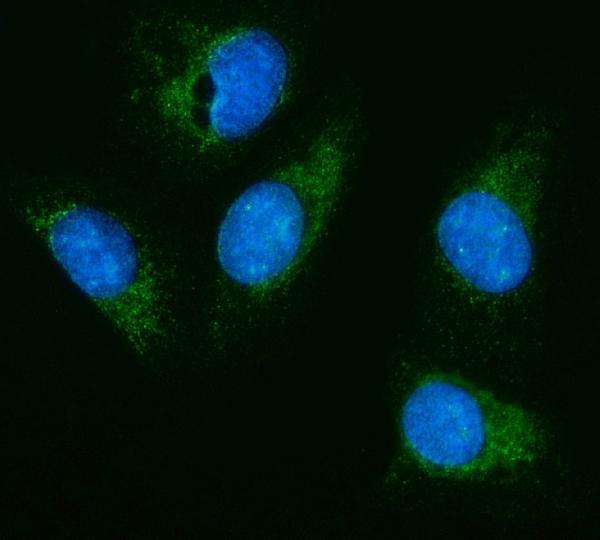
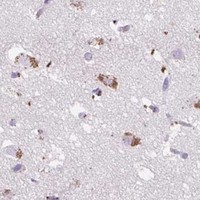
Two anti-HEXA antibodies have pictured results in human intestinal cancer paraffin sections (PA1787 image caption) and U20S cells (A00692-1 image caption); both list Human, Mouse and Rat reactivity (catalog).

Real IHC data Anti-HEXA antibody, PA1787, IHC(P) IHC(P):Human Intestinal Cancer Tissue
Anti-HEXA Antibody ®
Cat # PA1787
Real IF data IF analysis of HEXA using anti-HEXA antibody (A00692-1). HEXA was detected in an immunocytochemical section of U20S cells. Enzyme antigen retrieval was performed using IHC enzyme antigen retrieval reagent (AR0022) for 15 mins. The cells were blocked with 10% goat serum. And then incubated with 5 μg/mL rabbit anti-HEXA Antibody (A00692-1) overnight at 4°C. DyLight®488 Conjugated Goat Anti-Rabbit IgG (BA1127) was used as secondary antibody at 1:100 dilution and incubated for 30 minutes at 37°C. The section was counterstained with DAPI. Visualize using a fluorescence microscope and filter sets appropriate for the label used.
Anti-HEXA Antibody ®
Cat # A00692-1

PA1787 is listed for IHC, with an image of human intestinal cancer tissue in paraffin sections (PA1787 catalog; PA1787 image caption). A00692-1 is listed for IF/ICC, with an image of HEXA staining in U20S cells (A00692-1 catalog; A00692-1 image caption).

Which to pick: Choose PA1787 for paraffin-section IHC at 0.5–1 μg/ml (PA1787 datasheet); its own image shows human intestinal cancer tissue, but the fixative is unreported (PA1787 image caption). Choose A00692-1 for IF/ICC at 5 μg/ml; its own image shows staining in U20S cells (A00692-1 datasheet; A00692-1 image caption). Both list Human, Mouse and Rat reactivity (catalog), while the pictured samples support the specific applications and samples stated above (image captions).

Each figure is that product's own IHC / IF validation image from its datasheet.

References

  1. UniProt Consortium. UniProt entry P06865 (HEXA_HUMAN, Beta-hexosaminidase subunit alpha).
  2. Human Protein Atlas. HEXA tissue IHC expression (reliability: Supported).
  3. Human Protein Atlas. HEXA subcellular location (ICC-IF): Mainly localized to vesicles. In addition localized to the cytosol..
  4. Human Protein Atlas. HEXA antibody validation summary (2 antibodies).
  5. Nucleus accumbens GLT-1a overexpression reduces glutamate efflux during reinstatement of cocaine-seeking but is not sufficient to attenuate reinstatement. Neuropharmacology 2018 — PMC6383073.
  6. Microglia-Specific Expression of HEXA and HEXB Leads to Poor Prognosis in Glioblastoma Patients. Frontiers in oncology 2021 — PMC8371334.
  7. Microsatellite instability in penile cancer: comparative analysis of primary tumors and metastases. Frontiers in urology 2026 — PMC13553218.
  8. Autophagic flux is impaired in the brain tissue of Tay-Sachs disease mouse model. PloS one 2023 — PMC10019743.
  9. PubMed PMID:2933746 — UniProt-cited evidence.
  10. PubMed PMID:2952641 — UniProt-cited evidence.
  11. PubMed PMID:1833974 — UniProt-cited evidence.