This website uses cookies to ensure you get the best experience on our website.


- Table of Contents
Plan chromogenic HGS IHC in paraffin sections using the catalog antibody at 2 µg/mL (datasheet M01174-1). Compare cytoplasmic staining across cell types, while treating the tissue profile as provisional because it awaits external verification (HPA tissue IHC).

Expected localisation, validated protocols, controls and antibodies — the at-a-glance facts below, then the full design guide.
| Expected localisation | Variable cytoplasmic tissue staining (HPA tissue IHC) | |
| Staining pattern | High cytoplasmic staining in pancreatic exocrine cells (HPA tissue IHC) | |
| Antigen retrieval | EDTA pH 8.0 HIER, heat-mediated (datasheet M01174-1) | |
| Positive control | Pancreas+4 more · see all | |
| Negative control | Adipose tissue+4 more · see all |
| Fixation | Keep fixation consistent across paraffin sections. (standard IHC practice; not target-specific) | |
| Caveat | Cell-type variation: exocrine pancreas high, adipocytes undetected (HPA tissue IHC) | |
| Regulation | Higher in testis and blood leukocytes (UniProt) | |
| Isoform / epitope | Two isoforms; epitope coverage is unspecified (UniProt) |
The catalog antibody’s IHC-P protocol is accompanied by one published HGS IHC workflow using mouse liver sections (PMC4592276).
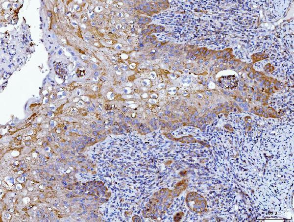
| Sample | Paraffin-embedded human laryngeal squamous cell carcinomas tissue; fixative not specified (datasheet M01174-1) |
| Fixation | Image fixative and duration unreported (datasheet M01174-1); verify before use. |
| Sectioning | 4–5 µm sections on charged slides (standard) |
| Deparaffinisation | Xylene, graded ethanol series to water (standard) |
| Antigen retrieval | Heat retrieval: EDTA pH 8.0 (datasheet M01174-1); 20 min, 95–100 °C (standard) |
| Peroxidase block | 3% H2O2, 10 min, room temperature (standard) |
| Blocking | 10% goat serum (datasheet M01174-1) |
| Primary antibody | Mouse monoclonal (clone 6C2E1) anti-HGS, 2 μg/ml (datasheet M01174-1) |
| Primary incubation | Overnight at 4 °C (datasheet M01174-1) |
| Detection | Streptavidin-biotin complex (SABC), DAB chromogen (datasheet M01174-1) |
| Counterstain | Hematoxylin, blue, dehydrate and mount (standard) |
| Expected result | HGS-positive staining in exocrine glandular cells of pancreas (HPA tissue IHC: High). HPA tissue profile: Cytoplasmic expression at variable levels in most tissues. No signal in the no-primary control. |
HGS is cytoplasmic and associates with early endosome and multivesicular body membranes without a transmembrane segment (UniProt O14964 localization and topology). In paraffin sections, expect cytoplasmic staining in glandular cells, especially pancreatic exocrine cells (HPA: high in pancreatic exocrine cells; medium in several other glandular tissues). Treat this as a guide, not a guaranteed pattern: HPA rates the tissue IHC evidence uncertain, pending external verification (HPA: tissue reliability).
| Cytoplasmic staining in pancreatic exocrine cells, with variable staining across other tissues. | This fits HPA’s reported high pancreatic exocrine signal and variable cytoplasmic expression in most tissues (HPA: tissue IHC). Compare cells within each section; the HPA tissue assessment remains uncertain (HPA: tissue reliability). |
| Predominantly nuclear staining, or a crisp outline of the entire plasma membrane. | These compartments conflict with the reported cytoplasmic and endosomal localization (UniProt O14964 localization; HPA: subcellular). Review morphology, staining distribution and controls before interpreting this as HGS (general IHC practice). |
| Strong adipocyte or cholangiocyte staining despite weak staining in expected positive cells. | HPA reports HGS as not detected in those cell types (HPA: tissue IHC). Check cross-reactivity and endogenous detection activity with appropriate controls (general IHC practice); HPA’s uncertain reliability limits any definitive negative call (HPA: tissue reliability). |
| Haze across tissue, stroma and blank regions, obscuring cell boundaries. | A diffuse field cannot establish the reported cytoplasmic pattern (HPA: tissue IHC). Review nonspecific binding, detection background and washing against control sections (general IHC practice). |
| No convincing signal in pancreatic exocrine cells. | That differs from HPA’s high reported staining in this cell type (HPA: tissue IHC). Check the assay controls and tissue preservation before concluding absence; the reference pattern itself awaits external verification (HPA: tissue reliability). |
| Cell type and tissue context | HPA reports high pancreatic exocrine staining, medium staining in several glandular cell types, and undetected staining in adipocytes and cholangiocytes (HPA: tissue IHC). Interpret each compartment within identified cells, since these observations carry an uncertain tissue reliability rating (HPA: tissue reliability). |
| Intracellular distribution | UniProt places HGS in the cytoplasm and on early endosome and multivesicular body membranes, with no transmembrane segment (UniProt O14964 localization and topology). HPA ICC-IF supports endosomes and cytosol; its lysosome assignment is uncertain (HPA: subcellular). Chromogenic IHC may resolve cytoplasmic staining without resolving individual vesicles (general IHC practice). |
| Antibody evidence for paraffin IHC | Both listed antibodies, HPA004872 and HPA007728, have uncertain IHC status and supported ICC status (HPA: antibody validation). Supported ICC staining does not independently validate their paraffin-section pattern (HPA: antibody validation; general IHC practice). Seek agreement with morphology and controls before assigning specific HGS staining (general IHC practice). |
| IF/ICC Q&A: Should its puncta be required in IHC? | No. HPA ICC-IF places HGS mainly at endosomes, additionally in cytosol, with lysosomal localization uncertain (HPA: subcellular). Use that evidence to assess compartment plausibility; do not require visible puncta in chromogenic paraffin sections, whose resolution and presentation differ (general IHC practice). |
| Isoforms and antigen recognition | UniProt lists 2 isoforms, but the supplied evidence does not map antibody epitopes to either one (UniProt O14964 isoforms; HPA: antibody entries). An isoform-specific explanation for discordant staining therefore cannot be assigned from these records. Compare antibodies only after checking their documented antigen information (general IHC practice). |
| Situation | Likely cause | Next action |
|---|---|---|
| Pancreatic exocrine cells show no staining. | The result conflicts with HPA’s reported high signal, although that IHC assessment is uncertain (HPA: tissue IHC and reliability). | Confirm tissue identity and positive assay controls; review retrieval and detection steps as general IHC checks before interpreting absence (general IHC practice). |
| Strong nuclear staining dominates. | Nuclear localization is outside the supplied cytoplasmic and endosomal pattern (UniProt O14964 localization; HPA: subcellular). | Check counterstain separation, secondary-only control and antibody-dependent background, then compare cytoplasmic signal in the same cells (general IHC practice). |
| Adipocytes or cholangiocytes stain strongly. | These cell types are reported as not detected, subject to HPA’s uncertain tissue reliability (HPA: tissue IHC and reliability). | Inspect the negative reagent control for endogenous detection activity and compare staining with pancreatic exocrine cells (general IHC practice; HPA: high in pancreatic exocrine cells). |
| A diffuse brown haze hides cell boundaries. | Background prevents a reliable reading of HPA’s cytoplasmic pattern (HPA: tissue IHC; general IHC practice). | Review blocking, antibody concentration, washing and detection controls as general workflow variables; rescore only where cellular detail is clear (general IHC practice). |
| Only scattered puncta appear, with little diffuse cytoplasm. | Endosomal enrichment is plausible, but ICC-IF also supports cytosolic HGS and does not establish a required paraffin-section appearance (HPA: subcellular; HPA: antibody validation). | Check whether puncta sit within the expected cells and recur across fields; interpret alongside the broader cytoplasmic pattern (HPA: tissue IHC; general IHC practice). |
| Two antibodies give different cell patterns. | Both listed antibodies have uncertain IHC validation, and their supplied entries do not identify isoform-specific epitopes (HPA: antibody validation; UniProt O14964 isoforms). | Compare reagent controls, cell morphology and documented antigen information; avoid attributing the difference to an isoform without further evidence (general IHC practice). |
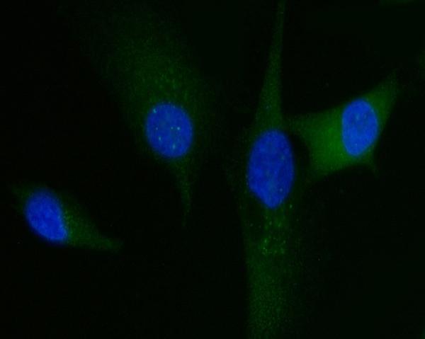
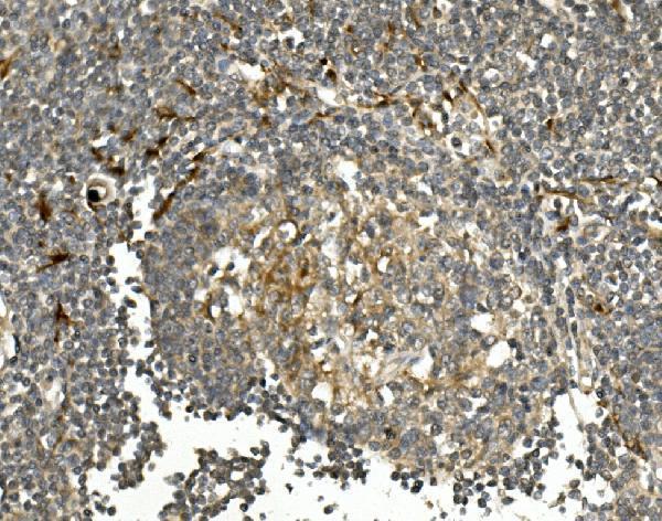
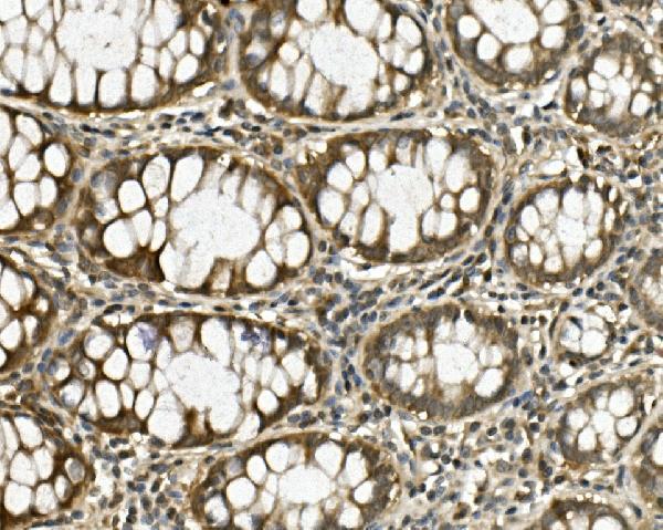
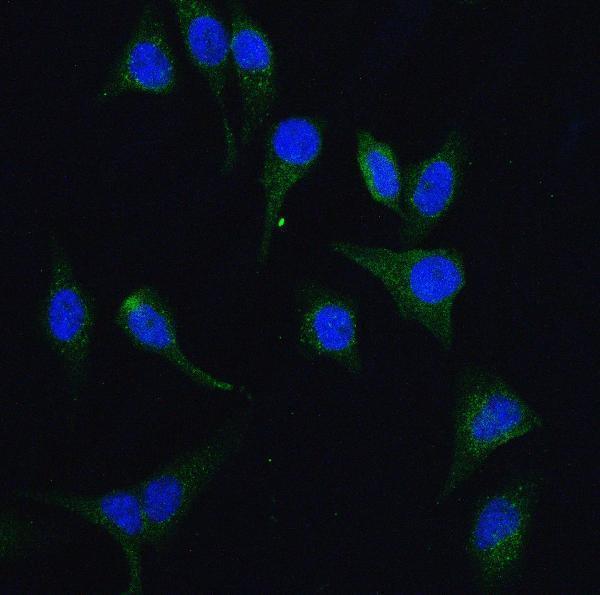
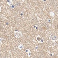
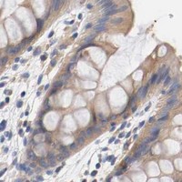


Comprehensive Human Protein Atlas IHC scoring per tissue (reliability: Uncertain — Pending external verification.). Rows are taken directly from the HPA tissue chart — click any row's HPA link to view the source.
| Tissue | Cell type | Level | Evidence | Source |
|---|---|---|---|---|
| Pancreas | Exocrine glandular cells | High | Protein (IHC) | HPA → |
| Adrenal gland | Glandular cells | Medium | Protein (IHC) | HPA → |
| Appendix | Glandular cells | Medium | Protein (IHC) | HPA → |
| Breast | Glandular cells | Medium | Protein (IHC) | HPA → |
| Cerebellum | Purkinje cells | Medium | Protein (IHC) | HPA → |
| Tissue | Cell type | Level | Evidence | Source |
|---|---|---|---|---|
| Adipose tissue | Adipocytes | Not detected | Protein (IHC) | HPA → |
| Liver | Cholangiocytes | Not detected | Protein (IHC) | HPA → |
| Lymph node | Germinal center cells | Not detected | Protein (IHC) | HPA → |
| Soft tissue | Fibroblasts | Not detected | Protein (IHC) | HPA → |
| Spleen | Cells in red pulp | Not detected | Protein (IHC) | HPA → |
Troubleshoot HGS staining in paraffin sections by checking retrieval, signal location, controls, and scoring before interpreting chromogenic IHC results.
The catalog shows human paraffin-section IHC for three HGS antibodies and cell IF images for two; all three list human, mouse and rat reactivity (catalog image captions; catalog reactivity).
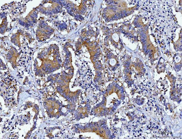
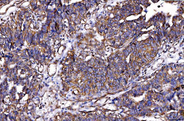
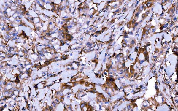
M01174-1 has IHC images in human laryngeal squamous cell carcinoma, colorectal adenocarcinoma, placenta and hepatocellular carcinoma (M01174-1 IHC captions); M01174-2 has IHC images in human bladder, breast, colorectal and endometrial cancers, plus IF in U87 cells (M01174-2 image captions). A01174-1 has IHC images in human rectal and testis cancers and an IF image in HeLa cells (A01174-1 image captions).
Which to pick: For tissue IHC, choose mouse monoclonal M01174-1 or M01174-2, or rabbit A01174-1; each has its own human paraffin-section image using EDTA retrieval at pH 8.0 (catalog host and clone fields; respective IHC captions). For IF/ICC, choose M01174-2 or A01174-1: both list those applications and have cell IF images (catalog applications; respective IF captions). For mouse or rat samples, all three list reactivity, though their tissue IHC images show human samples; the IHC captions do not report a fixative (catalog reactivity; respective IHC captions).