This website uses cookies to ensure you get the best experience on our website.


- Table of Contents
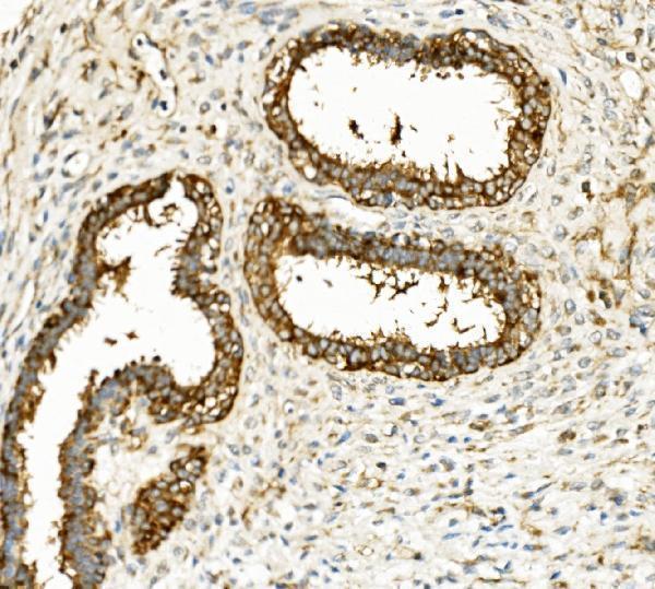
Plan HK1 paraffin IHC around granular cytoplasmic staining (HPA tissue IHC) and its molecular association with the mitochondrial outer membrane (UniProt). Neuronal cells in the cerebral cortex show high staining and can serve as a positive reference (HPA tissue IHC).

Expected localisation, validated protocols, controls and antibodies — the at-a-glance facts below, then the full design guide.
| Expected localisation | Observed granular cytoplasm (HPA tissue IHC); molecular outer mitochondrial membrane association (UniProt) | |
| Staining pattern | Ubiquitous granular cytoplasmic staining (HPA tissue IHC) | |
| Antigen retrieval | EDTA pH 8.0 HIER, heat-mediated (datasheet M01504-2) | |
| Positive control | Appendix+4 more · see all | |
| Negative control | Adipose tissue+1 more · see all |
| Fixation | Keep fixation conditions consistent across sections. (standard IHC practice; not target-specific) | |
| Caveat | Adipocytes may lack signal despite the broad tissue profile (HPA tissue IHC) | |
| Regulation | GlcNAc shifts HK1 to cytosol (UniProt) | |
| Isoform / epitope | 4 isoforms; 2 is erythrocyte-specific, 3–4 testis-specific; epitope coverage unknown (UniProt) |
The catalog antibody’s IHC-P protocol (datasheet: M01504-2) is accompanied by published chromogenic HK1 methods for mouse tissue sections and mink uterus (PMC7658079; PMC4284318).
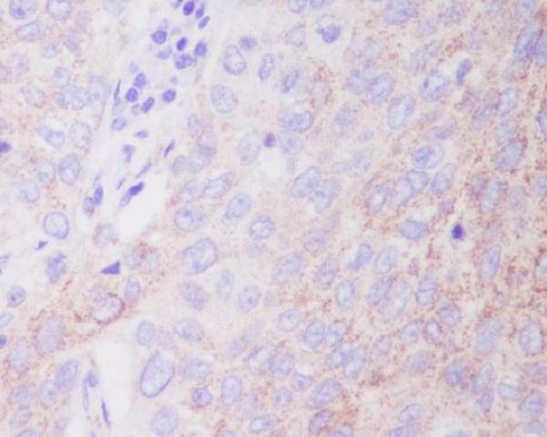
| Sample | Paraffin-embedded human lung cancer tissue; fixative not specified (datasheet M01504-2) |
| Fixation | Image fixative and duration unreported (datasheet M01504-2); verify before use. |
| Sectioning | 4–5 µm sections on charged slides (standard) |
| Deparaffinisation | Xylene, graded ethanol series to water (standard) |
| Antigen retrieval | Heat retrieval: EDTA pH 8.0 (datasheet M01504-2); 20 min, 95–100 °C (standard) |
| Peroxidase block | 3% H2O2, 10 min, room temperature (standard) |
| Blocking | 10% goat serum (datasheet M01504-2) |
| Primary antibody | Mouse monoclonal (clone 4B7) anti-HK1, 0.5-1μg/ml (datasheet M01504-2) |
| Primary incubation | Overnight at 4 °C (datasheet M01504-2) |
| Detection | Streptavidin-biotin complex (SABC), DAB chromogen (datasheet M01504-2) |
| Counterstain | Hematoxylin, blue, dehydrate and mount (standard) |
| Expected result | HK1-positive staining in endocrine cells of appendix (HPA tissue IHC: High). HPA tissue profile: Ubiquitous cytoplasmic expression with a granular pattern. No signal in the no-primary control. |
HK1 should appear as granular cytoplasmic staining in many cell types, with especially strong signal in selected endocrine, epithelial, and neuronal cells (HPA tissue IHC: Supported; ubiquitous granular cytoplasmic profile). Its mitochondrial outer membrane association and cytosolic pool explain the cytoplasmic location; HK1 has no transmembrane segment (UniProt P19367: subcellular location and topology). HPA reports medium agreement between antibody staining and RNA expression (HPA tissue IHC: reliability description).
| Granular cytoplasmic staining is strong in cerebral cortex neurons, bronchial respiratory epithelium, or appendix endocrine cells (HPA tissue IHC: High in these cells). | This matches the reported tissue pattern and is a useful positive-slide check (HPA tissue IHC: ubiquitous granular cytoplasmic profile; High in these cells). Judge the signal in the named cells, since a mixed tissue contains cells with different reported staining levels (HPA tissue IHC: cell-level annotations). |
| Signal appears mainly nuclear, with little cytoplasmic staining. | A predominantly nuclear result conflicts with the reported mitochondrial and cytosolic locations and granular cytoplasmic IHC pattern (UniProt P19367: subcellular location; HPA tissue IHC: profile). Treat it as suspect staining and review the counterstain, detection background, and control slide before calling HK1 positive (standard IHC practice). |
| Adipocytes or chondrocytes stain as strongly as the selected positive cells. | HPA reports HK1 as not detected in adipocytes and chondrocytes (HPA tissue IHC: Not detected). Unexpected staining raises concern for cross-reactivity, endogenous detection activity, or slide background; it does not identify which cause is responsible (standard IHC practice). Compare the pattern with a no-primary control (standard IHC practice). |
| Color spreads evenly across tissue and empty slide areas, obscuring cell outlines. | A uniform haze does not resemble HK1's reported granular cytoplasmic distribution (HPA tissue IHC: profile). Excess detection background or insufficient blocking or washing can obscure a genuine signal (standard IHC practice). Interpret intensity only after the background is brought under control (standard IHC practice). |
| No signal is visible in a section containing cerebral cortex neurons or esophageal squamous epithelium. | Both cell populations are reported High by HPA, so a blank result calls for an assay check (HPA tissue IHC: High in cerebral cortex neuronal cells and esophageal squamous epithelial cells). Verify that those cells are present, then review antibody, retrieval, and detection performance (standard IHC practice). A blank slide alone does not establish absent HK1. |
| Compartment and topology (UniProt P19367: subcellular location and topology) | HK1 associates with the mitochondrial outer membrane through a mitochondrial-binding region and can also reside in the cytosol; no transmembrane segment is annotated (UniProt P19367). Expect a cytoplasmic readout in chromogenic IHC, where individual mitochondria may not be resolved (HPA tissue IHC: granular cytoplasmic profile; standard IHC practice). |
| Cell choice for controls (HPA tissue IHC: cell-level staining) | Use a reported High population, such as hippocampal neurons or duodenal endocrine cells, when checking for a positive pattern (HPA tissue IHC: High). Adipocytes and chondrocytes are reported Not detected and can help flag unexpected color, but absence in them cannot by itself validate antibody specificity (HPA tissue IHC: Not detected; standard IHC practice). |
| Isoform coverage (UniProt P19367: isoforms and tissue specificity) | Four HK1 isoforms are listed; isoform 2 is erythrocyte specific, while isoforms 3 and 4 are testis specific (UniProt P19367). The supplied evidence does not locate the catalog antibody's epitope, so do not infer which isoforms it detects or use isoform specificity to explain a discrepant slide. |
| IHC evidence strength (HPA tissue IHC: Supported reliability; HPA antibodies: IHC Supported) | HPA describes medium consistency between antibody staining and RNA expression, and lists three antibodies with Supported IHC status (HPA tissue IHC: reliability description; HPA antibodies: IHC status). These observations support a pattern comparison, while an unexpected specimen still needs its own controls (standard IHC practice). |
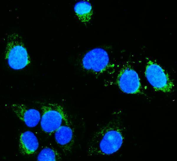
| IF/ICC Q: What location should I expect? (HPA subcellular ICC-IF) | A: HPA reports mitochondrial localization as the enhanced main location in ICC-IF (HPA subcellular ICC-IF: main location). This helps interpret organelle-scale fluorescence; the chromogenic tissue-IHC expectation remains granular cytoplasm (HPA tissue IHC: profile). |
| Situation | Likely cause | Next action |
|---|---|---|
| The known-positive cell population is blank (HPA tissue IHC: High annotations). | The relevant cells may be missing from the section, or the retrieval, primary-antibody, or detection step may have failed (standard IHC practice). | Confirm cell identity on the counterstained section and run a reported High control beside the test section; review retrieval and reagent steps if both are blank (HPA tissue IHC: High annotations; standard IHC practice). |
| Granular signal is faint across a reported High population (HPA tissue IHC: profile and High annotations). | Weak assay signal or excessive background can make the expected cytoplasmic granules hard to distinguish (standard IHC practice). | Compare with the matched positive control and local background, then check antibody dilution and detection conditions against the available IHC procedure (standard IHC practice). Do not assign a new target-specific dilution from this evidence. |
| The slide shows widespread diffuse brown haze. | Nonspecific reagent retention or detection background can overwhelm cell-level staining (standard IHC practice). | Inspect a no-primary control; review blocking, washes, and detection exposure, then reassess whether granular cytoplasmic staining remains (HPA tissue IHC: profile; standard IHC practice). |
| Adipocytes or chondrocytes show conspicuous color (HPA tissue IHC: Not detected). | The finding conflicts with HPA's cell-level observations; cross-reactivity or endogenous detection activity are possibilities, not diagnoses (HPA tissue IHC: Not detected; standard IHC practice). | Compare a no-primary control and neighboring cell populations, and check endogenous enzyme blocking if an enzyme-based detection system is used (standard IHC practice). |
| Signal is predominantly nuclear. | That compartment differs from the reported mitochondrial and cytosolic locations (UniProt P19367: subcellular location). Counterstain or background may be mistaken for target signal (standard IHC practice). | Check the no-primary control and compare nuclear color with cytoplasmic granules in a reported High population before scoring the section (HPA tissue IHC: profile and High annotations; standard IHC practice). |
| One tissue looks weaker than another despite otherwise acceptable staining. | HK1 staining varies by cell population: HPA reports Low in liver cholangiocytes and High in hippocampal neurons (HPA tissue IHC: cell-level staining). | Score the specified cell type against its own background and an appropriate control. Do not treat lower intensity in a reported Low population as assay failure solely because a reported High population stains strongly (HPA tissue IHC: Low and High annotations; standard IHC practice). |


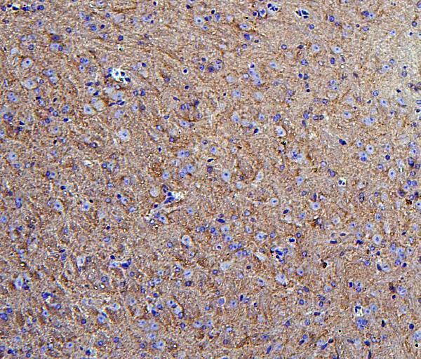
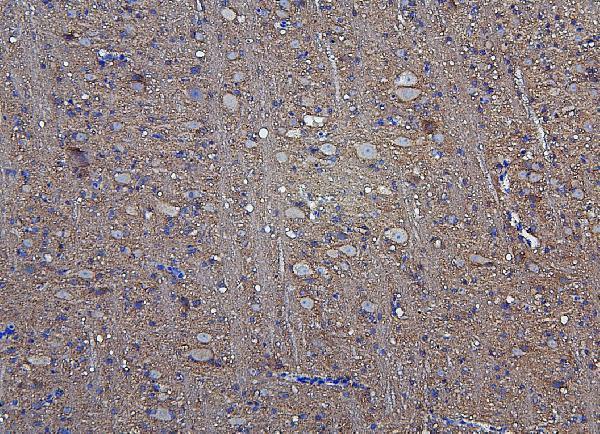
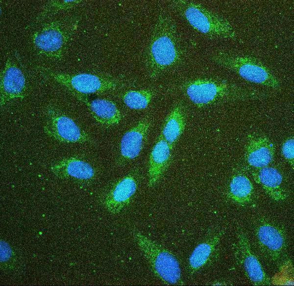
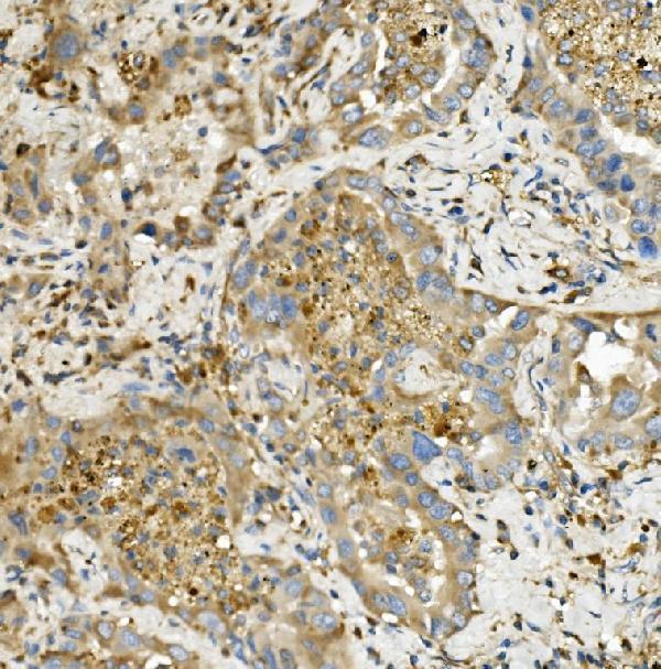
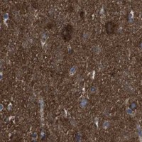


Comprehensive Human Protein Atlas IHC scoring per tissue (reliability: Supported — Medium consistency between antibody staining and RNA expression data.). Rows are taken directly from the HPA tissue chart — click any row's HPA link to view the source.
| Tissue | Cell type | Level | Evidence | Source |
|---|---|---|---|---|
| Appendix | Endocrine cells | High | Protein (IHC) | HPA → |
| Bronchus | Respiratory epithelial cells | High | Protein (IHC) | HPA → |
| Cerebellum | Cells in granular layer | High | Protein (IHC) | HPA → |
| Cerebral cortex | Neuronal cells | High | Protein (IHC) | HPA → |
| Colon | Endocrine cells | High | Protein (IHC) | HPA → |
Troubleshoot HK1 staining in paraffin sections by checking retrieval, controls and cell-level localisation before comparing signal across specimens.
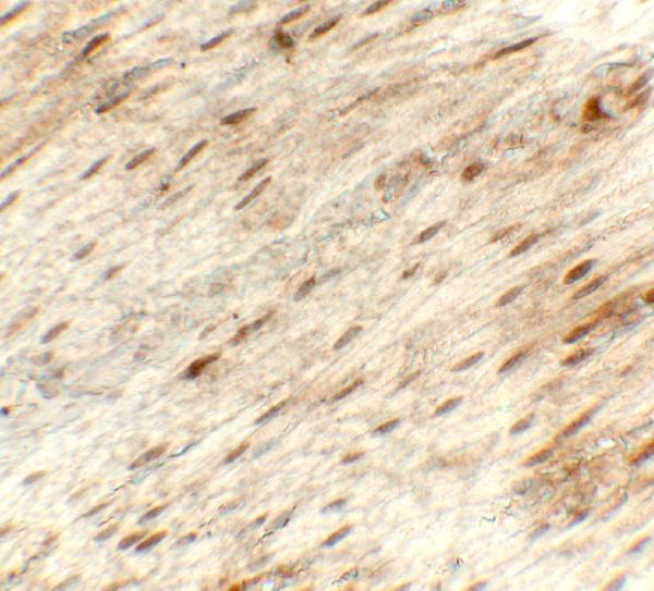
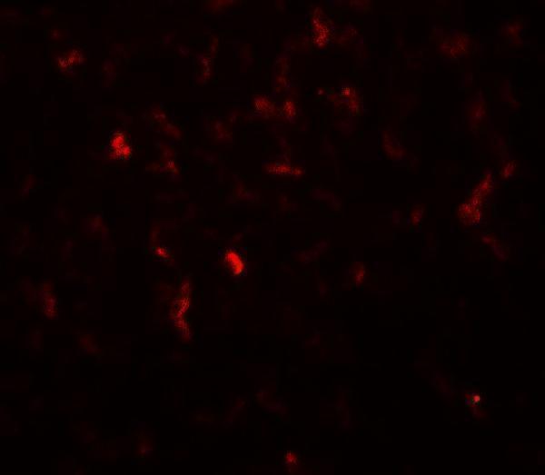
Anti-HK1 antibodies have IHC images from human lung tumors, mouse brain, and rat stomach, plus IF images from cells and tissue (catalog image captions).
M01504-2 has IHC images in human lung cancer and mouse intestine; A01504-1 in mouse and rat brain and human mammary and rectal cancers; M01504-1 in human lung, mammary, and testis cancers and frozen placenta (catalog IHC captions). A01504-2 has an IHC image in rat stomach, and M01504 in paraffin-embedded human lung carcinoma (catalog IHC captions).
Which to pick: For tissue IHC, start with M01504-2, a mouse monoclonal (4B7) with paraffin-section images in human lung cancer and mouse intestine and an IHC dilution of 0.5–1 μg/mL; its fixative is unreported (catalog host, clone, IHC captions and dilution). For IF/ICC, M01504-2 lists both applications and has IF images in U20S cells and human lung cancer sections at 2 μg/mL (catalog applications, IF captions and dilution). For cross-species IHC, A01504-1 lists human, mouse and rat reactivity and has paraffin-section images from all three species; its fixative is unreported (catalog reactivity and IHC captions).