This website uses cookies to ensure you get the best experience on our website.


- Table of Contents
Plan chromogenic HMGA1 IHC in paraffin sections with a catalog antibody validated at 1:50–1:200 (datasheet M01043). Use the nuclear tissue pattern and cell-specific staining examples to choose controls and interpret results (HPA tissue IHC).

Expected localisation, validated protocols, controls and antibodies — the at-a-glance facts below, then the full design guide.
| Expected localisation | Nuclear staining across tissues (HPA tissue IHC) | |
| Staining pattern | Nuclear signal across tissues, varying by cell type (HPA tissue IHC) | |
| Antigen retrieval | EDTA pH 8.0 HIER, heat-mediated (datasheet M01043) | |
| Positive control | Appendix+4 more · see all | |
| Negative control | Adipose tissue+1 more · see all |
| Fixation | Keep fixation consistent across sections (standard IHC practice; not target-specific) | |
| Caveat | Normal cells can also show nuclear staining (HPA tissue IHC) | |
| Regulation | Esophagus has tissue-enhanced RNA (HPA RNA specificity) | |
| Isoform / epitope | 3 isoforms (HMG-I/Y/R); epitope coverage unknown (UniProt) |
Compare the catalog antibody’s IHC-P protocol (datasheet: M01043) with published HMGA1 staining methods for pancreatic, urothelial, and breast cancer specimens (PMC3092591; PMC12984743; PMC5661362).
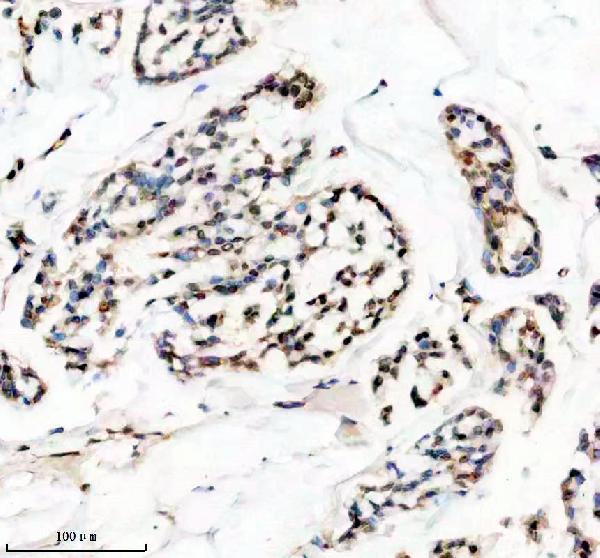
| Sample | Paraffin-embedded human breast cancer tissue; fixative not specified (datasheet M01043) |
| Fixation | Image fixative and duration unreported (datasheet M01043); verify before use. |
| Sectioning | 4–5 µm sections on charged slides (standard) |
| Deparaffinisation | Xylene, graded ethanol series to water (standard) |
| Antigen retrieval | Heat retrieval: EDTA pH 8.0 (datasheet M01043); 20 min, 95–100 °C (standard) |
| Peroxidase block | 3% H2O2, 10 min, room temperature (standard) |
| Blocking | 10% goat serum (datasheet M01043) |
| Primary antibody | Rabbit monoclonal (clone 18H23) anti-HMGA1, 1:50-1:200 (datasheet M01043) |
| Primary incubation | Overnight at 4 °C (datasheet M01043) |
| Detection | HRP-conjugated secondary, DAB chromogen (datasheet M01043) |
| Counterstain | Hematoxylin, blue, dehydrate and mount (standard) |
| Expected result | HMGA1-positive staining in endocrine cells of appendix (HPA tissue IHC: High). HPA tissue profile: Nuclear expression in all tissues. No signal in the no-primary control. |
HMGA1 is a nuclear and chromosome-associated protein without a transmembrane segment (UniProt P17096). In paraffin-section IHC, expect nuclear staining across tissues, with cell-dependent intensity (HPA tissue IHC: nuclear expression in all tissues; Enhanced reliability). High staining is documented in breast glandular cells, bronchial respiratory epithelial cells, and several endocrine cell populations (HPA tissue IHC). Interpret each cell population against its own reference pattern.
| Clear nuclear staining in breast glandular or bronchial respiratory epithelial cells, with little staining outside nuclei. | This fits the expected compartment (UniProt P17096: nucleus, chromosome) and documented High staining in those cell types (HPA tissue IHC). Nuclear staining in other tissues can also be expected; the HPA profile reports nuclear expression across tissues, while intensity varies by cell population (HPA tissue IHC). |
| Predominantly cytoplasmic or surface staining, with little nuclear signal, on a chromogenic IHC slide. | This is discordant with the dominant IHC pattern (HPA tissue IHC: nuclear expression). Review morphology and staining controls before calling it HMGA1. Cytosolic localisation is reported as an additional ICC-IF finding, so isolated cytosolic fluorescence should be judged in its own application context (HPA subcellular ICC-IF). |
| Strong staining in adipocytes or ovarian follicle cells, especially without convincing nuclear staining in adjacent cells. | Those cell populations are reported as Not detected by HPA tissue IHC. Investigate cross-reactivity or endogenous detection activity (standard IHC practice). The finding alone does not establish either cause; compare the stained slide with a reagent-omission control and an expected-positive cell population. |
| Diffuse brown colour across nuclei, cytoplasm, and tissue spaces, with little separation between cell populations. | A poorly resolved pattern cannot reliably establish nuclear HMGA1 localisation (UniProt P17096: nucleus; HPA tissue IHC: nuclear expression). Check the detection background and primary-antibody dilution using standard IHC controls (standard IHC practice), then assess whether nuclei remain distinguishable from surrounding tissue. |
| No nuclear signal in a breast glandular or bronchial respiratory epithelial population that is identifiable on the section. | This conflicts with the reported High staining for those populations (HPA tissue IHC). First check section quality, control tissue, retrieval, antibody incubation, and detection steps (standard IHC practice). A single negative slide does not show that the tissue lacks HMGA1. |
| Nuclear target and absence of a transmembrane segment | Score staining against nuclei and tissue morphology (UniProt P17096: nucleus, chromosome; no transmembrane segment). A membrane-only chromogenic pattern does not match the reported IHC distribution (HPA tissue IHC: nuclear expression). |
| Cell-specific reference intensity | Use the matching cell population as the comparator: endocrine cells in appendix, colon, and duodenum are High; smooth muscle cells are low; adipocytes and ovarian follicle cells are Not detected (HPA tissue IHC). |
| Antibody validation and limits | HPA lists IHC as Enhanced for HPA065612 and HPA068442, both rabbit polyclonals (HPA antibodies). This supports the reported pattern; it does not validate an unlisted catalog antibody or predict its staining at a particular dilution. |
| IF/ICC pattern? | In the separate IF/ICC application, HPA reports supported nucleoplasm and nuclear membrane localisation, plus approved additional cytosolic localisation (HPA subcellular ICC-IF). Treat that as an IF/ICC interpretation reference, not an IHC-P protocol or a reason to score cytoplasm-only IHC as expected. |
| Isoforms, modifications, and retrieval | UniProt lists 3 isoforms and multiple modified residues (UniProt P17096). Their effects on a given antibody's epitope or retrieval response are unspecified here; select retrieval conditions from the antibody's IHC-P instructions and assess them with controls (standard IHC practice). |
| Situation | Likely cause | Next action |
|---|---|---|
| Expected-positive nuclei are blank throughout the slide. | The slide may have failed at retrieval, primary incubation, or detection (standard IHC practice); this result conflicts with High reference staining in selected cell populations (HPA tissue IHC). | Confirm the expected-positive population is present, inspect the run control, then check each IHC-P step against the antibody instructions. Do not assign a target-specific fixation or retrieval sensitivity from the supplied evidence. |
| Nuclei stain weakly, while the tissue and detection controls appear usable. | Antibody concentration, incubation, or retrieval may yield insufficient contrast (standard IHC practice); the supplied HPA and UniProt records do not establish which variable is responsible. | Adjust one parameter at a time within the antibody's IHC-P guidance and compare nuclear contrast in the same reference cell population (standard IHC practice; HPA tissue IHC: cell-specific levels). |
| Brown signal coats tissue edges or fills spaces between cells. | Diffuse deposition or detection background can obscure localisation (standard IHC practice). This distribution cannot be read as the reported nuclear IHC pattern (HPA tissue IHC). | Review washing, blocking, and detection conditions; compare a reagent-omission control, then rescore only well-preserved cells whose nuclei can be identified (standard IHC practice). |
| Adipocytes or ovarian follicle cells stain strongly. | Cross-reactivity or endogenous detection activity is possible (standard IHC practice), because HPA reports these cells as Not detected (HPA tissue IHC). | Check a primary-antibody omission control and the location of signal within cells; compare with expected-positive nuclei on the same run before attributing the signal to HMGA1 (standard IHC practice; HPA tissue IHC). |
| Smooth muscle appears much fainter than nearby stained cells. | Low staining in smooth muscle cells is part of the reported tissue pattern (HPA tissue IHC); intensity differences alone do not establish an assay failure. | Judge nuclear localisation and the run control before changing conditions. Use a documented High cell population, such as bronchial respiratory epithelium, to assess assay performance (HPA tissue IHC; standard IHC practice). |
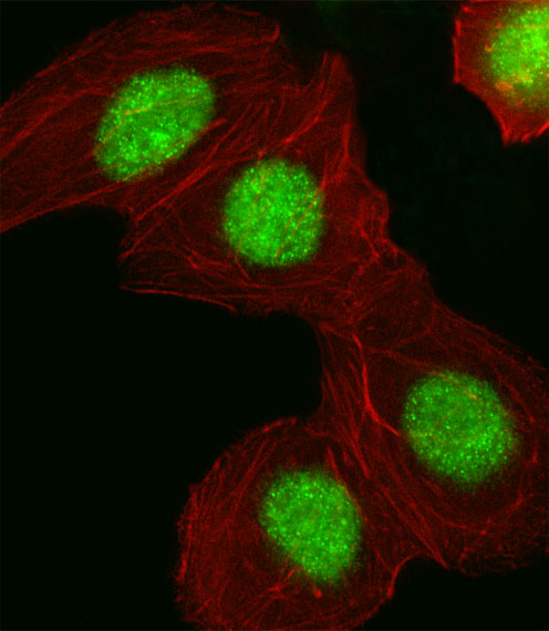
| IF/ICC shows some cytosolic fluorescence alongside nuclear signal. | HPA reports cytosol as an additional ICC-IF location, while nucleoplasm and nuclear membrane are the supported main locations (HPA subcellular ICC-IF). | Evaluate the fluorescence in the separate IF/ICC guide against its controls. For this chromogenic IHC-P guide, base the expected call on nuclear tissue staining (HPA tissue IHC). |

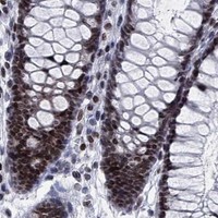


Comprehensive Human Protein Atlas IHC scoring per tissue (reliability: Enhanced — High consistency between antibody staining and RNA expression data.). Rows are taken directly from the HPA tissue chart — click any row's HPA link to view the source.
| Tissue | Cell type | Level | Evidence | Source |
|---|---|---|---|---|
| Appendix | Endocrine cells | High | Protein (IHC) | HPA → |
| Breast | Glandular cells | High | Protein (IHC) | HPA → |
| Bronchus | Respiratory epithelial cells | High | Protein (IHC) | HPA → |
| Caudate | Glial cells | High | Protein (IHC) | HPA → |
| Cerebellum | Granular cells - nucleus | High | Protein (IHC) | HPA → |
These troubleshooting questions focus on chromogenic HMGA1 IHC in paraffin sections, with one entry on IF/ICC (selected product caption: paraffin section; HPA: nuclear expression).
The catalog includes HMGA1 antibodies with IHC data from human breast cancer paraffin sections and IF data from human A549 cells; one antibody also lists mouse and rat reactivity (catalog; image captions).
M01043 lists IHC and ICC/IF applications, with an IHC image from a human breast cancer paraffin section (catalog; M01043 image caption). A01043-2 lists IF and shows nuclear staining in human A549 cells (catalog; A01043-2 image caption).
Which to pick: Choose M01043 for paraffin-section IHC: it is monoclonal, lists IHC at 1:50–1:200, and its IHC caption documents EDTA retrieval at pH 8.0; the fixative is unreported (catalog; M01043 image caption). Choose A01043-2 for IF in human cells based on its A549 image; for ICC specifically, M01043 lists ICC/IF while A01043-2 lists IF only (catalog; A01043-2 image caption). For mouse or rat samples, M01043 is the option with listed reactivity, although its supplied IHC image shows human tissue (catalog; M01043 image caption).