HMGCR / 3-hydroxy-3-methylglutaryl-coenzyme A reductase · IHC design guide

Design Immunohistochemistry for HMGCR

Plan paraffin-section HMGCR IHC around the cytoplasmic tissue pattern (HPA tissue IHC) and an IHC-validated antibody at 2–5 μg/ml (datasheet A00643-3). Use documented high-staining cells as positive references and account for sterol-accelerated degradation when comparing intensity (HPA tissue IHC; UniProt).

Evidence assembled Oct 2026 · For research use; verify linked source records and product datasheet before use
Immunohistochemistry protocol sheet for HMGCR (IHC for HMGCR): expected localisation Cytoplasmic tissue staining (HPA tissue IHC); ER/peroxisome membranes (UniProt), antibody A00643-3, validated IHC image, and IHC protocol steps
Printable HMGCR IHC protocol sheet — expected localisation Cytoplasmic tissue staining (HPA tissue IHC); ER/peroxisome membranes (UniProt), antibody A00643-3, controls and protocol steps. Open the full HMGCR IHC guide →

HMGCR Immunohistochemistry Experimental Design Guide

Expected localisation, validated protocols, controls and antibodies — the at-a-glance facts below, then the full design guide.

Must know before staining
Expected localisation Cytoplasmic tissue staining (HPA tissue IHC); ER/peroxisome membranes (UniProt)
Staining pattern Ubiquitous cytoplasmic staining across cell types (HPA tissue IHC)
Antigen retrieval EDTA pH 8.0 HIER, heat-mediated (datasheet A00643-3)
Positive control ⓘ Appendix+4 more · see all
Negative control ⓘ Ovary+1 more · see all
Important caveats
Reasons your staining may differ from the expected pattern.
Fixation Keep paraffin-section fixation consistent (standard IHC practice; not target-specific); Matched tissue-IHC evidence does not establish the fixation claim. Validate the specimen-specific method before use. (selected-SKU IHC image A00643-3)
Caveat Tissue staining and RNA show medium consistency (HPA tissue IHC)
Regulation Sterols accelerate HMGCR degradation (UniProt)
Isoform / epitope 3 isoforms; map epitopes to lumenal or cytoplasmic regions (UniProt)
Section 1

Recommended HMGCR IHC & IF Protocols

The catalog antibody’s IHC-P protocol (datasheet: A00643-3) appears alongside published HMGCR methods for ovarian carcinoma, colorectal cancer, and breast tumors (PMC8664653; PMC4000148; PMC6969108).

Recommended immunohistochemistry (IHC-P) protocol parameters
SampleParaffin-embedded human breast cancer tissue; fixative not specified (datasheet A00643-3)
FixationImage fixative and duration unreported (datasheet A00643-3); verify before use.
Sectioning4–5 µm sections on charged slides (standard)
DeparaffinisationXylene, graded ethanol series to water (standard)
Antigen retrievalHeat retrieval: EDTA pH 8.0 (datasheet A00643-3); 20 min, 95–100 °C (standard)
Peroxidase block3% H2O2, 10 min, room temperature (standard)
Blocking10% goat serum (datasheet A00643-3)
Primary antibodyRabbit anti-HMGCR, 2-5 μg/ml (datasheet A00643-3)
Primary incubationOvernight at 4 °C (datasheet A00643-3)
DetectionHRP-conjugated secondary, DAB chromogen (datasheet A00643-3)
CounterstainHematoxylin, blue, dehydrate and mount (standard)
Expected resultHMGCR-positive staining in glandular cells of appendix (HPA tissue IHC: High). HPA tissue profile: Ubiquitous cytoplasmic expression. No signal in the no-primary control.
💡Decision noteStart with heat-mediated EDTA, pH 8.0 (datasheet: A00643-3). Citrate, pH 6, is a published alternative for breast tumor sections (PMC6969108).
Section 2

What Is the Expected HMGCR Staining Pattern?

HMGCR is an endoplasmic reticulum and peroxisome membrane protein with eight transmembrane segments and a large cytoplasmic region (UniProt P04035 topology). In paraffin IHC, expect cytoplasmic staining in many cell types (HPA: ubiquitous cytoplasmic expression). HPA reports high staining in selected glandular, neuronal, alveolar type II, and trophoblastic cells; its tissue IHC reliability is Supported, with medium consistency between staining and RNA data (HPA tissue IHC).

What am I looking at on my slide?
Cytoplasmic chromogen in appendix glandular cells or cerebral cortex neurons, with discernible cell boundaries (HPA: High in both cell types).This matches the reported tissue pattern and is compatible with HMGCR membrane localisation (HPA tissue IHC; UniProt P04035). Judge the relevant cell population, since a whole-section average can obscure staining restricted to particular cells (general IHC practice).
Predominantly nuclear staining, with little or no cytoplasmic signal in cells expected to stain (HPA tissue IHC).A nuclear-dominant pattern conflicts with HPA's cytoplasmic tissue profile and UniProt's membrane localisation (HPA tissue IHC; UniProt P04035). Treat it as suspect until controls support specificity; neither source establishes a characteristic nuclear HMGCR pattern.
Strong signal in ovarian stromal cells or vaginal squamous epithelium (HPA: Not detected in these cell types).This disagrees with the reported cell-level IHC pattern and warrants checks for cross-reactivity or endogenous detection activity (HPA tissue IHC; general IHC practice). It is not proof of an artefact: HPA reports reference observations, not an absolute absence in every specimen.
Uniform haze across cells and surrounding tissue, without a resolvable cytoplasmic pattern (general IHC practice).Diffuse deposition is difficult to assign to HMGCR-positive cells (general IHC practice). Compare a no-primary control and review blocking, wash conditions, and detection background before scoring it as positive (general IHC practice).
No signal in appendix glandular cells or hippocampal neurons in a technically adequate section (HPA: High in both cell types).An absent expected control signal raises a technical or reagent concern, though HPA's Supported rating does not guarantee every section will stain (HPA tissue IHC; general IHC practice). Check control performance before calling other cells negative.
💡Expected HMGCR appearanceCall a section positive when its expected cell population shows interpretable cytoplasmic chromogen—potentially strong in HPA High populations—while nuclear-dominant staining or uniform haze remains suspect (HPA tissue IHC; UniProt P04035; general IHC practice).
How each factor affects the staining
Compartment and topology (UniProt P04035 topology).Eight transmembrane segments occupy residues 10–339; residues 340–888 are cytoplasmic (UniProt P04035 topology). This supports interpreting cytoplasmic staining, but does not establish the catalog antibody's epitope or an antigen-retrieval requirement.
Choice of positive tissue and cell population (HPA tissue IHC).Appendix glandular cells, hippocampal neurons, and lung alveolar type II cells are reported High (HPA tissue IHC). Select a stated cell population for comparison; signal in neighboring cells alone does not verify that population (general IHC practice).
Tissue and RNA distinctions (HPA tissue IHC).HPA lists liver RNA as tissue enhanced, yet liver cholangiocyte protein staining as Low (HPA tissue IHC). Do not translate a tissue-wide RNA label into a predicted strong chromogenic result for that cell type.
Antibody evidence (HPA antibody validation).HPA008338, CAB016797, and CAB078688 each have Supported IHC status (HPA antibodies). HPA describes overall tissue staining as medium-consistency relative to RNA (HPA tissue IHC); match the catalog antibody and its own controls when assessing a result (general IHC practice).
Isoforms and processing (UniProt P04035).UniProt lists three isoforms and a full-length 1–888 chain, with no signal peptide or propeptide (UniProt P04035). These entries alone do not identify which isoforms the catalog antibody detects or justify predicting a distinct IHC pattern.
IF/ICC Q: What localisation can this guide predict? (HPA subcellular; UniProt P04035).A: HPA summarises the location as membrane but supplies no ICC-IF images or main location; UniProt lists ER and peroxisome membranes (HPA subcellular; UniProt P04035). A finer IF distribution cannot be confirmed from this payload.
Why is my staining missing, weak or wrong?
SituationLikely causeNext action
Expected positive cells show no chromogen (HPA: High in appendix glandular cells).The staining run or detection system may have failed; a negative section alone cannot distinguish technical failure from specimen variation (general IHC practice; HPA tissue IHC).Run the catalog antibody with a known-positive cell population and check detection controls, reagent preparation, and the chosen retrieval conditions against its validated IHC-P protocol (general IHC practice).
Signal is nuclear-dominant (HPA: ubiquitous cytoplasmic expression).The compartment disagrees with the HPA tissue profile and UniProt membrane localisation; off-target staining is possible (HPA tissue IHC; UniProt P04035).Compare the same run with a known-positive control and a no-primary control; review whether cytoplasmic signal is independently present before scoring (general IHC practice).
Ovarian stromal cells or vaginal squamous cells stain strongly (HPA: Not detected).Cross-reactivity or endogenous detection activity is possible; HPA's reference result alone cannot identify the mechanism (HPA tissue IHC; general IHC practice).Check a no-primary control and appropriate endogenous-activity blocking for the chosen chromogenic system; compare staining with HPA's specified cell types (general IHC practice; HPA tissue IHC).
The section has widespread diffuse color (general IHC practice).Excess background can prevent cell-level interpretation and may reflect blocking, wash, or detection conditions (general IHC practice).Inspect the no-primary control, then optimise blocking and washes within the validated IHC-P workflow; score only resolvable cellular staining (general IHC practice).
Liver cholangiocytes appear weak despite the liver RNA label (HPA tissue IHC).HPA reports liver tissue-enhanced RNA but Low protein staining in cholangiocytes; the measures describe different observations (HPA tissue IHC).Use an HPA High cell population as the positive reference and interpret cholangiocytes against their reported Low IHC level (HPA tissue IHC).
Retrieval changes the apparent signal between runs (general IHC practice).The payload supplies no HMGCR-specific fixation or retrieval-sensitivity evidence; the cause of the change is undetermined (HPA tissue IHC; UniProt P04035).Keep section processing and controls comparable, and follow the catalog antibody's validated IHC-P retrieval instructions when evaluating the difference (general IHC practice).

Sample controls for HMGCR IHC & IF

🧪Run appendix first: its glandular cells must stain (HPA: High in appendix glandular cells). Run ovary as a negative tissue, assessing ovarian stroma cells (HPA: Not detected in ovarian stroma cells); use other cells on the appendix slide as internal background references only after confirming they are HMGCR negative.
Positive control tissue: Appendix (Glandular cells, HPA High)
Negative control tissue: Ovary (HPA Not detected)
ICC-IF cell lines (HPA subcellular resource): HPA carries no ICC-IF cell line for HMGCR; derive a cell-line control from the positive tissue's cell type (Glandular cells) and confirm it by RNA or western blot first.
Technical controls: Include a no-primary (secondary-only) slide, a species- and clonality-matched rabbit IgG isotype control (selected-SKU caption: rabbit primary), and an HMGCR-knockout specimen or validated peptide-block control. For chromogenic detection, quench endogenous peroxidase and check appendix inflammatory cells for residual signal before interpreting DAB staining (standard IHC practice).
⚠️Feasibility: A target-specific fixation window or fixation effect is unreported, and the exact A00643-3 tissue-IHC caption does not state the fixative (selected-SKU caption: fixative unreported). Heat retrieval in EDTA at pH 8.0 is documented for its paraffin-section example, but retrieval dependency has not been established (selected-SKU caption: EDTA retrieval). The supplied evidence gives a paraffin IHC example but no frozen-section comparison or ICC-IF cell-line images, so it does not establish that frozen sections or IF are easier (selected-SKU caption: paraffin section; HPA subcellular: no ICC-IF image cell lines).

HPA tissue IHC evidence for HMGCR

Comprehensive Human Protein Atlas IHC scoring per tissue (reliability: Supported — Medium consistency between antibody staining and RNA expression data.). Rows are taken directly from the HPA tissue chart — click any row's HPA link to view the source.

Positive expression · recommended positive controls

TissueCell typeLevelEvidenceSource
Appendix Glandular cells High Protein (IHC) HPA →
Cerebral cortex Neuronal cells High Protein (IHC) HPA →
Epididymis Glandular cells High Protein (IHC) HPA →
Gallbladder Glandular cells High Protein (IHC) HPA →
Hippocampus Neuronal cells High Protein (IHC) HPA →

Undetected expression · recommended negative controls

TissueCell typeLevelEvidenceSource
Ovary Ovarian stroma cells Not detected Protein (IHC) HPA →
Vagina Squamous epithelial cells Not detected Protein (IHC) HPA →
Section 3

Advanced HMGCR IHC Tips

Troubleshoot HMGCR staining in paraffin sections by checking retrieval, cytoplasmic localisation, cell identity and matched controls before comparing signal intensity.

Which retrieval conditions should I start with for HMGCR in paraffin sections?
Start with heat-mediated retrieval in EDTA at pH 8.0 for the catalog antibody (datasheet A00643-3). The selected paraffin-section example used that retrieval before 2 μg/ml primary antibody overnight at 4°C; its fixative was not reported (caption A00643-3). If staining is weak, compare a short and a longer heating interval while holding antibody concentration, detection and section thickness constant (standard IHC practice). Keep an established positive section in each run, and inspect morphology as well as signal before choosing an interval (standard IHC practice). Record the heating and cooling conditions so subsequent sections receive equivalent treatment (standard IHC practice).
How should I assess whether fixation is affecting HMGCR staining?
Target-specific fixation sensitivity is unknown from the supplied evidence; the catalog example identifies a paraffin section but does not state its fixative (caption A00643-3). Document the fixative and processing history for each specimen, then compare sections processed alike before attributing an intensity difference to HMGCR (standard IHC practice). Start with EDTA retrieval at pH 8.0 and the reported 2 μg/ml antibody concentration when assessing the catalog method (datasheet A00643-3; caption A00643-3). Use an established positive section in the same run to check whether the staining procedure worked (standard IHC practice). Do not infer a fixation effect from tissue-expression patterns or membrane topology alone (HPA tissue IHC; UniProt P04035 topology).
What staining pattern fits HMGCR's membrane topology?
Expect predominantly cytoplasmic staining by chromogenic IHC, consistent with the reported ubiquitous cytoplasmic tissue profile (HPA tissue IHC). HMGCR resides in the endoplasmic reticulum and peroxisome membranes, so a perinuclear or reticular cytoplasmic pattern can be biologically plausible when section detail permits (UniProt P04035 subcellular location; standard IHC interpretation). Its 8 transmembrane segments occupy residues 10–339, while residues 340–888 face the cytoplasm (UniProt P04035 topology). Compare staining within morphologically identified cells rather than treating all cytoplasmic colour as equivalent (standard IHC practice). Predominantly isolated nuclear staining warrants an artefact check because it conflicts with the recorded membrane locations (UniProt P04035 subcellular location; standard IHC interpretation).
Can this antibody distinguish HMGCR isoforms or membrane-facing epitopes?
Do not assign an IHC signal to a particular isoform without an antibody epitope or isoform-specific validation: the record lists 3 isoforms (UniProt P04035 isoforms). HMGCR has multiple membrane spans near its N terminus and a cytoplasm-facing region at residues 340–888 (UniProt P04035 topology). Therefore, retrieval and antibody access should be evaluated against the mapped epitope once that information is available (UniProt P04035 topology; standard IHC practice). Glycosylation sites at residues 281 and 296 and phosphoserines at 504 and 872 are recorded, but their effect on this antibody's tissue staining is unreported (UniProt P04035 modifications; caption A00643-3).
How can I adapt this HMGCR IHC evidence to multiplex IF?
Treat IF as a separately validated application: the supplied catalog example documents chromogenic paraffin-section IHC, and the HPA subcellular record supplies no ICC/IF images (caption A00643-3; HPA subcellular). Multiplex HMGCR with a marker for the cell population under study, such as a neuronal marker when examining cerebral cortex, where neuronal staining is reported high (HPA tissue IHC; standard IF practice). Choose fluorophores after inspecting unstained tissue autofluorescence, and include single-label controls to assess spectral bleed-through (standard IF practice). If the antibody epitope lies within cytoplasmic residues 340–888, optimise permeabilisation for access to that membrane-facing region; a lumenal epitope requires separate access testing (UniProt P04035 topology; standard IF practice). Do not transfer the IHC retrieval or antibody concentration directly into IF without testing (caption A00643-3; standard IF practice).
How do I distinguish HMGCR signal from diffuse DAB background?
Check a no-primary control alongside the HMGCR section to assess secondary-reagent and detection background (standard IHC practice). The selected example used 10% goat serum blocking, 2 μg/ml primary antibody overnight at 4°C, and DAB detection (caption A00643-3). Keep a peroxidase-blocking step in the chromogenic workflow and examine whether residual colour follows endogenous enzyme activity rather than cell morphology (standard IHC practice). If diffuse colour persists, titrate primary antibody and detection time while retaining the same positive control and counterstain (standard IHC practice). Interpret faint cytoplasmic signal cautiously because widespread cytoplasmic HMGCR expression is reported (HPA tissue IHC).
What is a defensible way to score HMGCR in tissue sections? ⚠ ANSWER MARKED FOR VERIFICATION
Define the cell population and compartment before scoring: the reported tissue profile is broadly cytoplasmic, with high staining in several specified cell types (HPA tissue IHC). Within that population, record the percentage of positive cells and staining intensity, then calculate an H-score if graded intensity is reproducible (standard IHC practice). Alternatively, report positive-cell density per mm² of viable tissue when cell abundance differs across sections (standard IHC practice). Normalise to the number of eligible cells or viable tissue area, and apply the same threshold and imaging settings across samples (standard IHC practice). Include a same-run reference section because the HPA tissue IHC reliability rating is Supported with medium RNA–protein staining consistency (HPA tissue IHC).
Which findings support a true HMGCR-positive cell rather than artefact?
A credible positive is cytoplasmic staining in an identifiable viable cell, consistent with the HPA tissue pattern and HMGCR's ER and peroxisome membrane locations (HPA tissue IHC; UniProt P04035 subcellular location). Neuronal cells in cerebral cortex and glandular cells in appendix are reported high, providing cell-specific comparison points (HPA tissue IHC). Question strong isolated nuclear colour, section-edge staining or necrotic-area deposits before calling a cell positive (UniProt P04035 subcellular location; standard IHC interpretation). Check a no-primary control for residual peroxidase or detection signal, and compare the same cell type across matched sections (standard IHC practice). Interpret differences cautiously because tissue IHC support has only medium consistency with RNA expression data (HPA tissue IHC).
Boster reagents

Best HMGCR / 3-hydroxy-3-methylglutaryl-coenzyme A reductase IHC Antibodies

A00643-3 has human paraffin-section IHC and IF images (catalog image captions); M00643-1 lists IHC for human, mouse and rat (catalog applications/reactivity).

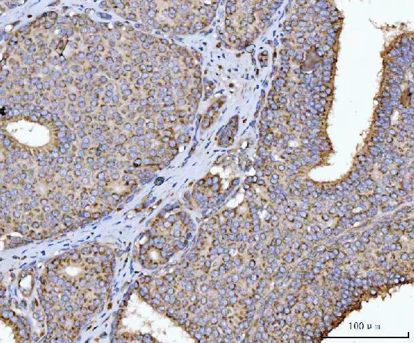
Real IHC data IHC analysis of HMGCR using anti-HMGCR antibody (A00643-3). HMGCR was detected in a paraffin-embedded section of human breast cancer tissue. Heat mediated antigen retrieval was performed in EDTA buffer (pH 8.0, epitope retrieval solution). The tissue section was blocked with 10% goat serum. The tissue section was then incubated with 2 μg/ml rabbit anti-HMGCR Antibody (A00643-3) overnight at 4°C. Peroxidase Conjugated Goat Anti-rabbit IgG was used as secondary antibody and incubated for 30 minutes at 37°C. The tissue section was developed using HRP Conjugated Rabbit IgG Super Vision Assay Kit (Catalog # SV0002) with DAB as the chromogen.
Anti-HMGCR Antibody ®
Cat # A00643-3

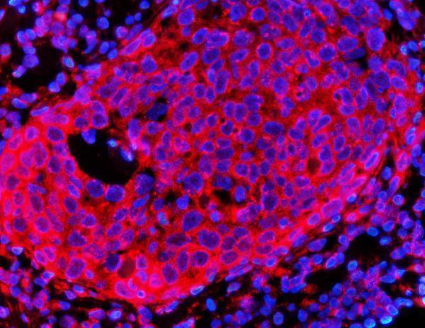
A00643-3 will render with its own IHC figure from a human breast cancer paraffin section; its catalog also includes IF imaging of that sample type (A00643-3 image captions). M00643-1 lists IHC for human, mouse and rat, but has no supplied IHC or IF figure and will not render a card (catalog applications/reactivity/image captions).

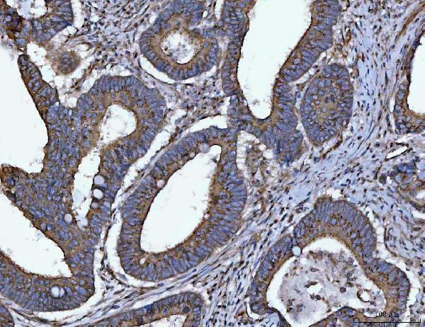
Which to pick: For tissue IHC, pick A00643-3: its IHC captions document human breast, colorectal and testicular cancer paraffin sections; the fixative is unreported (A00643-3 IHC captions). For IF, pick A00643-3 based on its human breast cancer paraffin-section image; ICC validation is unreported (A00643-3 IF caption; catalog applications). For mouse or rat IHC, consider rabbit monoclonal M00643-1, clone 33H19, which lists those species and IHC, while noting that no IHC image is supplied (catalog host/clone/applications/reactivity/image captions).

Each figure is that product's own IHC / IF validation image from its datasheet.

References

  1. UniProt Consortium. UniProt entry P04035 (HMDH_HUMAN, 3-hydroxy-3-methylglutaryl-coenzyme A reductase).
  2. Human Protein Atlas. HMGCR tissue IHC expression (reliability: Supported).
  3. Human Protein Atlas. HMGCR subcellular location (ICC-IF): Membrane.
  4. Human Protein Atlas. HMGCR antibody validation summary (3 antibodies).
  5. A conserved eIF1A(+) luminal cell-centered hypoxic and "cold" tumor microenvironment promotes pan-subtype prostate cancer progression. Cell reports. Medicine 2026 — PMC12923978.
  6. Low Density Lipoprotein Receptor (LDLR) and 3-Hydroxy-3-Methylglutaryl Coenzyme a Reductase (HMGCR) Expression are Associated with Platinum-Resistance and Prognosis in Ovarian Carcinoma Patients. Cancer management and research 2021 — PMC8664653.
  7. HMG-CoA reductase expression in primary colorectal cancer correlates with favourable clinicopathological characteristics and an improved clinical outcome. Diagnostic pathology 2014 — PMC4000148.
  8. Statin use, HMGCR expression, and breast cancer survival - The Malmö Diet and Cancer Study. Scientific reports 2020 — PMC6969108.
  9. PubMed PMID:2991281 — UniProt-cited evidence.
  10. PubMed PMID:14702039 — UniProt-cited evidence.
  11. PubMed PMID:15372022 — UniProt-cited evidence.