This website uses cookies to ensure you get the best experience on our website.


- Table of Contents
Plan chromogenic IHC-P for HMGCS2 using liver hepatocytes as a positive control (HPA tissue IHC). The guide covers expected cytoplasmic staining across positive cell types (HPA tissue IHC) and the catalog antibody’s IHC-P working range of 2–5 μg/ml (datasheet A04371-2).

Expected localisation, validated protocols, controls and antibodies — the at-a-glance facts below, then the full design guide.
| Expected localisation | Cytoplasmic tissue staining (HPA tissue IHC) | |
| Staining pattern | Hepatocytes, GI endocrine cells and enterocytes: cytoplasmic (HPA tissue IHC) | |
| Antigen retrieval | EDTA pH 8.0 HIER, heat-mediated (datasheet A04371-2) | |
| Positive control | Appendix+4 more · see all | |
| Negative control | Adipose tissue+4 more · see all |
| Fixation | Keep fixation consistent across paraffin sections. (standard IHC practice; not target-specific) | |
| Caveat | GI staining varies by cell type and site (HPA tissue IHC) | |
| Regulation | Liver expression 200-fold higher (UniProt) | |
| Isoform / epitope | 3 isoforms; mature chain 38–508; check epitope coverage (UniProt) |
The catalog antibody protocol specifies heat-mediated EDTA pH 8.0 retrieval (datasheet A04371-2). Published IHC options cover colon tissue (PMC11023725) and breast apocrine carcinoma (PMC4229141).
| Sample | Paraffin-embedded human gastric cancer tissue; fixative not specified (datasheet A04371-2) |
| Fixation | Image fixative and duration unreported (datasheet A04371-2); verify before use. |
| Sectioning | 4–5 µm sections on charged slides (standard) |
| Deparaffinisation | Xylene, graded ethanol series to water (standard) |
| Antigen retrieval | Heat retrieval: EDTA pH 8.0 (datasheet A04371-2); 20 min, 95–100 °C (standard) |
| Peroxidase block | 3% H2O2, 10 min, room temperature (standard) |
| Blocking | 10% goat serum (datasheet A04371-2) |
| Primary antibody | Rabbit anti-HMGCS2, 2-5 μg/ml (datasheet A04371-2) |
| Primary incubation | Overnight at 4 °C (datasheet A04371-2) |
| Detection | Streptavidin-biotin complex (SABC), DAB chromogen (datasheet A04371-2) |
| Counterstain | Hematoxylin, blue, dehydrate and mount (standard) |
| Expected result | HMGCS2-positive staining in endocrine cells of appendix (HPA tissue IHC: High). HPA tissue profile: Cytoplasmic expression in hepatocytes, gastrointestinal tract, urinary bladder, gallbladder and cells in seminiferous tubules. No signal in the no-primary control. |
In paraffin-section IHC, expect HMGCS2 staining in hepatocyte cytoplasm and in selected gastrointestinal, gallbladder and kidney cell populations (HPA: tissue IHC, Enhanced). Its mitochondrial localization supports a cytoplasmic, sometimes granular pattern; a precise mitochondrial outline may not resolve with chromogenic detection (UniProt P54868: mitochondrion; HPA: subcellular ICC-IF). HMGCS2 has no transmembrane segment, so a membrane-rim pattern is unexpected (UniProt P54868: topology).
| Clear cytoplasmic staining in hepatocytes, with low background in the surrounding section. | This fits the strongest reference tissue: HPA records high hepatocyte staining and tissue-enriched liver RNA (HPA: liver tissue IHC; HPA: RNA specificity). Compare cell-localized signal with the counterstain and nearby unstained areas; a uniform tint across the section is less persuasive than staining confined to hepatocytes. |
| Cytoplasmic staining in selected nonhepatic cells, including intestinal endocrine cells or kidney collecting ducts. | These can be valid positives: HPA reports high staining in appendix, colon, duodenal and rectal endocrine cells, small-intestinal enterocytes, gallbladder glandular cells, and kidney collecting ducts (HPA: tissue IHC). UniProt describes low kidney and colon expression at the tissue level, so bulk expression should not be used to dismiss a cell-restricted IHC signal (UniProt P54868: tissue specificity). |
| Predominantly nuclear staining or a sharp plasma-membrane rim in otherwise positive cells. | Treat this as a localization mismatch requiring investigation: UniProt places HMGCS2 in mitochondria and annotates no transmembrane segment (UniProt P54868: subcellular location and topology). Check whether the signal follows tissue edges, nuclei or the detection reagents before scoring it as HMGCS2. |
| Staining concentrated in an unexpected cell population, such as adipocytes, while expected cells are weak. | Consider cross-reactivity or endogenous detection activity before interpreting the result biologically. HPA reports HMGCS2 as not detected in adipocytes, while hepatocytes stain highly (HPA: adipose tissue and liver IHC). An HPA negative is a comparison for this assay, not proof that every sample must be negative. |
| Diffuse color throughout cells, stroma or the whole section, or no signal in hepatocytes. | Diffuse staining lacks the cell and compartment restriction expected from HPA tissue IHC and mitochondrial localization (HPA: tissue IHC; UniProt P54868: mitochondrion). A blank liver section is inconclusive until a working positive control, detection reagents and the antibody's IHC-P conditions have been checked (HPA: high hepatocyte staining; general IHC practice). |
| Cell distribution across tissues | Liver is the strongest reference because HPA reports high hepatocyte staining and liver-enriched RNA (HPA: liver IHC and RNA specificity). Other high calls are cell-specific; do not require every cell in a positive organ to stain (HPA: tissue IHC). |
| Compartment and optical resolution | HMGCS2 is mitochondrial, and HPA ICC-IF mainly localizes it to mitochondria (UniProt P54868: subcellular location; HPA: subcellular ICC-IF). In chromogenic IHC, score a cell-restricted cytoplasmic pattern without requiring individual mitochondria to resolve (general IHC microscopy practice). |
| Protein forms and antibody coverage | UniProt lists three isoforms and a mature chain spanning residues 38–508 (UniProt P54868: isoforms and processing). The supplied evidence gives no epitope or isoform coverage for the catalog antibody; staining alone cannot identify an isoform or establish recognition of the processed chain. |
| Strength of the reference pattern | HPA rates the tissue pattern Enhanced and lists three antibodies with Enhanced IHC validation (HPA: tissue reliability and antibody validation). That supports use of the documented distribution as a comparator, while an unexpected result in a new specimen still needs its own controls. |
| Situation | Likely cause | Next action |
|---|---|---|
| Liver positive control has no visible hepatocyte staining. | The expected high hepatocyte signal is missing; the supplied sources do not identify a target-specific fixation failure (HPA: liver IHC). | Check the catalog antibody's IHC-P instructions, reagent activity, detection sequence and a concurrent positive control; review retrieval conditions as a general IHC workflow check, without assuming HMGCS2-specific retrieval sensitivity. |
| Most of the section shows uniform brown color. | A tissue-wide deposit does not match HPA's selected cell pattern and can arise from nonspecific detection or residual endogenous enzyme activity (HPA: tissue IHC; general chromogenic IHC practice). | Inspect no-primary and detection-only controls, then review blocking, washing and detection timing. Reassess only cell-localized signal after the background is controlled (general IHC practice). |
| Apparent signal is strongest in nuclei or along cell borders. | That compartment conflicts with mitochondrial HMGCS2 and its lack of a transmembrane segment (UniProt P54868: location and topology). | Compare with the counterstain and a no-primary control; check for edge deposits or chromogen precipitate before calling the compartment positive (general IHC practice). |
| Adipocytes stain strongly while hepatocytes remain weak. | The distribution conflicts with the HPA comparison: adipocytes are not detected and hepatocytes are high (HPA: adipose tissue and liver IHC). Cross-reactivity or detection background is plausible, but the image alone cannot distinguish them. | Repeat with appropriate negative detection controls and a liver positive control. Confirm that the signal belongs to cells rather than extracellular deposits before interpreting a new distribution (general IHC practice). |
| Kidney collecting ducts stain despite a low kidney expression summary. | These observations use different scales: HPA calls collecting-duct staining high, whereas UniProt describes low kidney expression overall (HPA: kidney IHC; UniProt P54868: tissue specificity). | Score the collecting ducts separately from other renal cells and compare the location and cytoplasmic pattern with the HPA reference; do not reject a localized positive solely because whole-tissue expression is low. |
| An IF image appears more punctate than the chromogenic IHC section. | HPA approves mitochondrial localization by ICC-IF, while individual organelles may be unresolved in chromogenic paraffin sections (HPA: subcellular ICC-IF; general IHC microscopy practice). | Use the mitochondrial IF finding as a localization check and assess this IHC section for the expected cell-restricted cytoplasmic signal; consult the separate IF/ICC guide for that application's workflow. |
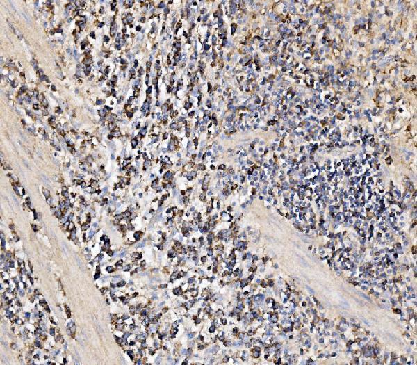
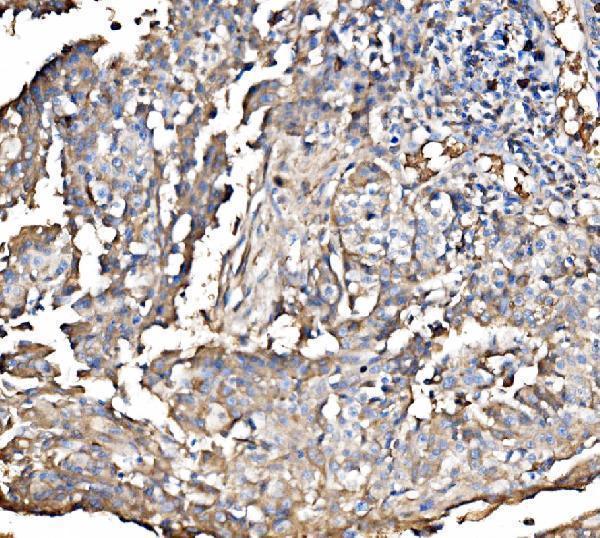
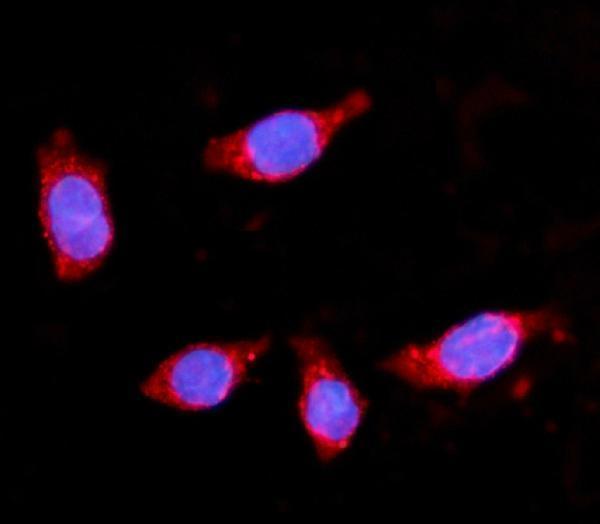
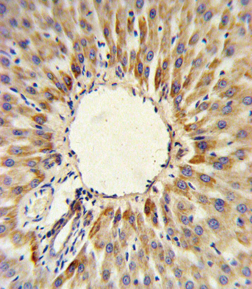


Comprehensive Human Protein Atlas IHC scoring per tissue (reliability: Enhanced — High consistency between antibody staining and RNA expression data.). Rows are taken directly from the HPA tissue chart — click any row's HPA link to view the source.
| Tissue | Cell type | Level | Evidence | Source |
|---|---|---|---|---|
| Appendix | Endocrine cells | High | Protein (IHC) | HPA → |
| Colon | Endocrine cells | High | Protein (IHC) | HPA → |
| Duodenum | Endocrine cells | High | Protein (IHC) | HPA → |
| Gallbladder | Glandular cells | High | Protein (IHC) | HPA → |
| Kidney | Collecting ducts | High | Protein (IHC) | HPA → |
| Tissue | Cell type | Level | Evidence | Source |
|---|---|---|---|---|
| Adipose tissue | Adipocytes | Not detected | Protein (IHC) | HPA → |
| Adrenal gland | Glandular cells | Not detected | Protein (IHC) | HPA → |
| Bone marrow | Hematopoietic cells | Not detected | Protein (IHC) | HPA → |
| Caudate | Glial cells | Not detected | Protein (IHC) | HPA → |
| Cerebellum | Cells in granular layer | Not detected | Protein (IHC) | HPA → |
Troubleshoot HMGCS2 staining in paraffin sections by checking retrieval, cellular location, detection background, and scoring against defined tissue controls.
Two anti-HMGCS2 antibodies have human paraffin-section IHC images (catalog IHC captions); A04371-2 also has IF data in CACO-2 cells (catalog IF caption) and lists human, mouse, and rat reactivity (catalog reactivity).
A04371-1 has an IHC image from formalin-fixed, paraffin-embedded human hepatocarcinoma (A04371-1 IHC caption). A04371-2 has IHC images from paraffin-embedded human gastric, laryngeal, liver, and gall bladder cancers, plus IF data in CACO-2 cells (A04371-2 IHC and IF captions).
Which to pick: For tissue IHC, choose A04371-1 when documented fixation matters: its polyclonal antibody is listed for human IHC-P and imaged in paraffin-section, paraffin-embedded hepatocarcinoma (A04371-1 catalog applications, reactivity, dilution data, and IHC caption). For IF/ICC, choose A04371-2, which lists both applications and has a CACO-2 IF image; its IHC captions document paraffin sections but do not report the fixative (A04371-2 catalog applications, IF caption, and IHC captions). For cross-species work, A04371-2 lists human, mouse, and rat reactivity, while its pictured IHC examples are human (A04371-2 catalog reactivity and IHC captions). The selected A04371-2 tissue-IHC caption documents paraffin sections, but does not specify the fixative (selected-SKU IHC image A04371-2).