HMGN2 / Non-histone chromosomal protein HMG-17 · IHC design guide

Design Immunohistochemistry for HMGN2

Plan HMGN2 staining in paraffin sections around its general nuclear tissue pattern (HPA tissue IHC). Use bone marrow hematopoietic cells with high staining and parathyroid glandular cells with low staining as comparison tissues (HPA tissue IHC).

Evidence assembled Oct 2026 · For research use; verify linked source records and product datasheet before use
Immunohistochemistry protocol sheet for HMGN2 (IHC for HMGN2): expected localisation General nuclear tissue staining (HPA tissue IHC), antibody A04839-2, validated IHC image, and IHC protocol steps
Printable HMGN2 IHC protocol sheet — expected localisation General nuclear tissue staining (HPA tissue IHC), antibody A04839-2, controls and protocol steps. Open the full HMGN2 IHC guide →

HMGN2 Immunohistochemistry Experimental Design Guide

Expected localisation, validated protocols, controls and antibodies — the at-a-glance facts below, then the full design guide.

Must know before staining
Expected localisation General nuclear tissue staining (HPA tissue IHC)
Staining pattern Nuclear staining across many tissue cell types (HPA tissue IHC)
Antigen retrieval EDTA pH 8.0 HIER, heat-mediated (datasheet A04839-2)
Positive control ⓘ Bone marrow+4 more · see all
Negative control ⓘ None in HPA (detected in all 45 tissues); use no-primary + isotype controls
Important caveats
Reasons your staining may differ from the expected pattern.
Fixation Keep fixation consistent across sections (standard IHC practice; not target-specific)
Caveat Phosphorylation can enrich cytoplasmic HMGN2 (UniProt)
Regulation No expression regulator annotated (UniProt)
Isoform / epitope No annotated isoforms; chain spans residues 2–90 (UniProt)
Section 1

Recommended HMGN2 IHC & IF Protocols

The catalog antibody’s IHC-P protocol is followed by published HMGN2 staining protocols for esophageal tissue (PMC7675040) and paraffin-embedded tissue sections (PMC11907455).

Recommended immunohistochemistry (IHC-P) protocol parameters
SampleParaffin-embedded human rectal cancer tissue; fixative not specified (datasheet A04839-2)
FixationImage fixative and duration unreported (datasheet A04839-2); verify before use.
Sectioning4–5 µm sections on charged slides (standard)
DeparaffinisationXylene, graded ethanol series to water (standard)
Antigen retrievalHeat retrieval: EDTA pH 8.0 (datasheet A04839-2); 20 min, 95–100 °C (standard)
Peroxidase block3% H2O2, 10 min, room temperature (standard)
Blocking10% goat serum (datasheet A04839-2)
Primary antibodyRabbit anti-HMGN2, 0.5-1μg/ml recommended; image 2 μg/ml (datasheet A04839-2)
Primary incubationOvernight at 4 °C (datasheet A04839-2)
DetectionHRP-conjugated secondary, DAB chromogen (datasheet A04839-2)
CounterstainHematoxylin, blue, dehydrate and mount (standard)
Expected resultHMGN2-positive staining in hematopoietic cells of bone marrow (HPA tissue IHC: High). HPA tissue profile: General nuclear expression. No signal in the no-primary control.
💡Decision noteStart with EDTA pH 8.0 heat retrieval for the catalog antibody (datasheet: A04839-2); citrate retrieval is a published alternative for esophageal sections (PMC7675040).
Section 2

What Is the Expected HMGN2 Staining Pattern?

Expect HMGN2 staining chiefly in nuclei: HPA reports general nuclear expression in tissue IHC, while ICC-IF places it mainly in the nucleoplasm, with additional nucleolar signal (HPA: tissue IHC; HPA: ICC-IF). UniProt also lists cytoplasm, with enrichment upon phosphorylation (UniProt P05204: localization). Staining can span cell types; HPA rates the tissue IHC profile Supported, with medium consistency against RNA data (HPA: tissue IHC). HMGN2 has no transmembrane segment (UniProt P05204: topology).

What am I looking at on my slide?
Distinct nuclear chromogen in hematopoietic cells, glia or glandular cells.This fits the general nuclear profile (HPA: tissue IHC). HPA reports High staining in bone-marrow hematopoietic cells, caudate glia, and epididymal, gallbladder and thyroid glandular cells (HPA: tissue IHC). Identify the stained cell population against tissue morphology and the counterstain before scoring intensity.
Cytoplasm dominates while nuclei are pale, or staining outlines cell membranes.A membrane-dominant pattern conflicts with the nuclear tissue profile and absence of a transmembrane segment (HPA: tissue IHC; UniProt P05204: topology). Check for artefact and compare a known-positive section. Do not automatically discard cytoplasmic signal: phosphorylation can enrich HMGN2 there (UniProt P05204: localization).
Strong colour appears in an unexpected cell population, with little nuclear signal in a matched positive area.Consider cross-reactivity or endogenous detection activity before assigning that colour to HMGN2 (general IHC practice). HPA reports Low staining in esophageal squamous cells and bladder urothelial cells, but lists no negative tissue; Low is not proof of absence (HPA: tissue IHC).
Weak, diffuse colour covers nuclei, cytoplasm and tissue spaces alike.Poor compartment contrast limits interpretation. Uneven reagent removal, excess antibody or endogenous detection activity can produce background (general IHC practice). Compare a primary-antibody omission control and inspect whether nuclei remain distinguishable; diffuse colour alone does not demonstrate HMGN2.
No nuclear signal appears in bone-marrow hematopoietic cells or another selected high-staining population.A negative run is difficult to interpret when a matched known-positive section also lacks signal. HPA reports High staining in bone-marrow hematopoietic cells, among other populations (HPA: tissue IHC). First verify section quality and the detection run (general IHC practice); tissue variation remains possible because the HPA profile is Supported, with medium RNA-staining consistency (HPA: tissue IHC).
💡Expected HMGN2 appearanceCall a result positive when identifiable cells show distinct, predominantly nuclear chromogen, especially High staining in an HPA high-staining population (HPA: tissue IHC); isolated membrane colour or uniform diffuse colour without nuclear contrast is suspect (UniProt P05204: topology; general IHC practice).
How each factor affects the staining
Cell population and intensityHPA reports High staining in lung alveolar type I and pancreatic endocrine cells, Medium in adipocytes, and Low in several listed populations (HPA: tissue IHC). Select and score the intended cell type; whole-section colour can conceal that variation.
Compartment and modificationNuclear staining anchors tissue interpretation (HPA: tissue IHC). UniProt reports cytoplasmic enrichment upon phosphorylation, but the supplied record does not establish how often this appears in paraffin IHC (UniProt P05204: localization).
Antibody evidenceCAB018371 is Supported for IHC; HPA077364 has no listed IHC status (HPA: antibodies). HPA rates the tissue staining profile Supported, with medium RNA-staining consistency (HPA: tissue IHC). These labels support interpretation without proving every cell-level signal is specific.
Processing and membrane associationUniProt lists no signal peptide, propeptide or transmembrane segment, and records a chain spanning residues 2–90 (UniProt P05204: processing; topology). Those annotations provide no basis for expecting a membrane or secreted staining pattern.
Q: What should IF/ICC show?A: Mainly nucleoplasm, with additional nucleolar signal (HPA: ICC-IF). The HPA subcellular assessment is Supported; it describes ICC-IF localization, while this section's primary readout is chromogenic tissue IHC (HPA: ICC-IF; HPA: tissue IHC).
Why is my staining missing, weak or wrong?
SituationLikely causeNext action
Known-positive tissue has no visible nuclear staining.The detection run or section may have failed (general IHC practice); HPA reports High staining in bone-marrow hematopoietic cells (HPA: tissue IHC).Confirm the tissue contains the expected cells, review reagent performance and compare a concurrent known-positive section (general IHC practice). If signal remains absent, revisit the catalog antibody's IHC-P instructions; the supplied evidence gives no target-specific retrieval setting.
Chromogen is widespread and nuclear boundaries are unclear.Background from reagent carryover, excessive primary antibody or endogenous detection activity can obscure compartments (general IHC practice).Compare a primary-antibody omission control; review blocking, washing and detection conditions, then score only interpretable nuclear signal (general IHC practice). No HMGN2-specific dilution is supplied.
Staining appears almost entirely cytoplasmic.This diverges from general nuclear tissue staining (HPA: tissue IHC), although phosphorylation-associated cytoplasmic enrichment is reported (UniProt P05204: localization).Check nuclear counterstaining and a known-positive section, then assess whether the cytoplasmic pattern is cell-specific or diffuse background (general IHC practice). Do not infer phosphorylation from chromogen location alone.
Unexpectedly strong colour appears in an HPA Low population.Cell identification, cross-reactivity or endogenous detection activity may explain the discrepancy (general IHC practice); HPA Low does not mean absent (HPA: tissue IHC).Confirm cell identity with morphology, compare a high-staining population and examine an omission control (general IHC practice). Treat the result as unresolved if specificity cannot be distinguished from background.
Two sections give different nuclear intensity.Section and run variation can change apparent chromogen intensity (general IHC practice). HPA also reports varying levels across cell populations and medium consistency with RNA data (HPA: tissue IHC).Compare the same cell type, counterstain and concurrent controls under consistent scoring criteria (general IHC practice). Report tissue and cell identity alongside intensity; do not infer a fixation effect specific to HMGN2 from these data.

Sample controls for HMGN2 IHC & IF

🧪Run bone marrow first: hematopoietic cells should stain (HPA: High in bone marrow hematopoietic cells). HPA detects HMGN2 in all 45 scored tissues, so there is no supported negative tissue; use no-primary and isotype controls, and treat cells showing counterstain without nuclear chromogen as internal background references rather than proven HMGN2-negative cells (HPA: no negative tissue rows; UniProt P05204: nucleus).
Positive control tissue: Bone marrow (Hematopoietic cells, HPA High)
Negative control tissue: None in HPA: HMGN2 is detected in all 45 scored tissues. Use a no-primary (secondary-only) and an isotype control instead.
ICC-IF cell lines (HPA subcellular resource): HPA ICC-IF images show HMGN2 in A-431, U-251MG, U2OS, Rh30, SiHa, with annotated localisation: Nucleoplasm (supported) (HPA subcellular).
Technical controls: Include a no-primary, secondary-only slide and a concentration-matched rabbit IgG isotype control matched to the primary antibody’s clonality, plus HMGN2 knockout material processed in parallel as a biological negative (selected caption: rabbit anti-HMGN2 antibody). Quench endogenous peroxidase in bone marrow before HRP/DAB detection because hematopoietic cells can produce peroxidase background (HPA: bone marrow hematopoietic cells High; selected caption: HRP/DAB detection).
⚠️Feasibility: A target-specific fixation window or fixation effect is unreported, and the selected A04839-2 paraffin-section caption does not state a fixative. The caption reports heat retrieval in EDTA at pH 8.0, but does not establish whether retrieval is required (selected A04839-2 tissue-IHC caption). HPA supports nucleoplasmic and additional nucleolar ICC/IF localization, but the supplied evidence gives no frozen-section or IF protocol from which to judge relative ease (HPA subcellular: supported nucleoplasm and nucleoli).

HPA tissue IHC evidence for HMGN2

Comprehensive Human Protein Atlas IHC scoring per tissue (reliability: Supported — Medium consistency between antibody staining and RNA expression data.). Rows are taken directly from the HPA tissue chart — click any row's HPA link to view the source.

Positive expression · recommended positive controls

TissueCell typeLevelEvidenceSource
Bone marrow Hematopoietic cells High Protein (IHC) HPA →
Caudate Glial cells High Protein (IHC) HPA →
Epididymis Glandular cells High Protein (IHC) HPA →
Gallbladder Glandular cells High Protein (IHC) HPA →
Lung Alveolar cells type I High Protein (IHC) HPA →

Undetected expression · recommended negative controls

TissueCell typeLevelEvidenceSource
None in HPA: HMGN2 is detected in all 45 scored tissues. Use a no-primary (secondary-only) and an isotype control instead.
Section 3

Advanced HMGN2 IHC Tips

Troubleshoot chromogenic HMGN2 staining in paraffin sections using the catalog image, protein annotations, and tissue expression evidence.

Which retrieval conditions should I start with for weak HMGN2 nuclear staining?
Start with heat mediated retrieval in EDTA at pH 8.0 (datasheet A04839-2). The selected paraffin section image used those conditions before overnight incubation with 2 μg/ml primary antibody at 4°C (datasheet A04839-2). If nuclei remain weak, compare a small series of retrieval heating times while holding buffer, antibody concentration, and detection constant (standard IHC practice). Score nuclear signal alongside tissue damage and diffuse background, because stronger staining alone does not establish improved specificity (standard IHC practice). Include a previously staining section in each run; HMGN2 is generally nuclear, providing a compartment check (HPA: general nuclear expression).
Could fixation explain weak or uneven HMGN2 staining?
Target specific fixation sensitivity is unknown: the selected paraffin section caption does not state a fixative (datasheet A04839-2). Record the specimen fixative and duration, then compare sections with matched processing and the same pH 8.0 EDTA retrieval (datasheet A04839-2; standard IHC practice). Inspect tissue preservation and nuclear counterstain before changing antibody concentration, since damaged or poorly preserved nuclei complicate interpretation (standard IHC practice). Use a previously staining control section to separate a specimen processing problem from a run wide detection problem (standard IHC practice). Do not infer fixation tolerance from tissue expression patterns or protein modifications (HPA: tissue IHC; UniProt P05204: modified residues).
How should I assess HMGN2 staining outside the nucleus?
Expect predominantly nuclear staining: HPA reports general nuclear expression, with supported nucleoplasmic and additional nucleolar localisation (HPA: tissue IHC and subcellular). Cytoplasmic enrichment can occur upon phosphorylation, so a cytoplasmic pattern merits assessment rather than automatic rejection (UniProt P05204: subcellular location). Compare cytoplasmic staining with intact nuclear signal, cell morphology, and a matched negative control; diffuse staining across tissue compartments suggests background (standard IHC practice). HMGN2 has no annotated transmembrane segment, so a crisp membrane only pattern is unexpected (UniProt P05204: topology). Document nuclear and cytoplasmic staining separately instead of combining them into one positive score (standard IHC practice).
Could an epitope or protein variant explain a missing nuclear signal?
No isoforms are annotated for HMGN2 in the supplied record, so an isoform specific explanation lacks support here (UniProt P05204: isoforms). The record lists modifications including phosphoserine at position 25 and alternate modifications at positions 29 and 82 (UniProt P05204: modified residues). Without an epitope map for this antibody, their effect on recognition cannot be assigned (datasheet A04839-2: epitope not supplied). Compare retrieval and staining against a previously positive section before attributing loss to a modification (standard IHC practice). Report the antibody identity and processing conditions with the result so another laboratory can assess epitope related uncertainty (standard IHC practice).
How can I investigate an IHC pattern with multiplex IF?
Treat IF as a separate assay to validate; the selected antibody evidence describes chromogenic staining of a paraffin section (datasheet A04839-2). Pair HMGN2 with a validated marker for the cell population being examined, such as hematopoietic cells in bone marrow (HPA: high in bone marrow hematopoietic cells; standard IF practice). Choose fluorophores after checking unstained tissue autofluorescence, and include single stain controls to assess channel bleed through (standard IF practice). Because the expected signal is nuclear and HMGN2 has no transmembrane segment, permeabilise sufficiently for nuclear access, while avoiding assumptions about the unmapped antibody epitope (HPA: subcellular; UniProt P05204: topology; standard IF practice).
What should I check when DAB appears throughout the section?
The selected image used peroxidase detection and DAB after incubation with 2 μg/ml antibody overnight at 4°C (datasheet A04839-2). Check a no primary control, apply an appropriate endogenous peroxidase block, and inspect whether colour appears outside intact cells (standard IHC practice). If background persists, optimise blocking and washes, then titrate primary antibody around the documented concentration while keeping development time consistent (datasheet A04839-2; standard IHC practice). Interpret signal against the expected general nuclear pattern, rather than treating diffuse cytoplasmic or extracellular colour as positive (HPA: tissue IHC; standard IHC practice). Record any changes alongside the control results (standard IHC practice).
How should I score HMGN2 across heterogeneous tissue sections? ⚠ ANSWER MARKED FOR VERIFICATION
Define the cell population and anatomical region before scoring, then exclude folds, necrosis, and poorly preserved areas (standard IHC practice). For nuclear staining, record the percentage of positive nuclei and intensity, or calculate an H score from percentages at each intensity grade (HPA: general nuclear expression; standard IHC practice). Normalise positive counts to all evaluable nuclei in the same region, and report the number of cells or sampled area used (standard IHC practice). If cell density matters, report positive nuclei per mm² alongside the percentage, with identical thresholds across sections (standard IHC practice). Score cytoplasmic staining separately because phosphorylation may shift HMGN2 enrichment (UniProt P05204: subcellular location).
When is an apparent HMGN2 positive cell likely an artefact?
A convincing result shows staining in intact nuclei and the expected cell population, with a clean negative control (HPA: general nuclear expression; standard IHC practice). HPA reports high staining in bone marrow hematopoietic cells and lung alveolar type I cells, but its tissue IHC reliability is Supported with medium staining to RNA consistency (HPA: tissue IHC). Recheck isolated membrane only colour, section edge accentuation, necrotic debris, and signal in the no primary control before calling a cell positive (UniProt P05204: topology; standard IHC practice). Residual endogenous peroxidase can mimic DAB positivity, so compare the suspect area with an appropriately blocked control (standard IHC practice). Cytoplasmic enrichment remains biologically possible upon phosphorylation (UniProt P05204: subcellular location).
Boster reagents

Best HMGN2 / Non-histone chromosomal protein HMG-17 IHC Antibodies

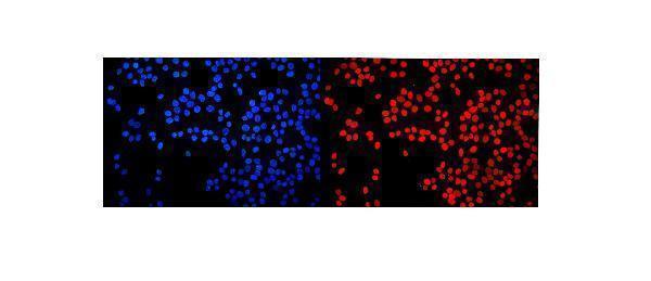
A04839-2 has IHC images from human paraffin sections and an IF image from MCF7 cells (catalog image captions); M04839 lists human IHC reactivity without an IHC image (catalog applications and images).

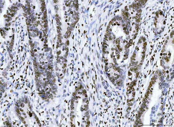
Real IHC data IHC analysis of HMGN2 using anti-HMGN2 antibody (A04839-2). HMGN2 was detected in a paraffin-embedded section of human rectal cancer tissue. Heat mediated antigen retrieval was performed in EDTA buffer (pH 8.0, epitope retrieval solution). The tissue section was blocked with 10% goat serum. The tissue section was then incubated with 2 μg/ml rabbit anti-HMGN2 Antibody (A04839-2) overnight at 4°C. Peroxidase Conjugated Goat Anti-rabbit IgG was used as secondary antibody and incubated for 30 minutes at 37°C. The tissue section was developed using HRP Conjugated Rabbit IgG Super Vision Assay Kit (Catalog # SV0002) with DAB as the chromogen.
Anti-HMGN2 Antibody ®
Cat # A04839-2

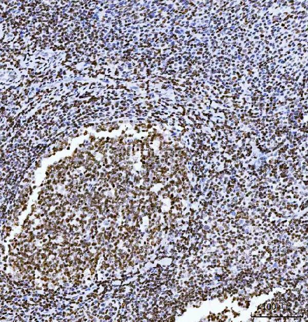
A04839-2 has IHC images from human rectal cancer, tonsil, thyroid cancer and liver cancer paraffin sections, plus an IF image from MCF7 cells (A04839-2 image captions). M04839 lists human IHC as an application but has no IHC or IF image in the payload (M04839 catalog applications and images).

Which to pick: For tissue IHC, choose A04839-2 when an image from a paraffin section is useful; its caption does not report the fixative (A04839-2 IHC image caption). For IF/ICC, choose A04839-2 because both applications are listed and an IF image is provided; M04839 is a rabbit monoclonal with IHC listed but no IHC image (catalog applications, clone and image captions). Neither SKU has stated cross-species reactivity beyond human (catalog reactivity).

Each figure is that product's own IHC / IF validation image from its datasheet.

References

  1. UniProt Consortium. UniProt entry P05204 (HMGN2_HUMAN, Non-histone chromosomal protein HMG-17).
  2. Human Protein Atlas. HMGN2 tissue IHC expression (reliability: Supported).
  3. Human Protein Atlas. HMGN2 subcellular location (ICC-IF): Mainly localized to the nucleoplasm. In addition localized to the nucleoli..
  4. Human Protein Atlas. HMGN2 antibody validation summary (2 antibodies).
  5. Spatial transcriptomic-metabolic features of tumor foci and tumor capsule in microvascular invasion with hepatocellular carcinoma: A spatial multi-omics study. PLoS medicine 2026 — PMC13178920.
  6. Multi-Omics Analysis to Characterize Cigarette Smoke Induced Molecular Alterations in Esophageal Cells. Frontiers in oncology 2020 — PMC7675040.
  7. HMGN2 accelerates the proliferation and cell cycle progression of glioblastoma by regulating CDC20 expression. Genes & diseases 2025 — PMC11907455.
  8. Identification and multi-layered validation of seven diagnostic biomarkers for dilated cardiomyopathy via integrative machine learning, single-cell transcriptomics, and Mendelian randomization. Frontiers in cell and developmental biology 2026 — PMC13286953.
  9. PubMed PMID:3754870 — UniProt-cited evidence.
  10. PubMed PMID:2565024 — UniProt-cited evidence.
  11. PubMed PMID:16710414 — UniProt-cited evidence.