This website uses cookies to ensure you get the best experience on our website.


- Table of Contents
Plan HNRNPF paraffin IHC around nuclear staining across tissues (HPA tissue IHC). Start with the catalog antibody's 0.5–1 μg/mL IHC range (datasheet A05806) and compare staining with the tissue profile (HPA tissue IHC).

Expected localisation, validated protocols, controls and antibodies — the at-a-glance facts below, then the full design guide.
| Expected localisation | Ubiquitous nuclear staining (HPA tissue IHC) | |
| Staining pattern | Nuclei across many tissue cell types (HPA tissue IHC) | |
| Antigen retrieval | Citrate pH 6 HIER, heat-mediated (datasheet A05806) | |
| Positive control | Adrenal gland+4 more · see all | |
| Negative control | Heart muscle |
| Fixation | Keep tissue fixation consistent across sections. (standard IHC practice; not target-specific) | |
| Caveat | Staining may reflect proteins from more than one gene (HPA tissue IHC) | |
| Regulation | Low tissue specificity (HPA tissue RNA) | |
| Isoform / epitope | No annotated isoforms; N-terminal processing at residue 2 (UniProt) |
The catalog antibody uses citrate pH 6 retrieval (datasheet A05806). These published chromogenic IHC protocols cover Merkel cell carcinoma (PMC4181674) and gastric cancer (PMC10590441).
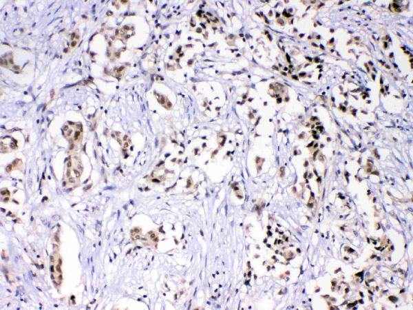
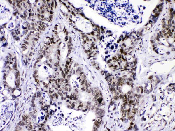
| Sample | Paraffin-embedded human mammary cancer tissues; fixative not specified (datasheet A05806) |
| Fixation | Image fixative and duration unreported (datasheet A05806); verify before use. |
| Sectioning | 4–5 µm sections on charged slides (standard) |
| Deparaffinisation | Xylene, graded ethanol series to water (standard) |
| Antigen retrieval | Heat retrieval: Citrate pH 6, 20 min (datasheet A05806) |
| Peroxidase block | 3% H2O2, 10 min, room temperature (standard) |
| Blocking | 10% goat serum (datasheet A05806) |
| Primary antibody | Rabbit anti-HNRNPF, 0.5-1μg/ml (datasheet A05806) |
| Primary incubation | Overnight at 4 °C (datasheet A05806) |
| Detection | Streptavidin-biotin complex (SABC), DAB chromogen (datasheet A05806) |
| Counterstain | Hematoxylin, blue, dehydrate and mount (standard) |
| Expected result | HNRNPF-positive staining in glandular cells of adrenal gland (HPA tissue IHC: High). HPA tissue profile: Ubiquitous nuclear expression. No signal in the no-primary control. |
HNRNPF is a nucleoplasmic protein with no transmembrane segment (UniProt P52597: location and topology). Expect nuclear staining across many cell types, with intensity varying by tissue and cell type (HPA tissue IHC: ubiquitous nuclear expression). HPA rates its tissue IHC evidence Supported, with medium agreement between staining and RNA data, and cautions that the staining may include protein from more than one gene (HPA tissue IHC: reliability).
| Distinct nuclear chromogen in glandular cells, respiratory epithelium, or marrow hematopoietic cells. | This fits the expected compartment (UniProt P52597: nucleoplasm) and cell patterns (HPA tissue IHC: High in adrenal, appendix and breast glandular cells; bronchial respiratory epithelial cells; and bone marrow hematopoietic cells). Score the proportion and intensity of stained nuclei within the relevant cell population. Nuclear staining supports the expected pattern, but the HPA warning about staining protein from more than one gene limits a claim of HNRNPF specificity (HPA tissue IHC: reliability). |
| Predominantly cytoplasmic or membrane staining with little nuclear staining. | This conflicts with the expected nucleoplasmic location and lack of a transmembrane segment (UniProt P52597: location and topology; HPA subcellular: mainly nucleoplasm). Treat it as a possible artefact or nonspecific signal. Review staining controls and whether nuclei can be clearly identified before calling the slide positive. |
| Strong cardiomyocyte staining, especially without a matching nuclear pattern. | Cardiomyocytes were not detected in the supplied tissue IHC record (HPA tissue IHC: heart muscle). Consider cross-reactivity or endogenous detection activity before interpreting this as HNRNPF. Compare the subcellular pattern and a detection-only control; the HPA finding is an observed pattern, not proof that every cardiomyocyte sample must be negative. |
| Diffuse chromogen obscures nuclear boundaries or appears across the section. | A diffuse deposit cannot establish the expected nuclear pattern (UniProt P52597: nucleoplasm; HPA tissue IHC: ubiquitous nuclear expression). Evaluate detection-only background, blocking, washing and chromogen development as general IHC checks. Do not score diffuse color as positive nuclei merely because HPA describes broad tissue expression (HPA tissue IHC: profile). |
| No nuclear signal in a tissue expected to stain strongly. | Check the sampled cell population first: HPA reports High staining in several defined populations, including adrenal glandular cells and bone marrow hematopoietic cells (HPA tissue IHC: positive tissue entries). Then examine section quality, retrieval and detection controls using general IHC practice. A failed control or absent expected cells prevents a confident biological negative call. |
| Cell population and expected intensity | Choose the population before scoring: HPA reports High staining in the listed glandular, respiratory epithelial, hematopoietic and selected brain cell populations, Low in hepatocytes, and Not detected in cardiomyocytes (HPA tissue IHC: positive, low and negative entries). A weak liver signal therefore carries a different expectation from a weak adrenal glandular signal. |
| IHC evidence and antibody specificity | The tissue profile is Supported, with medium agreement between staining and RNA data, and HPA cautions that the signal may include protein from more than one gene (HPA tissue IHC: reliability). HPA016884 has Supported IHC validation; HPA069667 has no IHC status in the supplied list (HPA antibodies: validation). Interpret staining as pattern evidence, with this specificity limit. |
| Retrieval and epitope information | The supplied records give no antibody epitope location or HNRNPF-specific retrieval response (UniProt P52597: supplied record; HPA tissue IHC: supplied record). If optimizing an IHC run, compare retrieval conditions against a suitable positive tissue and detection control as general IHC practice. Do not infer fixation sensitivity from topology, modifications or tissue staining levels. |
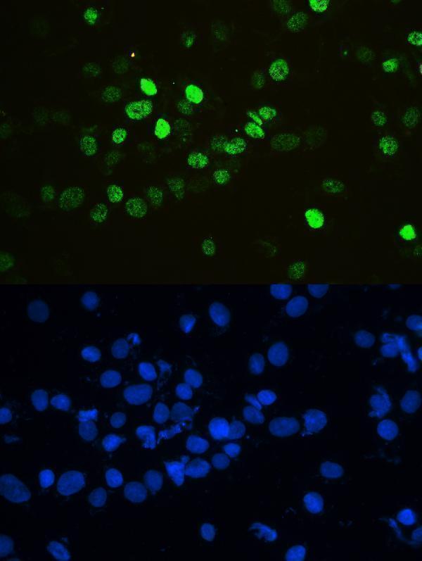
| IF/ICC Q&A: where should signal appear? | Predominantly in the nucleoplasm (HPA subcellular: mainly nucleoplasm; UniProt P52597: location). HPA lists ICC/IF images in A-431, U-251MG, U2OS, A-549 and HaCaT; HPA016884 is ICC Enhanced and HPA069667 is ICC Supported (HPA subcellular: image cell lines; HPA antibodies: validation). Those observations guide localisation, while IF/ICC methods belong in its separate guide. |
| Situation | Likely cause | Next action |
|---|---|---|
| No signal in adrenal glandular cells or marrow hematopoietic cells. | Both are reported High, so the result may reflect a run or sampling problem (HPA tissue IHC: adrenal gland and bone marrow). | Confirm that the named cells are present, then check the positive control, retrieval record, antibody application and detection reagents. These are general IHC checks; the supplied sources do not identify an HNRNPF-specific retrieval condition. |
| Only cytoplasmic or membrane color is visible. | The compartment conflicts with nucleoplasmic HNRNPF and its lack of a transmembrane segment (UniProt P52597: location and topology). | Use the counterstain to locate nuclei; compare detection-only and positive-control sections. Reassess the staining run before scoring cells positive. |
| Cardiomyocytes show conspicuous staining. | This departs from the supplied Not detected observation; cross-reactivity or endogenous detection activity is possible (HPA tissue IHC: cardiomyocytes). | Check whether color is truly nuclear. Inspect a detection-only control and the detection blocking steps as general chromogenic IHC practice; report the observed discrepancy if it persists. |
| Background obscures otherwise plausible nuclear staining. | Diffuse color prevents a reliable compartment call against the expected nuclear pattern (HPA tissue IHC: ubiquitous nuclear expression). | Inspect detection-only background, washing and chromogen development; adjust the run using general IHC controls, then score only nuclei whose boundaries remain distinguishable. |
| Hepatocytes stain weakly beside stronger cells. | Low hepatocyte staining is reported, so lower intensity there may fit the tissue profile (HPA tissue IHC: Low in hepatocytes). | Score hepatocytes separately from adjacent cell types and compare with a high-staining control population. Avoid treating low intensity alone as an assay failure. |
| A clean nuclear pattern is being treated as definitive HNRNPF identity. | HPA warns that its tissue staining may include protein from more than one gene (HPA tissue IHC: reliability). | Report the nuclear pattern and antibody used, including the specificity caveat. If gene-level attribution is essential, seek independent validation before making that claim. |


Comprehensive Human Protein Atlas IHC scoring per tissue (reliability: Supported — Medium consistency between antibody staining and RNA expression data. Caution, targets protein from more than one gene.). Rows are taken directly from the HPA tissue chart — click any row's HPA link to view the source.
| Tissue | Cell type | Level | Evidence | Source |
|---|---|---|---|---|
| Adrenal gland | Glandular cells | High | Protein (IHC) | HPA → |
| Appendix | Glandular cells | High | Protein (IHC) | HPA → |
| Bone marrow | Hematopoietic cells | High | Protein (IHC) | HPA → |
| Breast | Glandular cells | High | Protein (IHC) | HPA → |
| Bronchus | Respiratory epithelial cells | High | Protein (IHC) | HPA → |
| Tissue | Cell type | Level | Evidence | Source |
|---|---|---|---|---|
| Heart muscle | Cardiomyocytes | Not detected | Protein (IHC) | HPA → |
Troubleshoot HNRNPF staining by checking nuclear localisation, retrieval conditions, antibody specificity and cell level scoring (UniProt P52597; HPA tissue IHC).
A05806 has IHC images from paraffin sections of human mammary and intestinal cancers and mouse and rat intestine (catalog IHC captions), plus IF data in SKOV-3 cells (catalog IF caption).
A05806 is listed for IHC and has images from human mammary cancer, human intestinal cancer, mouse intestine, and rat intestine paraffin sections (catalog applications; catalog IHC captions). A05806 is also listed for IF and ICC, with an IF image from SKOV-3 cells (catalog applications; catalog IF caption).
Which to pick: Choose A05806 for paraffin-section IHC because its own captions document that preparation across the listed human, mouse, and rat samples (catalog IHC captions). Choose A05806 for IF/ICC because those applications are listed and its IF caption documents staining in SKOV-3 cells (catalog applications; catalog IF caption). For cross-species IHC, A05806 lists human, mouse, and rat reactivity and shows IHC images from all three; it is rabbit-hosted, its clonality is unreported, and the IHC captions do not report the fixative (catalog reactivity; catalog IHC captions; catalog host and clone fields).