This website uses cookies to ensure you get the best experience on our website.


- Table of Contents
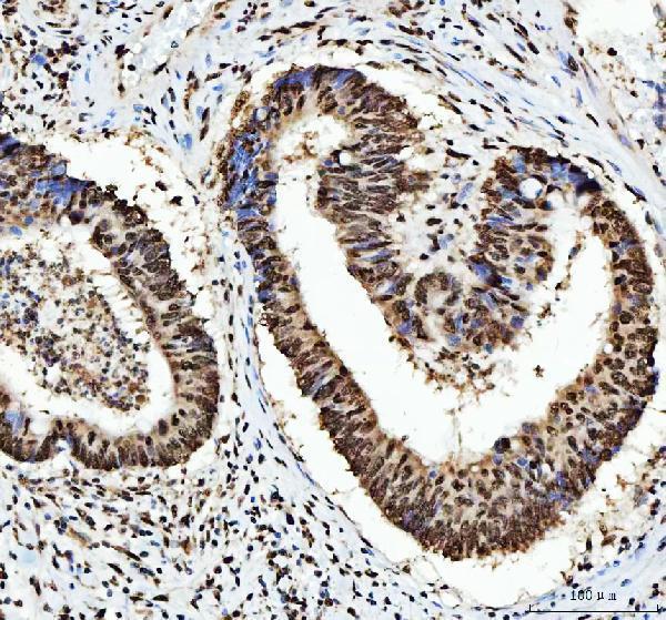
Plan HNRNPL paraffin IHC around its ubiquitous nuclear tissue pattern (HPA tissue IHC). The guide covers fixation consistency, antibody concentration, chromogenic detection and cell-level scoring; the catalog antibody is used at 2–5 μg/mL for IHC-P (datasheet A03769-4).

Expected localisation, validated protocols, controls and antibodies — the at-a-glance facts below, then the full design guide.
| Expected localisation | Ubiquitous nuclear tissue staining (HPA tissue IHC) | |
| Staining pattern | Widespread nuclear staining across tissue cell types (HPA tissue IHC) | |
| Antigen retrieval | EDTA pH 8.0 HIER, heat-mediated (datasheet A03769-4) | |
| Positive control | Adrenal gland+4 more · see all | |
| Negative control | None in HPA (detected in all 45 tissues); use no-primary + isotype controls |
| Fixation | Keep fixation conditions consistent across sections. (standard IHC practice; not target-specific) | |
| Caveat | Cell composition can shift scores; selected cells stain high (HPA tissue IHC) | |
| Regulation | Low tissue specificity; no inducer reported (HPA tissue IHC; UniProt) | |
| Isoform / epitope | 2 isoforms; epitope coverage unresolved (UniProt) |
Start with the catalog antibody’s IHC-P protocol, then compare the published paraffin-section procedures in the two articles below (datasheet A03769-4; PMC12103590; PMC6433863).
| Sample | Paraffin-embedded human breast cancer tissue; fixative not specified (datasheet A03769-4) |
| Fixation | Image fixative and duration unreported (datasheet A03769-4); verify before use. |
| Sectioning | 4–5 µm sections on charged slides (standard) |
| Deparaffinisation | Xylene, graded ethanol series to water (standard) |
| Antigen retrieval | Heat retrieval: EDTA pH 8.0 (datasheet A03769-4); 20 min, 95–100 °C (standard) |
| Peroxidase block | 3% H2O2, 10 min, room temperature (standard) |
| Blocking | 10% goat serum (datasheet A03769-4) |
| Primary antibody | Rabbit anti-HNRNPL, 2-5 μg/ml (datasheet A03769-4) |
| Primary incubation | Overnight at 4 °C (datasheet A03769-4) |
| Detection | HRP-conjugated secondary, DAB chromogen (datasheet A03769-4) |
| Counterstain | Hematoxylin, blue, dehydrate and mount (standard) |
| Expected result | HNRNPL-positive staining in glandular cells of adrenal gland (HPA tissue IHC: High). HPA tissue profile: Ubiquitous nuclear expression. No signal in the no-primary control. |
HNRNPL should stain nuclei across many cell types in paraffin tissue sections: HPA reports ubiquitous nuclear expression with a Supported tissue IHC profile (HPA: tissue IHC). UniProt places it mainly in the nucleoplasm and also in cytoplasmic mRNP granules; it has no transmembrane segment (UniProt P14866: subcellular location and topology). Interpret chromogenic staining primarily by its nuclear distribution, using the tissue’s expected cell populations as context (HPA: tissue IHC).
| Clear nuclear staining in glandular, hematopoietic, respiratory epithelial, glial, granular-layer, endothelial, or adipose cells. | This fits the reported ubiquitous nuclear pattern; HPA rates staining High in these named cell populations (HPA: tissue IHC). Compare nuclei within the same tissue rather than treating High as a universal numeric intensity threshold (HPA: tissue IHC; general IHC practice). |
| Predominantly cytoplasmic chromogen with little or no nuclear staining. | This conflicts with the primary nuclear pattern in tissue IHC and nucleoplasmic localization (HPA: tissue IHC; UniProt P14866: subcellular location). UniProt also reports cytoplasmic mRNP granules, so a limited cytoplasmic component alone does not prove an artefact (UniProt P14866: subcellular location). |
| Strong staining appears only in a surprising cell population, while expected nuclei remain unstained. | Investigate cross-reactivity or endogenous detection activity before calling a new cell-specific pattern (general IHC practice). HPA supplies no negative cell populations here, so it cannot establish that the surprising population truly lacks HNRNPL (HPA: tissue IHC). |
| Broad haze covers nuclei, cytoplasm, or empty tissue spaces without clear cell boundaries. | Treat the haze as background until control sections establish specific nuclear staining (general IHC practice). The supported HPA pattern is nuclear, and a diffuse deposit cannot be assigned to HNRNPL from appearance alone (HPA: tissue IHC; general IHC practice). |
| No nuclear signal appears in a tissue with an HPA High cell population. | Check the staining run before interpreting absence of expression (general IHC practice). HPA lists High staining in specific cells, including adrenal glandular cells and bone-marrow hematopoietic cells, but does not establish that every section or assay will score High (HPA: tissue IHC). |
| Tissue and cell choice | HPA reports Low tissue specificity and ubiquitous nuclear expression, with High staining in the listed cell populations (HPA: tissue IHC). These populations provide practical positive references, while the supplied HPA record identifies no negative tissue comparator (HPA: tissue IHC). |
| Antibody validation | Three listed antibodies have Supported IHC status: HPA051748, HPA052661, and CAB016326 (HPA: antibodies). Supported describes the reported IHC validation; it does not specify a dilution, retrieval condition, or performance for an unlisted catalog antibody (HPA: antibodies). |
| Molecular form and location | UniProt lists a 589-aa chain, no signal peptide, no propeptide, and no transmembrane segment (UniProt P14866: processing and topology). These annotations support interpreting the principal signal within cells, especially nuclei; they do not establish an epitope-specific staining outcome (UniProt P14866; HPA: tissue IHC). |
| Isoforms and modified residues | UniProt lists 2 isoforms and multiple modified residues (UniProt P14866: isoforms and modified residues). Without an antibody epitope or isoform-specific staining result in the supplied evidence, do not attribute a change in intensity or compartment to either feature (UniProt P14866). |
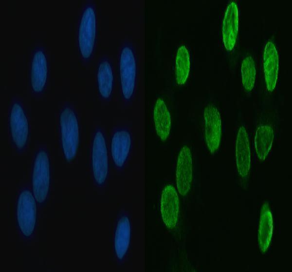
| IF/ICC Q: What localization is expected? | A: Nucleoplasmic signal is the HPA enhanced main location in cell images; UniProt also reports cytoplasmic mRNP granules (HPA: subcellular ICC-IF; UniProt P14866: subcellular location). The cell-image finding informs localization but is separate from tissue IHC validation (HPA: subcellular ICC-IF; HPA: antibodies). |
| Situation | Likely cause | Next action |
|---|---|---|
| Expected nuclei are blank across the slide. | The run may have failed, or the tested assay may not reveal its intended target under those conditions (general IHC practice). | Review a concurrently stained positive reference and the detection sequence; use an HPA-listed High cell population to assess whether nuclear signal can be recovered (HPA: tissue IHC; general IHC practice). |
| Only cytoplasm stains strongly. | The distribution departs from supported nuclear tissue staining, although UniProt allows cytoplasmic mRNP granules (HPA: tissue IHC; UniProt P14866: subcellular location). | Check nuclear counterstain and compartment boundaries, then compare an antibody-omission control and an HPA-listed positive tissue (general IHC practice; HPA: tissue IHC). Do not score cytoplasm alone as the expected IHC result. |
| Acellular areas or unexpected cells carry strong chromogen. | Nonspecific antibody binding or endogenous detection activity can create apparent positives (general IHC practice). HPA provides no negative-cell list for proving a cell-specific exception (HPA: tissue IHC). | Inspect antibody-omission and detection controls, apply the detection system’s appropriate endogenous-activity control, and require coherent nuclear localization before assigning signal to HNRNPL (general IHC practice; HPA: tissue IHC). |
| The whole section has a uniform brown haze. | Background may obscure the supported nuclear pattern (HPA: tissue IHC; general IHC practice). | Compare control sections, inspect reagent exposure and washes, and adjust general blocking or detection conditions as needed (general IHC practice). Judge improvement by clearer nuclear contrast, not simply darker tissue (HPA: tissue IHC; general IHC practice). |
| Nuclear staining varies among sections. | HPA’s High category describes observed cell populations; it does not give a fixed intensity for every preparation (HPA: tissue IHC). | Compare sections stained together using the same cell population and scoring criteria (general IHC practice). Record distribution and nuclear contrast separately from intensity; avoid inferring an HNRNPL-specific fixation effect, which the supplied sources do not report. |
| An IF/ICC image and tissue IHC section appear different. | HPA reports enhanced nucleoplasmic localization in cell images and a supported ubiquitous nuclear tissue profile, while UniProt also describes cytoplasmic granules (HPA: subcellular ICC-IF; HPA: tissue IHC; UniProt P14866: subcellular location). | Compare the nuclear compartment first and interpret any cytoplasmic signal within its assay context (HPA: tissue IHC; HPA: subcellular ICC-IF; general IHC practice). Do not transfer cell-image intensity expectations directly to chromogenic tissue scoring. |
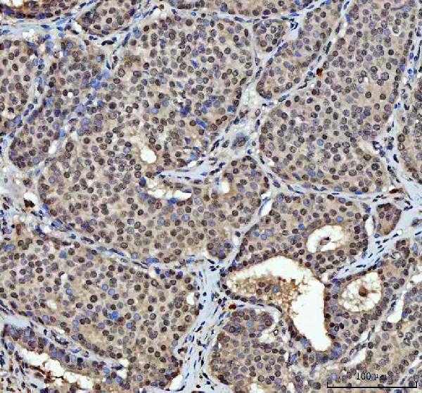
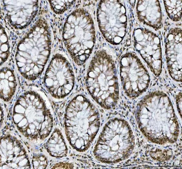
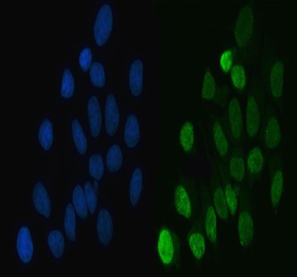
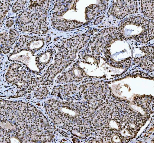


Comprehensive Human Protein Atlas IHC scoring per tissue (reliability: Supported — High consistency between antibody staining and RNA expression data.). Rows are taken directly from the HPA tissue chart — click any row's HPA link to view the source.
| Tissue | Cell type | Level | Evidence | Source |
|---|---|---|---|---|
| Adrenal gland | Glandular cells | High | Protein (IHC) | HPA → |
| Appendix | Glandular cells | High | Protein (IHC) | HPA → |
| Bone marrow | Hematopoietic cells | High | Protein (IHC) | HPA → |
| Breast | Adipocytes | High | Protein (IHC) | HPA → |
| Bronchus | Respiratory epithelial cells | High | Protein (IHC) | HPA → |
| Tissue | Cell type | Level | Evidence | Source |
|---|---|---|---|---|
| None in HPA: HNRNPL is detected in all 45 scored tissues. Use a no-primary (secondary-only) and an isotype control instead. | ||||
Troubleshoot HNRNPL staining in paraffin sections by checking retrieval, nuclear localisation, background and scoring against the catalog protocol and reference expression patterns (datasheet A03769-4; HPA).
Both antibodies have human paraffin-section IHC images and MCF-7 IF images (catalog image captions). A03769-4 also has human colorectal adenocarcinoma tissue IF images (A03769-4 IF captions).
A03769 has IHC images from human breast, colorectal, laryngeal, and liver cancer paraffin sections, plus IF data from MCF-7 cells (A03769 image captions). A03769-4 has IHC images from human breast, colorectal, gastric, and hepatocellular cancer paraffin sections, plus IF data from MCF-7 cells and human colorectal adenocarcinoma paraffin sections (A03769-4 image captions).
Which to pick: For tissue IHC, either SKU has its own paraffin-section images; choose the one with an image matching your tissue when available (A03769 IHC captions; A03769-4 IHC captions). For IF/ICC, both list those applications and show MCF-7 IF data, while A03769-4 also shows tissue IF (catalog applications; A03769 IF caption; A03769-4 IF captions). Both list human, mouse, and rat reactivity, but the supplied images show human samples only; the fixative used for the paraffin sections is unreported (catalog reactivity; catalog image captions).