This website uses cookies to ensure you get the best experience on our website.


- Table of Contents
Plan HNRNPM chromogenic IHC in paraffin sections around its widespread nuclear staining (HPA tissue IHC). Start with the catalog antibody’s IHC-P conditions (datasheet M06017-1), then compare nuclear staining across specimens processed consistently (standard IHC practice).

Expected localisation, validated protocols, controls and antibodies — the at-a-glance facts below, then the full design guide.
| Expected localisation | Ubiquitous nuclear staining (HPA tissue IHC) | |
| Staining pattern | Nuclear staining across many tissue cell types (HPA tissue IHC) | |
| Antigen retrieval | EDTA pH 8.0 HIER, heat-mediated (datasheet M06017-1) | |
| Positive control | Adipose tissue+4 more · see all | |
| Negative control | None in HPA (detected in all 45 tissues); use no-primary + isotype controls |
| Fixation | Matched tissue-IHC evidence does not establish the fixation claim. Validate the specimen-specific method before use. (selected-SKU IHC image M06017-1) | |
| Caveat | Widespread nuclear staining can limit cell-type contrast (HPA tissue IHC) | |
| Regulation | Low tissue specificity (HPA RNA) | |
| Isoform / epitope | Three isoforms; epitope coverage is unspecified (UniProt) |
The catalog antibody protocol and 2 published HNRNPM IHC protocols provide starting conditions for paraffin sections (datasheet M06017-1; PMC9372219; PMC8938476).
| Sample | Paraffin-embedded human rectal cancer tissue; fixative not specified (datasheet M06017-1) |
| Fixation | Image fixative and duration unreported (datasheet M06017-1); verify before use. |
| Sectioning | 4–5 µm sections on charged slides (standard) |
| Deparaffinisation | Xylene, graded ethanol series to water (standard) |
| Antigen retrieval | Heat retrieval: EDTA pH 8.0 (datasheet M06017-1); 20 min, 95–100 °C (standard) |
| Peroxidase block | 3% H2O2, 10 min, room temperature (standard) |
| Blocking | 10% goat serum (datasheet M06017-1) |
| Primary antibody | Rabbit monoclonal (clone 29H77) anti-HNRNPM, 1:50-1:200 (datasheet M06017-1) |
| Primary incubation | Overnight at 4 °C (datasheet M06017-1) |
| Detection | HRP-conjugated secondary, DAB chromogen (datasheet M06017-1) |
| Counterstain | Hematoxylin, blue, dehydrate and mount (standard) |
| Expected result | HNRNPM-positive staining in adipocytes of adipose tissue (HPA tissue IHC: High). HPA tissue profile: Ubiquitous nuclear expression. No signal in the no-primary control. |
HNRNPM should produce nuclear staining across many cell types in paraffin tissue sections: HPA describes its tissue IHC pattern as ubiquitous nuclear expression, with Supported reliability and low tissue RNA specificity (HPA: tissue IHC). Expect a nucleoplasmic pattern, while nucleolar localisation is also annotated (HPA: nucleoplasm approved; UniProt P52272: nucleus, nucleolus). HNRNPM has no transmembrane segment (UniProt P52272: topology).
| Distinct chromogenic signal lies within cell nuclei, including nuclei of glandular, respiratory epithelial, or hematopoietic cells. | This fits the expected compartment and several observed positive populations (HPA: ubiquitous nuclear expression; High in adrenal glandular, bronchial respiratory epithelial, and bone marrow hematopoietic cells). Compare nuclear signal with the counterstain before scoring. |
| Color is predominantly cytoplasmic or outlines cell membranes while nuclei remain pale. | Treat this as a compartment mismatch that warrants investigation, rather than a convincing HNRNPM positive result (HPA: nucleoplasm approved; UniProt P52272: nucleus, nucleolus; no transmembrane segment). Check the detection and counterstain controls (general IHC practice). |
| Strong color appears in acellular material or isolated cells without a convincing nuclear profile. | Consider nonspecific binding or endogenous detection activity (general IHC practice). HPA reports broad nuclear expression, so an unlisted cell type alone cannot establish cross-reactivity; judge its staining by nuclear localisation and appropriate controls (HPA: ubiquitous nuclear expression; low tissue RNA specificity). |
| Weak, diffuse color covers nuclei and surrounding tissue with little compartment contrast. | This is difficult to score as specific nuclear HNRNPM staining (HPA: ubiquitous nuclear expression; nucleoplasm approved). Excess detection signal or incomplete blocking can produce diffuse background; inspect a control lacking primary antibody and optimise the general IHC workflow (general IHC practice). |
| A known positive population shows no nuclear signal. | A negative result conflicts with the reported High staining in that population (HPA: High in adipocytes, adrenal glandular cells, or bone marrow hematopoietic cells). First check section integrity, detection controls, and the antibody’s validated IHC conditions (general IHC practice); do not infer biological absence from one failed section. |
| Breadth of expression (HPA: tissue IHC) | HPA reports ubiquitous nuclear expression and low tissue RNA specificity, with High staining in adipocytes, glandular cells, hematopoietic cells, respiratory epithelial cells, glial cells, and cerebellar granular layer cells (HPA: tissue IHC). Lack of a tissue listing does not make its nuclei a negative control. |
| Subnuclear interpretation (HPA: subcellular; UniProt P52272) | The approved ICC-IF location is nucleoplasm, while UniProt also annotates nucleolus (HPA: nucleoplasm approved; UniProt P52272: nucleus, nucleolus). Use nuclear localisation as the main IHC decision criterion; these sources do not define a required nucleolar chromogenic pattern in tissue sections. |
| IHC antibody evidence (HPA: antibody validation) | HPA rates IHC staining Supported for both HPA024344 and CAB016113 (HPA: antibody validation). This supports comparison with the tissue pattern, but the supplied record gives no antibody-specific epitope, dilution, or retrieval condition; use the catalog antibody’s IHC-P instructions for those choices. |
| Isoforms and target structure (UniProt P52272) | UniProt lists 3 isoforms, RNA recognition motifs, and no transmembrane segment or signal peptide (UniProt P52272: isoforms, domains, topology, processing). The supplied sources do not locate the IHC antibody epitope or show whether it detects every isoform, so do not explain a staining difference as isoform-selective detection without further evidence. |
| IF/ICC Q&A (HPA: subcellular; antibody validation) | Q: What should IF/ICC show? A: Predominantly nucleoplasmic signal (HPA: nucleoplasm approved). HPA lists HPA024344 as ICC Enhanced and CAB016113 as ICC Approved (HPA: antibody validation). Those ICC ratings describe that application; they do not supply an IF protocol for this IHC-P guide. |
| Situation | Likely cause | Next action |
|---|---|---|
| No signal in a section containing an HPA High population. | The result conflicts with reported staining for that population (HPA: High in listed tissue cells). A failed detection step or unsuitable general IHC conditions may also remove visible signal (general IHC practice). | Confirm tissue morphology and a working detection control, then follow the IHC-validated antibody’s documented IHC-P conditions, including its recommended retrieval and dilution if supplied (general IHC practice). |
| Nuclei are pale but cytoplasm or cell borders are dark. | The observed compartment conflicts with nuclear localisation (HPA: ubiquitous nuclear expression; nucleoplasm approved; UniProt P52272: nucleus, nucleolus). Nonspecific staining or a detection artefact is possible (general IHC practice). | Review the nuclear counterstain and a control lacking primary antibody; adjust general blocking, washing, or detection conditions if the controls indicate background (general IHC practice). |
| The entire section has diffuse chromogenic haze. | Background obscures the nuclear pattern expected for HNRNPM (HPA: ubiquitous nuclear expression). Incomplete blocking, excess reagent, or residual endogenous enzyme activity can contribute to haze in chromogenic IHC (general IHC practice). | Inspect the control lacking primary antibody; verify the relevant endogenous activity block, then optimise antibody concentration, washes, and detection exposure using the catalog antibody’s IHC-P instructions (general IHC practice). |
| Some unlisted cell types show clear nuclear staining. | This need not indicate cross-reactivity: HPA describes ubiquitous nuclear expression and low tissue RNA specificity, while its supplied High examples are a subset of tissues (HPA: tissue IHC). | Assess the nuclear pattern and control staining in those cells before calling them unexpected; compare their intensity with a listed High population on a suitable section (HPA: tissue IHC; general IHC practice). |
| A cell population appears negative beside stained neighbors. | HPA reports broad nuclear expression but does not assign a staining level to every cell type or establish a universal minimum intensity (HPA: ubiquitous nuclear expression; supplied tissue IHC entries). | Score only interpretable nuclei with adequate morphology and counterstain; check that the positive population and detection controls worked before assigning a negative result (general IHC practice). |
| A punctate nuclear signal seems confined to nucleoli. | UniProt annotates nucleolar localisation, but HPA approves nucleoplasm as the ICC-IF main location; the sources do not specify a required nucleolar IHC pattern (UniProt P52272: nucleus, nucleolus; HPA: nucleoplasm approved). | Confirm that staining remains nuclear and exceeds background, then report the subnuclear appearance as observed without treating nucleolar enrichment alone as proof of specificity (general IHC practice). |


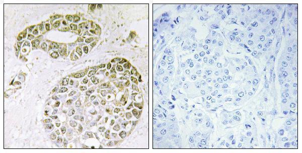
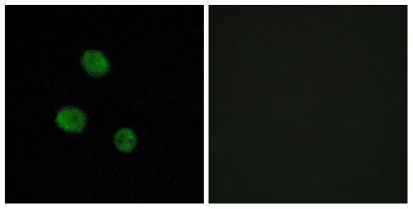
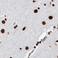


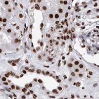


Comprehensive Human Protein Atlas IHC scoring per tissue (reliability: Supported — High consistency between antibody staining and RNA expression data.). Rows are taken directly from the HPA tissue chart — click any row's HPA link to view the source.
| Tissue | Cell type | Level | Evidence | Source |
|---|---|---|---|---|
| Adipose tissue | Adipocytes | High | Protein (IHC) | HPA → |
| Adrenal gland | Glandular cells | High | Protein (IHC) | HPA → |
| Appendix | Glandular cells | High | Protein (IHC) | HPA → |
| Bone marrow | Hematopoietic cells | High | Protein (IHC) | HPA → |
| Breast | Adipocytes | High | Protein (IHC) | HPA → |
| Tissue | Cell type | Level | Evidence | Source |
|---|---|---|---|---|
| None in HPA: HNRNPM is detected in all 45 scored tissues. Use a no-primary (secondary-only) and an isotype control instead. | ||||
Use nuclear staining, matched controls and the catalog antibody’s tissue workflow to troubleshoot HNRNPM chromogenic IHC in paraffin sections.
Two anti-HNRNPM antibodies have paraffin-section IHC images (catalog IHC captions); A06017 also has an IF image in MCF7 cells (catalog IF caption).
M06017-1 will render with IHC data from human rectal carcinoma, human stomach carcinoma, mouse liver and rat liver paraffin sections (M06017-1 IHC captions). A06017 will render with IHC data from human breast carcinoma paraffin sections and has IF data from MCF7 cells (A06017 image captions).
Which to pick: For tissue IHC across human, mouse and rat samples, choose monoclonal M06017-1: its IHC captions document paraffin sections from all three species (M06017-1 catalog; M06017-1 IHC captions). For IF/ICC, choose polyclonal A06017 when an IF image is useful: its catalog lists IF/ICC and shows IF staining in MCF7 cells (A06017 catalog; A06017 IF caption). Both IHC captions specify paraffin sections; neither reports the fixative (catalog IHC captions).