This website uses cookies to ensure you get the best experience on our website.


- Table of Contents
Plan HOXA10 paraffin-section IHC around the nuclear staining reported in several tissues (HPA tissue IHC). Use the tissue and cell examples to choose controls, while accounting for the reported low consistency between antibody staining and RNA expression (HPA tissue IHC).

Expected localisation, validated protocols, controls and antibodies — the at-a-glance facts below, then the full design guide.
| Expected localisation | Nuclear staining observed in several tissues (HPA tissue IHC) | |
| Staining pattern | Nuclear staining in several tissue cell types (HPA tissue IHC) | |
| Antigen retrieval | Tris-EDTA pH 9.0 HIER, heat-mediated (datasheet A02288-1) | |
| Positive control | Breast+4 more · see all | |
| Negative control | Caudate+4 more · see all |
| Fixation | Keep section fixation consistent. (standard IHC practice; not target-specific) | |
| Caveat | Staining has low consistency with RNA expression (HPA tissue IHC) | |
| Regulation | Expression regulation not annotated (UniProt) | |
| Isoform / epitope | Two isoforms; epitope coverage is unknown (UniProt) |
The catalog antibody protocol uses Tris-EDTA pH 9.0 heat retrieval (datasheet A02288-1). The published HOXA10 IHC protocols below provide four tissue-based alternatives (PMC6966495; PMC6745829; PMC8165781; PMC11622399).
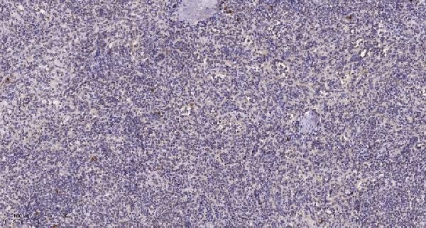
| Sample | Paraffin-embedded human spleen tissue; fixative not specified (datasheet A02288-1) |
| Fixation | Image fixative and duration unreported (datasheet A02288-1); verify before use. |
| Sectioning | 4–5 µm sections on charged slides (standard) |
| Deparaffinisation | Xylene, graded ethanol series to water (standard) |
| Antigen retrieval | Heat retrieval: Tris-EDTA pH 9.0 (datasheet A02288-1); 20 min, 95–100 °C (standard) |
| Peroxidase block | 3% H2O2, 10 min, room temperature (standard) |
| Blocking | 10% normal serum of the secondary host, 30 min, room temperature (standard) |
| Primary antibody | Rabbit anti-HOXA10, 1:50 (datasheet A02288-1) |
| Primary incubation | Overnight at 4 °C (standard) |
| Detection | HRP-polymer secondary, DAB chromogen 5–10 min (standard) |
| Counterstain | Hematoxylin, blue, dehydrate and mount (standard) |
| Expected result | HOXA10-positive staining in adipocytes of breast (HPA tissue IHC: Medium). HPA tissue profile: Nuclear expression in several tissues. No signal in the no-primary control. |
HOXA10 is a nuclear, sequence-specific transcription factor with no transmembrane segment (UniProt P31260: function, location, topology). In paraffin-section IHC, expect staining in nuclei of selected cells, including cervical glandular cells and endometrial stromal cells, both reported at medium intensity (HPA: tissue IHC). HPA describes nuclear staining in several tissues but rates its IHC profile “Approved” with low consistency between staining and RNA expression; interpret a positive field alongside cell identity and controls (HPA: tissue IHC reliability).
| Distinct nuclear chromogen in cervical glandular cells or endometrial stromal cells, with limited surrounding stain (HPA: Medium in both cell types). | This fits the reported cell distribution and nuclear profile (HPA: tissue IHC). Judge intensity relative to controls: “Medium” is an HPA observation, not a required numeric score or a guarantee for every specimen (HPA: tissue IHC). |
| Predominantly cytoplasmic, membranous, or extracellular chromogen instead of nuclear staining (UniProt P31260: nucleus; HPA: nuclear tissue profile). | Treat this as discordant localization and investigate nonspecific binding or detection artefact before assigning HOXA10 positivity (UniProt P31260: nucleus; HPA: nuclear tissue profile). |
| Strong signal in a cell population HPA reports as not detected, such as fallopian-tube glandular cells (HPA: Not detected in fallopian-tube glandular cells). | Check cell identification, primary-antibody specificity, and endogenous detection activity. A discrepancy raises concern for cross-reactivity or artefact; HPA’s tissue observations alone cannot establish the cause (HPA: tissue IHC; standard IHC practice). |
| Diffuse color across tissue, stroma, and blank areas with little nuclear contrast (HPA: expected nuclear tissue profile). | Do not score diffuse color as HOXA10. Check the no-primary control, blocking, washes, and detection chemistry to locate background sources (standard IHC practice); assess nuclear signal separately (HPA: tissue IHC). |
| No convincing nuclear signal in a section containing cervical glandular cells or endometrial stromal cells (HPA: Medium in these cell types). | An absent result conflicts with those HPA examples but does not prove absent HOXA10 in the specimen. Review tissue identity, run controls, and inspect the antibody’s IHC-P conditions before interpreting a negative (HPA: tissue IHC; standard IHC practice). |
| Tissue and cell context | HPA reports medium staining in several cell types, including cervical glandular cells, and no detection in fallopian-tube glandular cells; a glandular label alone does not predict positivity (HPA: tissue IHC). |
| IHC antibody evidence | CAB019384 has an IHC “Approved” designation; HPA071918 has no IHC designation in the supplied record. Neither entry is marked IHC “Enhanced” (HPA: antibody validation). |
| Isoforms and epitope coverage | UniProt lists two HOXA10 isoforms, but the supplied record gives no antibody epitope or isoform-detection data; do not infer that a stain detects both (UniProt P31260: isoforms; HPA: antibody record). |
| IF/ICC: where should signal appear? | Mainly in nucleoplasm, with additional nuclear bodies (HPA: supported ICC-IF localization). That cell-image observation supports a nuclear expectation but does not set an IHC-P protocol (HPA: ICC-IF; HPA: tissue IHC). |
| Fixation sensitivity | Target-specific fixation sensitivity is unreported in the supplied UniProt and HPA records; these data do not predict a retrieval condition or a fixation-driven change in staining (UniProt P31260; HPA: tissue IHC). |
| Situation | Likely cause | Next action |
|---|---|---|
| No staining in an HPA medium-positive cell population (HPA: cervical glandular or endometrial stromal cells). | The stain may have failed, or the sampled cells may differ from the HPA examples; the cause cannot be assigned from HPA intensity alone (HPA: tissue IHC). | Confirm cell identity and positive-control performance, then review the IHC-validated antibody’s stated retrieval and dilution conditions (standard IHC practice; HPA: antibody validation). |
| Signal is mainly cytoplasmic or membranous (UniProt P31260: nucleus; HPA: nuclear tissue profile). | The compartment conflicts with the supplied localization evidence; nonspecific binding or detection artefact is possible (UniProt P31260; HPA: tissue IHC). | Compare the no-primary control and repeat with the IHC-validated antibody under its documented IHC-P conditions before scoring nuclear positivity (standard IHC practice; HPA: antibody validation). |
| Chromogen appears in an HPA not-detected cell type (HPA: fallopian-tube glandular cells, Not detected). | Cell misidentification, cross-reactivity, or endogenous detection activity may explain the discrepancy; HPA does not distinguish these causes (HPA: tissue IHC; standard IHC practice). | Recheck morphology and compare no-primary and detection controls; score only interpretable cellular staining (standard IHC practice). |
| Diffuse tissue color obscures nuclei (HPA: expected nuclear tissue profile). | Background can arise from incomplete blocking, washing, or detection-system controls (standard IHC practice). | Check no-primary background, blocking, washes, and chromogen development; repeat until nuclei can be assessed separately from background (standard IHC practice). |
| Nuclear signal is weak in a cell type HPA lists as low, such as skeletal-muscle myocytes (HPA: Low). | Low reported staining offers less visual separation from background; weak color alone is inconclusive (HPA: tissue IHC; standard IHC practice). | Compare a documented medium-positive cell population and the negative control on the same run before calling the weak field negative or positive (HPA: tissue IHC; standard IHC practice). |
| Different antibodies yield different IHC patterns (HPA: CAB019384 IHC Approved; HPA071918 no IHC designation). | The supplied validation statuses differ, and epitope coverage is unspecified; the records do not identify which discrepancy is biological (HPA: antibody validation; UniProt P31260: isoforms). | Prioritize the IHC-validated antibody, verify its documented conditions, and compare cell-specific nuclear staining with controls (HPA: antibody validation; standard IHC practice). |


Comprehensive Human Protein Atlas IHC scoring per tissue (reliability: Approved — Low consistency between antibody staining and RNA expression data.). Rows are taken directly from the HPA tissue chart — click any row's HPA link to view the source.
| Tissue | Cell type | Level | Evidence | Source |
|---|---|---|---|---|
| Breast | Adipocytes | Medium | Protein (IHC) | HPA → |
| Bronchus | Respiratory epithelial cells | Medium | Protein (IHC) | HPA → |
| Cervix | Glandular cells | Medium | Protein (IHC) | HPA → |
| Colon | Glandular cells | Medium | Protein (IHC) | HPA → |
| Endometrium | Cells in endometrial stroma | Medium | Protein (IHC) | HPA → |
| Tissue | Cell type | Level | Evidence | Source |
|---|---|---|---|---|
| Caudate | Glial cells | Not detected | Protein (IHC) | HPA → |
| Cerebellum | Cells in granular layer | Not detected | Protein (IHC) | HPA → |
| Cerebral cortex | Endothelial cells | Not detected | Protein (IHC) | HPA → |
| Duodenum | Glandular cells | Not detected | Protein (IHC) | HPA → |
| Esophagus | Squamous epithelial cells | Not detected | Protein (IHC) | HPA → |
Troubleshooting guidance for chromogenic HOXA10 IHC on paraffin sections, with one Q&A on IF multiplexing.
A02288-1 has an IHC image of paraffin-embedded human spleen (IHC image caption). IF is listed for this human- and mouse-reactive antibody, but no IF image is supplied (catalog applications/reactivity/IF image alts).
A02288-1 will render with an IHC figure from paraffin-embedded human spleen (IHC image caption). Its listed applications include IHC and IF, and its listed reactivity covers human and mouse; no IF figure is supplied (catalog applications/reactivity/IF image alts).
Which to pick: For tissue IHC, choose A02288-1: its own image caption documents paraffin-embedded human spleen, Tris-EDTA pH 9 retrieval and a 1:200 primary dilution; the fixative is unreported (IHC image caption). For IF, A02288-1 is listed at 1:50, but no IF image is supplied; ICC is not listed and requires local validation (catalog applications/if_dilution/IF image alts). For human or mouse samples, A02288-1 is a rabbit polyclonal with both species listed as reactive, although its IHC image shows human tissue only (catalog host/dilution_raw/reactivity; IHC image caption).