HPCAL1 / Hippocalcin-like protein 1 · IHC design guide

Design Immunohistochemistry for HPCAL1

Plan HPCAL1 chromogenic IHC on paraffin sections using cerebellar Purkinje cells as a high-staining reference (HPA tissue IHC). The guide covers fixation consistency, antibody dilution and interpretation of cytoplasmic staining, with cross-gene detection as a validation concern (HPA tissue IHC).

Evidence assembled Oct 2026 · For research use; verify linked source records and product datasheet before use
Immunohistochemistry protocol sheet for HPCAL1 (IHC for HPCAL1): expected localisation Cytoplasmic staining in several tissues (HPA tissue IHC), antibody A08911-1, validated IHC image, and IHC protocol steps
Printable HPCAL1 IHC protocol sheet — expected localisation Cytoplasmic staining in several tissues (HPA tissue IHC), antibody A08911-1, controls and protocol steps. Open the full HPCAL1 IHC guide →

HPCAL1 Immunohistochemistry Experimental Design Guide

Expected localisation, validated protocols, controls and antibodies — the at-a-glance facts below, then the full design guide.

Must know before staining
Expected localisation Cytoplasmic staining in several tissues (HPA tissue IHC)
Staining pattern Purkinje cells show cytoplasmic staining in cerebellum (HPA tissue IHC)
Antigen retrieval EDTA pH 8.0 HIER, heat-mediated (datasheet A08911-1)
Positive control ⓘ Caudate+4 more · see all
Negative control ⓘ Adipose tissue+4 more · see all
Important caveats
Reasons your staining may differ from the expected pattern.
Fixation Matched tissue-IHC evidence does not establish the fixation claim. Validate the specimen-specific method before use. (selected-SKU IHC image A08911-1)
Caveat Staining may detect protein from more than one gene (HPA tissue IHC)
Regulation Brain-enriched RNA; regulation unknown (HPA tissue IHC/RNA)
Isoform / epitope No isoforms listed; chain 2–193; epitope unspecified (UniProt)
Section 1

Recommended HPCAL1 IHC & IF Protocols

The catalog antibody’s IHC-P protocol is accompanied by four published HPCAL1 IHC protocols covering liver, colorectal and cholangiocarcinoma tissues (PMC11800645; PMC13316108; PMC9844623; PMC9691343).

Recommended immunohistochemistry (IHC-P) protocol parameters
SampleParaffin-embedded human liver cancer tissue; fixative not specified (datasheet A08911-1)
FixationImage fixative and duration unreported (datasheet A08911-1); verify before use.
Sectioning4–5 µm sections on charged slides (standard)
DeparaffinisationXylene, graded ethanol series to water (standard)
Antigen retrievalHeat retrieval: EDTA pH 8.0 (datasheet A08911-1); 20 min, 95–100 °C (standard)
Peroxidase block3% H2O2, 10 min, room temperature (standard)
Blocking10% goat serum (datasheet A08911-1)
Primary antibodyRabbit anti-HPCAL1, 1:50 recommended; image 1:100 (datasheet A08911-1)
Primary incubationOvernight at 4 °C (datasheet A08911-1)
DetectionHRP-conjugated secondary, DAB chromogen (datasheet A08911-1)
CounterstainHematoxylin, blue, dehydrate and mount (standard)
Expected resultHPCAL1-positive staining in neuronal cells of caudate (HPA tissue IHC: High). HPA tissue profile: Cytoplasmic expression in several tissues, most abundant in cerebellum. No signal in the no-primary control.
💡Decision noteStart with heat-mediated EDTA pH 8.0 retrieval (datasheet A08911-1); the colorectal protocol reports antigen repair at 37 °C for 30 min without naming a buffer (PMC13316108).
Section 2

What Is the Expected HPCAL1 Staining Pattern?

HPCAL1 staining should be predominantly cytoplasmic, especially in cerebellar Purkinje cells and caudate neuronal cells, with strong staining also reported in intestinal Paneth cells (HPA tissue IHC: High). UniProt places HPCAL1 at membranes but reports no transmembrane segment (UniProt P37235: subcellular location and topology). Treat this pattern as a guide, because HPA rates the tissue evidence Supported, notes medium agreement with RNA, and cautions that staining may detect proteins from more than one gene (HPA tissue IHC: reliability).

What am I looking at on my slide?
Strong cytoplasmic staining in Purkinje cells, caudate neurons, or Paneth cells.This matches reported high staining in those cell types (HPA tissue IHC: High). Score the named cells and compartment; staining alone does not resolve the HPA cross-gene caution (HPA tissue IHC: reliability).
Cytoplasmic staining is accompanied by a membrane-associated edge or processes.Compare the full pattern with tissue architecture. HPA describes cytoplasmic tissue staining (HPA tissue IHC: profile); UniProt lists membrane association without a transmembrane segment (UniProt P37235: location and topology). A sharp membrane outline alone is insufficient confirmation.
Predominantly nuclear staining in the expected positive cells.Nuclear localisation is outside the supplied HPA cytoplasmic profile and UniProt membrane annotation (HPA tissue IHC: profile; UniProt P37235: location). Check morphology, counterstain, and a detection-only control before interpreting it as target signal (general IHC practice).
Staining appears in adipocytes, cardiomyocytes, or other cells reported as undetected.HPA reports no detection in adipocytes and cardiomyocytes (HPA tissue IHC: Not detected). Reproducible staining there warrants a specificity check; possible explanations include cross-reactivity or endogenous detection activity (HPA tissue IHC: cross-gene caution; general IHC practice).
Diffuse chromogen covers cells and surrounding tissue without a clear cell pattern.This cannot be scored as the reported cell-selective, cytoplasmic pattern (HPA tissue IHC: profile and positive cells). Assess background with a detection-only control and review blocking, washes, and detection conditions (general IHC practice).
💡Expected HPCAL1 appearanceA convincing positive is strong, predominantly cytoplasmic staining in Purkinje cells, caudate neurons, or Paneth cells (HPA tissue IHC: High and cytoplasmic profile); broad nuclear, extracellular, or undifferentiated staining needs control-based review (HPA tissue IHC: profile; general IHC practice).
How each factor affects the staining
Which tissue gives the clearest positive reference?Cerebellar Purkinje cells are reported High, and the overall tissue profile is most abundant in cerebellum (HPA tissue IHC). Caudate neurons and Paneth cells are other reported High references (HPA tissue IHC: positive cells).
How should weaker staining be judged?HPA reports Medium staining in hippocampal neurons, cortical neuropil, bronchial respiratory epithelium, and fallopian tube glandular cells; lung macrophages are Low (HPA tissue IHC). Judge intensity against the specific cell type and tissue, rather than applying one threshold throughout a section.
What does antibody validation establish?HPA043245 is a rabbit polyclonal antibody with IHC status Supported (HPA antibodies: HPA043245). HPA also flags medium staining–RNA consistency and possible recognition of proteins from more than one gene; the tissue pattern is therefore a reference, not proof of unique HPCAL1 detection (HPA tissue IHC: reliability).
Does membrane annotation require membrane-only chromogen?No. The observed tissue profile is cytoplasmic (HPA tissue IHC: profile), while UniProt annotates membrane location and no transmembrane segment (UniProt P37235: location and topology). Evaluate any edge staining in the context of the cytoplasmic, cell-specific pattern.
Q: Can IF/ICC establish an independent compartment reference?A: The supplied HPA subcellular record has no main location and no cell lines with ICC-IF images (HPA subcellular: location and images). It provides no independent image-based compartment benchmark; use the tissue IHC pattern for this section (HPA tissue IHC: profile).
Why is my staining missing, weak or wrong?
SituationLikely causeNext action
No signal in a cerebellar positive section.The result conflicts with High Purkinje-cell staining (HPA tissue IHC: cerebellum); the record does not identify a target-specific fixation sensitivity.Confirm Purkinje cells are present, then review retrieval, antibody incubation, detection, and the run's positive control (general IHC practice).
Only faint staining in a presumed positive tissue.Expected intensity depends on the sampled cell type: hippocampal neurons are Medium, whereas Purkinje cells are High (HPA tissue IHC: positive cells).Identify and score the relevant cells first. Compare the same tissue and cell type under matched staining conditions before adjusting dilution or detection (general IHC practice).
Strong nuclear signal dominates the slide.The compartment differs from HPA's cytoplasmic profile and UniProt's membrane annotation (HPA tissue IHC: profile; UniProt P37235: location).Check counterstain and cell boundaries; compare a detection-only control and review primary-antibody concentration and washes (general IHC practice).
Many cell types stain uniformly, including expected negatives.That distribution does not match reported undetected adipocytes or cardiomyocytes (HPA tissue IHC: Not detected). Cross-reactivity remains plausible under HPA's cross-gene caution; endogenous detection activity is another general IHC possibility.Run a detection-only control and compare cell-level staining with the HPA reference; review blocking and detection steps (general IHC practice).
Chromogen deposits obscure cellular boundaries.Diffuse deposition prevents assessment of the reported cytoplasmic, cell-specific pattern (HPA tissue IHC: profile and positive cells); a cause cannot be assigned from appearance alone.Inspect a detection-only control and review wash and chromogen development conditions before rescoring (general IHC practice).
Paneth cells stain, but adjacent intestinal cells do not.High Paneth-cell staining is reported in duodenum and small intestine (HPA tissue IHC: positive cells); absence in neighboring cells alone does not invalidate that selective pattern.Score identifiable Paneth cells separately. If they are absent or unstained, verify tissue orientation and include a known-positive section in the run (general IHC practice).

Sample controls for HPCAL1 IHC & IF

🧪Run cerebellum first and require staining in Purkinje cells (High; HPA: cerebellum, Purkinje cells). Run adipose tissue as the negative, with adipocytes at background (Not detected; HPA: adipose tissue, adipocytes); on the cerebellar slide, use unstained cells outside the Purkinje layer as internal background comparators only after confirming that they lack signal (HPA: cerebellum, Purkinje cells High).
Positive control tissue: Caudate (Neuronal cells, HPA High)
Negative control tissue: Adipose tissue (HPA Not detected)
ICC-IF cell lines (HPA subcellular resource): HPA carries no ICC-IF cell line for HPCAL1; derive a cell-line control from the positive tissue's cell type (Neuronal cells) and confirm it by RNA or western blot first.
Technical controls: Include a no-primary, secondary-only control and a rabbit isotype control, plus HPCAL1 knockout tissue or peptide competition if the antibody immunogen is known (caption: rabbit primary; standard IHC controls). Quench endogenous peroxidase for HRP/DAB detection, and check neuronal autofluorescence if adapting the assay to IF (caption: HRP/DAB detection; standard IHC/IF practice).
⚠️Feasibility: The selected paraffin-section caption reports heat retrieval in EDTA at pH 8.0, but does not report a fixative; a target-specific fixation window or fixation effect is unreported (caption: A08911-1 tissue IHC). Use that retrieval condition as a starting point; whether retrieval is required, or whether frozen sections or IF are easier, is unestablished in the supplied evidence (caption: heat retrieval; HPA subcellular: no ICC-IF images). Neuronal autofluorescence can complicate cerebellar IF, so assess it with an unstained or no-primary section (standard IF practice).

HPA tissue IHC evidence for HPCAL1

Comprehensive Human Protein Atlas IHC scoring per tissue (reliability: Supported — Medium consistency between antibody staining and RNA expression data. Caution, targets protein from more than one gene. Pending external verification.). Rows are taken directly from the HPA tissue chart — click any row's HPA link to view the source.

Positive expression · recommended positive controls

TissueCell typeLevelEvidenceSource
Caudate Neuronal cells High Protein (IHC) HPA →
Cerebellum Purkinje cells High Protein (IHC) HPA →
Duodenum Paneth cells High Protein (IHC) HPA →
Small intestine Paneth cells High Protein (IHC) HPA →
Bronchus Respiratory epithelial cells Medium Protein (IHC) HPA →

Undetected expression · recommended negative controls

TissueCell typeLevelEvidenceSource
Adipose tissue Adipocytes Not detected Protein (IHC) HPA →
Appendix Endocrine cells Not detected Protein (IHC) HPA →
Bone marrow Hematopoietic cells Not detected Protein (IHC) HPA →
Cervix Glandular cells Not detected Protein (IHC) HPA →
Colon Endocrine cells Not detected Protein (IHC) HPA →
Section 3

Advanced HPCAL1 IHC Tips

Use the catalog IHC image as a starting point, then judge HPCAL1 staining by cell type, compartment and controls (datasheet A08911-1; HPA tissue IHC; UniProt P37235).

Which retrieval conditions should I start with for HPCAL1 paraffin IHC?
Start with heat-mediated EDTA retrieval at pH 8.0 for paraffin sections (datasheet A08911-1). The selected image used this retrieval before 1:100 primary antibody incubation overnight at 4°C (datasheet A08911-1). Keep section thickness, heating and cooling consistent across a staining run, and include a known positive section to assess retrieval (standard IHC practice; HPA: high staining in cerebellar Purkinje cells). If staining is weak, adjust retrieval duration on matched sections while monitoring tissue damage and nonspecific signal (standard IHC practice). Compare staining in the expected cells, since the catalog image alone does not establish retrieval performance across tissues (datasheet A08911-1; HPA tissue IHC).
How should I troubleshoot fixation-related loss of HPCAL1 staining?
The selected image identifies a paraffin section but does not state its fixative, so target-specific fixation sensitivity is unknown (datasheet A08911-1). Record fixative, fixation duration and processing history for each specimen before comparing signal intensity (standard IHC practice). On matched sections, compare the documented EDTA pH 8.0 retrieval and 1:100 primary condition while keeping detection constant (datasheet A08911-1; standard IHC practice). Examine morphology alongside staining, because poorly preserved or damaged tissue can complicate interpretation (standard IHC practice). Do not infer fixation tolerance from the HPA tissue pattern or from HPCAL1 membrane annotation, EF-hands or lack of a transmembrane segment (HPA tissue IHC; UniProt P37235 topology and domains).
Should HPCAL1 stain membranes or cytoplasm in paraffin sections?
Expect prominent cytoplasmic staining in several tissues, especially cerebellum, according to tissue IHC observations (HPA: cytoplasmic profile). UniProt assigns HPCAL1 to membranes but lists no transmembrane segment, so a membrane-associated pattern can be assessed without requiring a membrane-spanning outline (UniProt P37235 topology and subcellular annotation). Examine Purkinje cells or caudate neuronal cells as high-staining reference populations, and score their subcellular pattern separately from intensity (HPA: high in Purkinje and caudate neuronal cells; standard IHC practice). Diffuse nuclear-only staining warrants scrutiny because neither supplied localisation source supports a predominantly nuclear pattern (HPA tissue IHC; UniProt P37235 subcellular annotation). Use morphology and controls before assigning fine membrane localisation from DAB staining (standard IHC practice).
How can epitope uncertainty affect HPCAL1 IHC interpretation?
The record lists 0 isoforms, but the antibody epitope is not supplied, so isoform-specific staining cannot be claimed (UniProt P37235 isoforms; datasheet A08911-1). HPCAL1 has four annotated EF-hand regions spanning residues 41–179, which makes epitope position relevant when interpreting retrieval-dependent staining (UniProt P37235 domains; standard IHC practice). Its annotated chain spans residues 2–193, with no listed glycosylation sites or modified residues (UniProt P37235 processing and PTMs). Avoid assigning a stained compartment to a particular EF-hand or modification without epitope-mapping evidence (UniProt P37235 domains and PTMs; standard IHC practice). If staining changes with retrieval, confirm specificity using independent controls before proposing epitope exposure as the cause (standard IHC practice).
How should I adapt HPCAL1 localisation checks to multiplex IF?
Treat multiplex IF as a separate validation exercise: the supplied HPA subcellular record has no ICC/IF images, while the catalog example documents paraffin IHC (HPA subcellular; datasheet A08911-1). Pair HPCAL1 with a marker that identifies the expected cell population, such as Purkinje cells in cerebellum, and assess signal within that population (HPA: high in Purkinje cells; standard IF practice). Choose spectrally separated fluorophores and place the weaker signal in a channel with less tissue autofluorescence, using single-label controls to check bleed-through (standard IF practice). Because the antibody epitope and its membrane-facing side are unspecified, compare permeabilised and nonpermeabilised conditions before interpreting intracellular access (UniProt P37235 topology; standard IF practice). Keep IF conclusions separate from chromogenic IHC intensity scores (standard IF and IHC practice).
What should I check when HPCAL1 DAB staining is widespread?
Begin with a no-primary control to assess detection-system staining, and inspect tissue edges and damaged areas separately (standard IHC practice). The catalog image used 10% goat serum blocking, a peroxidase-conjugated secondary and DAB detection; those settings document one image, not specificity across tissues (datasheet A08911-1). Include a peroxidase block before HRP detection and assess background after DAB development as general chromogenic IHC practice (standard IHC practice). If signal persists broadly, check secondary-only staining, antibody concentration and wash consistency while retaining the documented 1:100 condition as a reference (datasheet A08911-1; standard IHC practice). Compare cellular distribution with high-staining reference populations before calling diffuse deposition HPCAL1 (HPA tissue IHC; standard IHC practice).
How should I quantify HPCAL1 staining across paraffin sections? ⚠ ANSWER MARKED FOR VERIFICATION
Define the cell population and analysis area before scoring, since HPCAL1 staining varies by tissue and cell type (HPA tissue IHC; standard IHC practice). For discrete cells, report percentage positive and an H-score combining positive-cell proportion with staining intensity; use positive-cell density per mm² when cell counts per area answer the question (standard IHC practice). Normalise each result to the number of eligible cells or the measured viable tissue area, and exclude folds, necrosis and section edges consistently (standard IHC practice). Score cytoplasmic and membrane-associated signal separately when both are visible, using the same threshold and imaging settings across sections (HPA: cytoplasmic profile; UniProt P37235 subcellular annotation; standard IHC practice). Report control performance alongside scores (standard IHC practice).
How can I distinguish true HPCAL1 staining from artefact?
Prioritise staining in expected cells and compartments: Purkinje and caudate neuronal cells show high tissue IHC staining, and the broader profile is cytoplasmic (HPA tissue IHC). Membrane-associated signal is biologically plausible, but a nuclear-only pattern lacks support in the supplied localisation records (UniProt P37235 subcellular annotation; HPA tissue IHC). Check whether staining concentrates at section edges, in necrotic regions or in the no-primary control before interpreting it as target signal (standard IHC practice). Investigate DAB signal attributable to endogenous peroxidase with an appropriate blocking and detection control (standard IHC practice). Treat a positive image as supporting evidence rather than definitive specificity: HPA rates tissue staining Supported but flags possible recognition of proteins from more than one gene (HPA reliability; datasheet A08911-1).
Boster reagents

Best HPCAL1 / Hippocalcin-like protein 1 IHC Antibodies

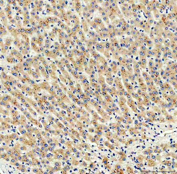
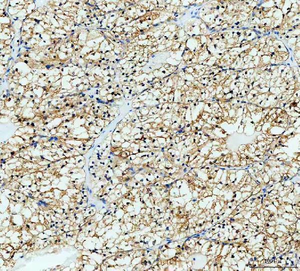
A08911-1 has IHC images from human liver cancer, human renal clear cell carcinoma, mouse cerebellum and rat cerebellum paraffin sections (catalog IHC captions); IF/ICC are listed applications, with no IF image supplied (catalog payload).

Real IHC data IHC analysis of VILIP3/HPCAL1 using anti-VILIP3/HPCAL1 antibody (A08911-1). VILIP3/HPCAL1 was detected in a paraffin-embedded section of human liver cancer tissue. Heat mediated antigen retrieval was performed in EDTA buffer (pH 8.0, epitope retrieval solution). The tissue section was blocked with 10% goat serum. The tissue section was then incubated with 1:100 rabbit anti-VILIP3/HPCAL1 Antibody (A08911-1) overnight at 4°C. Peroxidase Conjugated Goat Anti-rabbit IgG was used as secondary antibody and incubated for 30 minutes at 37°C. The tissue section was developed using HRP Conjugated Rabbit IgG Super Vision Assay Kit (Catalog # SV0002) with DAB as the chromogen.
Anti-VILIP3/HPCAL1 Antibody
Cat # A08911-1

A08911-1 has IHC images from paraffin sections of human liver cancer and renal clear cell carcinoma (catalog IHC captions). The same SKU has IHC images from mouse and rat cerebellum, and lists IF/ICC applications without an IF image (catalog IHC captions; catalog applications and image list).

Which to pick: Choose A08911-1 for paraffin section IHC because its images document staining in human, mouse and rat tissue; the captions do not report a fixative (catalog IHC captions). For IF/ICC, A08911-1 lists both applications and a 1:50 dilution, but has no supplied IF image (catalog applications, IF dilution and image list). It is a rabbit polyclonal antibody with listed human, mouse and rat reactivity for cross species work (catalog host, dilution record and reactivity).

Each figure is that product's own IHC / IF validation image from its datasheet.

References

  1. UniProt Consortium. UniProt entry P37235 (HPCL1_HUMAN, Hippocalcin-like protein 1).
  2. Human Protein Atlas. HPCAL1 tissue IHC expression (reliability: Supported).
  3. Human Protein Atlas. HPCAL1 subcellular location (ICC-IF): Highest expression in GAMG: 306.0 nTPM.
  4. Human Protein Atlas. HPCAL1 antibody validation summary (1 antibodies).
  5. Liver macrophage-derived exosomal miRNA-342-3p promotes liver fibrosis by inhibiting HPCAL1 in stellate cells. Human genomics 2025 — PMC11800645.
  6. HPCAL1 promotes colorectal cancer progression via TCF7/p65-mediated Wnt ligand upregulation and Wnt/β-catenin pathway activation. Oncogenesis 2026 — PMC13316108.
  7. Expressional and prognostic value of HPCAL1 in cholangiocarcinoma via integrated bioinformatics analyses and experiments. Cancer medicine 2023 — PMC9844623.
  8. Hippocalcin-Like 1 blunts liver lipid metabolism to suppress tumorigenesis via directly targeting RUVBL1-mTOR signaling. Theranostics 2022 — PMC9691343.
  9. PubMed PMID:8038222 — UniProt-cited evidence.
  10. PubMed PMID:15489334 — UniProt-cited evidence.
  11. PubMed PMID:21269460 — UniProt-cited evidence.