This website uses cookies to ensure you get the best experience on our website.


- Table of Contents
Plan HPSE2 IHC in paraffin sections using the reported mainly cytoplasmic tissue pattern and high staining in small-intestinal glandular cells (HPA tissue IHC). Interpret the pattern cautiously because at least one variant is secreted (HPA tissue IHC).

Expected localisation, validated protocols, controls and antibodies — the at-a-glance facts below, then the full design guide.
| Expected localisation | Tissue: mainly cytoplasmic (HPA tissue IHC); protein: secreted to extracellular matrix (UniProt) | |
| Staining pattern | Mainly cytoplasmic staining in most tissues (HPA tissue IHC) | |
| Antigen retrieval | Citrate pH 6.0 HIER, 95–98 °C, 20 min (rule: cytoplasmic / membrane antigen) | |
| Positive control | Duodenum+4 more · see all | |
| Negative control | Adipose tissue+4 more · see all |
| Fixation | Keep fixation consistent across sections (standard IHC practice; not target-specific) | |
| Caveat | A secreted variant may complicate tissue staining patterns (HPA tissue IHC) | |
| Regulation | No specific expression regulator reported (UniProt) | |
| Isoform / epitope | 4 isoforms; mature chain begins at residue 42 (UniProt) |
The catalog antibody’s IHC-P protocol is accompanied by published HPSE2 staining conditions for thyroid tissue (PMC4619411), colorectal adenomas (PMC4800254), and breast cancer samples (PMC7054484).
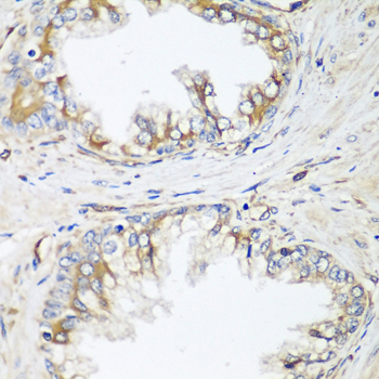
| Sample | Paraffin-embedded human prostate tissue; fixative not specified (datasheet A07511) |
| Fixation | Image fixative and duration unreported (datasheet A07511); verify before use. |
| Sectioning | 4–5 µm sections on charged slides (standard) |
| Deparaffinisation | Xylene, graded ethanol series to water (standard) |
| Antigen retrieval | Heat-induced epitope retrieval in citrate buffer, pH 6.0, 20 min at 95–98 °C (standard rule: cytoplasmic / membrane antigen) |
| Peroxidase block | 3% H2O2, 10 min, room temperature (standard) |
| Blocking | 10% normal serum of the secondary host, 30 min, room temperature (standard) |
| Primary antibody | Rabbit anti-HPSE2, 1:50-1:200 (datasheet A07511) |
| Primary incubation | Overnight at 4 °C (standard) |
| Detection | HRP-polymer secondary, DAB chromogen 5–10 min (standard) |
| Counterstain | Hematoxylin, blue, dehydrate and mount (standard) |
| Expected result | HPSE2-positive staining in glandular cells of duodenum (HPA tissue IHC: High). HPA tissue profile: Mainly cytoplasmic expression in most tissues. No signal in the no-primary control. |
HPSE2 is secreted and associated with extracellular space and matrix; it has no transmembrane segment (UniProt Q8WWQ2). In tissue IHC, HPA reports mainly cytoplasmic staining, including High staining in duodenal and small intestinal glandular cells, hepatocytes, and placental trophoblastic cells (HPA tissue IHC). Treat the pattern as provisional: HPA rates its tissue IHC reliability Uncertain and flags possible differences between RNA and protein location (HPA tissue IHC).
| Cytoplasmic chromogen in duodenal or small intestinal glandular cells, hepatocytes, or placental trophoblastic cells. | This matches the cell types and High levels reported by HPA (HPA tissue IHC). Score the named cells and their staining intensity rather than assigning the same score to the whole section. HPA's Uncertain reliability means agreement with this pattern supports interpretation but does not independently establish antibody specificity (HPA tissue IHC). |
| Extracellular staining near otherwise plausible positive cells, with or without cytoplasmic staining. | Extracellular signal is biologically plausible because HPSE2 is secreted and associated with extracellular space and matrix (UniProt Q8WWQ2). It is not the principal tissue pattern described by HPA, which reports mainly cytoplasmic staining (HPA tissue IHC). Assess whether the signal follows tissue structures and persists when the primary antibody is omitted (general IHC practice). |
| Strong, isolated nuclear staining or a continuous cell-surface outline with little expected cytoplasmic signal. | These distributions do not match HPA's mainly cytoplasmic tissue profile or UniProt's secreted, non-transmembrane annotation (HPA tissue IHC; UniProt Q8WWQ2). Treat them as suspect until a control or independent antibody supports them. A membrane outline alone should not be taken as evidence that HPSE2 is a transmembrane protein (UniProt Q8WWQ2). |
| Strong staining confined to adipocytes or oral squamous epithelial cells, while expected positive cells remain unstained. | HPA reports HPSE2 as Not detected in adipocytes of adipose tissue and breast, and in oral mucosal squamous epithelial cells (HPA tissue IHC). This reversal raises concern for nonspecific staining or detection background; it does not prove cross-reactivity. Check the negative control and the identity of the stained cells before interpreting the result (general IHC practice). |
| Uniform color across tissue compartments, or no signal in a section containing a known positive cell type. | Uniform color without a cell-resolved pattern favors background over a convincing HPSE2 result (general IHC practice). An absent signal in duodenal glandular cells or hepatocytes conflicts with HPA's High observations but is not, by itself, proof of HPSE2 absence (HPA tissue IHC). Review controls and detection conditions before scoring the specimen negative (general IHC practice). |
| Secreted location versus observed tissue pattern | UniProt places HPSE2 in extracellular space and matrix and annotates no transmembrane segment (UniProt Q8WWQ2). HPA nonetheless describes mainly cytoplasmic tissue IHC (HPA tissue IHC). Record both observations when scoring; do not require an exclusively extracellular pattern or interpret a membrane outline as established topology. |
| Tissue and cell selection | HPA reports High staining in duodenal, small intestinal, and stomach glandular cells, hepatocytes, and trophoblastic cells; prostate glandular cells are Low (HPA tissue IHC). Choose a reference section by its annotated cell population, since a low-level population offers a less clear visual comparison (HPA tissue IHC). |
| Antibody validation and transcript uncertainty | The supplied HPA antibody, HPA044603, has an Uncertain IHC status, and HPA notes possible splice or transcript discrepancies and complex RNA–protein correlation for a secreted variant (HPA antibody record; HPA tissue IHC). Interpret discordant staining cautiously; RNA enrichment alone does not predict where chromogenic protein signal must appear (HPA tissue IHC). |
| Processing, glycosylation, and isoforms | UniProt annotates a signal peptide at residues 1–41, a mature chain at 42–592, glycosylation sites at 254 and 392, and 4 isoforms (UniProt Q8WWQ2). These features can matter when assessing antibody epitope coverage, but the supplied evidence does not identify this antibody's epitope or establish an effect on IHC staining. |
| ICC/IF evidence boundary | HPA summarizes the subcellular location as secreted but supplies no main ICC/IF location or cell-line images, and the listed antibody has no ICC validation status (HPA subcellular record; HPA antibody record). Tissue IHC observations cannot establish an expected fluorescent cell-line pattern. Consult the separate IF/ICC guide for application-specific design. |
| Situation | Likely cause | Next action |
|---|---|---|
| No chromogenic signal in duodenal glandular cells or hepatocytes. | These cells are reported High by HPA, so a negative section may reflect assay failure, sampling, or true variation; the supplied evidence cannot distinguish them (HPA tissue IHC). | Confirm the relevant cells are present, then review the positive control, antibody dilution, retrieval conditions, and detection reagents (general IHC practice). Do not infer HPSE2-specific retrieval sensitivity from this result. |
| All tissue structures show similar diffuse color. | A section-wide pattern lacks the cell selectivity described in HPA tissue IHC and can arise from nonspecific reagent or detection background (HPA tissue IHC; general IHC practice). | Compare a section processed without primary antibody; review blocking, wash steps, and chromogen development (general IHC practice). Interpret only cell-resolved signal above the control background. |
| Signal is strongest in an HPA-negative cell population. | HPA lists adipocytes and oral squamous epithelial cells as Not detected; strong signal there may represent cross-reactivity or endogenous detection activity (HPA tissue IHC; general IHC practice). | Verify cell identity and compare primary-omission and detection controls. Reassess the pattern with independent antibody evidence if available (general IHC practice). |
| Staining appears only in nuclei or as a sharp cell-surface rim. | That pattern conflicts with HPA's mainly cytoplasmic tissue profile; UniProt annotates HPSE2 as secreted and lacking a transmembrane segment (HPA tissue IHC; UniProt Q8WWQ2). | Check whether the same pattern appears in the negative control and whether expected positive cells stain. Withhold a localization claim unless independent evidence supports it (general IHC practice). |
| RNA enrichment suggests a positive tissue, but IHC is weak. | HPA reports tissue-enhanced RNA in brain, cervix, and vagina, while some annotated cell populations there have Low protein staining; HPA also flags complex RNA–protein correlation (HPA tissue IHC). | Compare protein staining with the HPA annotation for the same cell type, such as Low vaginal squamous epithelial staining, rather than treating tissue-level RNA as a protein control (HPA tissue IHC). |
| Can this IHC pattern set the expected result for ICC/IF? | HPA provides no ICC/IF cell-line images or main subcellular location for HPSE2, and the listed antibody has no ICC validation status (HPA subcellular record; HPA antibody record). | Use the separate IF/ICC guide to design and assess that application. Treat the tissue IHC pattern as context, not as an established fluorescent reference (HPA tissue IHC; HPA subcellular record). |


Comprehensive Human Protein Atlas IHC scoring per tissue (reliability: Uncertain — At least one protein variant secreted, tissue location of RNA and protein might differ and correlation is complex. Caution, Splice and/or transcript discrepancy exists. Pending external verification.). Rows are taken directly from the HPA tissue chart — click any row's HPA link to view the source.
| Tissue | Cell type | Level | Evidence | Source |
|---|---|---|---|---|
| Duodenum | Glandular cells | High | Protein (IHC) | HPA → |
| Liver | Hepatocytes | High | Protein (IHC) | HPA → |
| Placenta | Trophoblastic cells | High | Protein (IHC) | HPA → |
| Small intestine | Glandular cells | High | Protein (IHC) | HPA → |
| Stomach | Glandular cells | High | Protein (IHC) | HPA → |
| Tissue | Cell type | Level | Evidence | Source |
|---|---|---|---|---|
| Adipose tissue | Adipocytes | Not detected | Protein (IHC) | HPA → |
| Breast | Adipocytes | Not detected | Protein (IHC) | HPA → |
| Caudate | Glial cells | Not detected | Protein (IHC) | HPA → |
| Oral mucosa | Squamous epithelial cells | Not detected | Protein (IHC) | HPA → |
| Salivary gland | Glandular cells | Not detected | Protein (IHC) | HPA → |
Troubleshoot HPSE2 staining in paraffin sections by checking retrieval, controls, compartment, and cell type before comparing signal intensity (UniProt Q8WWQ2; HPA tissue IHC).
A07511 has a paraffin-section human prostate IHC image (A07511 image caption) and lists human, mouse, and rat reactivity (catalog: reactivity).
A07511 will render as an IHC-listed antibody with human, mouse, and rat reactivity (catalog: applications and reactivity). Its image shows paraffin-embedded human prostate stained at 1:100 (A07511 image caption).
Which to pick: Choose A07511 for paraffin-section tissue IHC based on its human prostate image; the fixative is unreported (A07511 image caption). For cross-species IHC, A07511 lists human, mouse, and rat reactivity, although its IHC image shows human tissue only (catalog: reactivity; A07511 image caption). There is no IF/ICC-listed SKU here; A07511 has no IF image or dilution, and its rabbit host is reported while clonality is unreported (catalog: applications, IF images, IF dilution, host, and clone).