This website uses cookies to ensure you get the best experience on our website.


- Table of Contents
Plan HSD17B1 staining in paraffin sections using placental syncytiotrophoblast cytoplasm as the expected positive pattern (HPA tissue IHC). This guide covers fixation consistency, antibody titration, chromogenic detection, controls, and cell-specific scoring (standard IHC practice).

Expected localisation, validated protocols, controls and antibodies — the at-a-glance facts below, then the full design guide.
| Expected localisation | Cytoplasmic in placental trophoblasts (HPA tissue IHC) | |
| Staining pattern | Cytoplasmic syncytiotrophoblast cell-body staining (HPA tissue IHC) | |
| Antigen retrieval | EDTA pH 8.0 HIER, heat-mediated (datasheet PA1613-1) | |
| Positive control | Placenta | |
| Negative control | Adipose tissue+4 more · see all |
| Fixation | Matched tissue-IHC evidence does not establish the fixation claim. Validate the specimen-specific method before use. (selected-SKU IHC image PA1613-1) | |
| Caveat | Trophoblast content can affect overall section signal (HPA tissue IHC) | |
| Regulation | Placenta-enriched expression (HPA tissue RNA) | |
| Isoform / epitope | No isoforms annotated; aa 2–328 chain, no extracellular domain (UniProt) |
Compare the catalog antibody’s IHC-P protocol (datasheet PA1613-1) with four published chromogenic IHC protocols (PMC11906145; PMC9110888; PMC5481366; PMC12785116).
| Sample | Paraffin-embedded Rat Liver tissue; fixative not specified (datasheet PA1613-1) |
| Fixation | Image fixative and duration unreported (datasheet PA1613-1); verify before use. |
| Sectioning | 4–5 µm sections on charged slides (standard) |
| Deparaffinisation | Xylene, graded ethanol series to water (standard) |
| Antigen retrieval | Heat retrieval: EDTA pH 8.0 (datasheet PA1613-1); 20 min, 95–100 °C (standard) |
| Peroxidase block | 3% H2O2, 10 min, room temperature (standard) |
| Blocking | 10% goat serum (datasheet PA1613-1) |
| Primary antibody | Rabbit anti-HSD17B1, 0.5-1μg/ml (datasheet PA1613-1) |
| Primary incubation | Overnight at 4 °C (datasheet PA1613-1) |
| Detection | HRP-conjugated secondary, DAB chromogen (datasheet PA1613-1) |
| Counterstain | Hematoxylin, blue, dehydrate and mount (standard) |
| Expected result | HSD17B1-positive staining in syncytiotrophoblasts - cell body of placenta (HPA tissue IHC: High). HPA tissue profile: Cytoplasmic expression in placental trophoblasts. No signal in the no-primary control. |
HSD17B1 should stain the cytoplasm of placental trophoblasts, with high staining reported in syncytiotrophoblast cell bodies (HPA tissue IHC: Enhanced; HPA: High in syncytiotrophoblasts). UniProt places HSD17B1 in the cytoplasm and annotates no transmembrane segment (UniProt P14061). Read the result by both cell type and compartment; signal elsewhere needs control-based review.
| Strong cytoplasmic staining in placental syncytiotrophoblast cell bodies. | This matches the reported positive tissue and cell type (HPA: High in syncytiotrophoblasts), the tissue-level cytoplasmic profile (HPA tissue IHC), and UniProt localisation (UniProt P14061). Use it as the reference pattern for judging other sections. |
| Predominantly nuclear, membranous, or extracellular staining, with little cytoplasmic signal. | The dominant compartment conflicts with the reported cytoplasmic localisation (UniProt P14061; HPA tissue IHC). Review the negative reagent control and staining distribution before interpreting it as HSD17B1; compartment mismatch alone does not identify the artefact. |
| Prominent staining in a cell type reported as undetected, such as adrenal glandular cells or adipocytes. | HPA reports HSD17B1 as not detected in those specified cells (HPA tissue IHC). Check for cross-reactivity or endogenous detection activity with appropriate controls. A negative result for one listed cell type does not establish that every cell in its tissue is negative. |
| Diffuse chromogen over tissue, including areas without a defined cellular pattern. | A uniform deposit does not resemble the reported syncytiotrophoblast cytoplasmic pattern (HPA tissue IHC). In general IHC practice, inadequate blocking, reagent background, or endogenous enzyme activity can obscure cell-level interpretation; compare negative controls. |
| No staining in an otherwise evaluable placental syncytiotrophoblast positive control. | This conflicts with the high staining reported for that cell type (HPA: High in syncytiotrophoblasts). Treat an accompanying negative study section as inconclusive until the control, antibody performance, retrieval, and detection steps have been checked. |
| Which compartment should drive the IHC call? | Use cytoplasmic staining as the localisation criterion (UniProt P14061; HPA tissue IHC). HSD17B1 has no annotated transmembrane segment (UniProt P14061); the supplied sources do not establish a membrane staining pattern. |
| Which tissue observation supports a positive control? | Placental syncytiotrophoblast cell bodies are reported as High, and the tissue IHC profile is rated Enhanced for consistency with RNA data (HPA tissue IHC). These observations support a control choice, not a guarantee that every section will stain. |
| How should the antibody evidence be read? | HPA021032 and HPA065296 each have Enhanced IHC validation (HPA antibodies). That status supports the reported pattern; it does not establish the specificity of an untested antibody or explain a discrepant result in a new run. |
| IF/ICC Q&A: where should signal appear? | Cytosol is the supported main location (HPA subcellular ICC-IF), consistent with UniProt cytoplasmic localisation (UniProt P14061). HPA021032 has Supported ICC validation (HPA antibodies). Use the separate IF/ICC guide for assay design. |
| Situation | Likely cause | Next action |
|---|---|---|
| Placental syncytiotrophoblast control is blank. | The result conflicts with reported High staining (HPA tissue IHC); a failed IHC step is possible, but this observation does not pinpoint one. | Check section integrity and the run controls, then review retrieval, primary antibody application, and chromogenic detection in sequence (general IHC practice). |
| Signal is mainly nuclear or outlines cell membranes. | The distribution disagrees with cytoplasmic localisation (UniProt P14061; HPA tissue IHC). Non-specific staining or an interpretation error is possible. | Compare cellular detail and negative reagent controls; score HSD17B1 only when the expected cytoplasmic pattern is supported (general IHC practice; HPA tissue IHC). |
| Adrenal glandular cells or adipocytes stain strongly. | Those listed cell types are reported as Not detected (HPA tissue IHC). Cross-reactivity or endogenous detection activity is possible. | Check the negative reagent control and staining morphology; repeat with an independently validated antibody if available (general IHC practice). Do not infer that the entire tissue is negative. |
| Brown deposit covers many structures without cell boundaries. | Diffuse deposit can arise from background or endogenous chromogenic activity (general IHC practice); it is unlike the reported cytoplasmic cell-body pattern (HPA tissue IHC). | Inspect negative controls and review blocking, washes, detection reagents, and counterstain before assigning cellular positivity (general IHC practice). |
| A study section is negative while its placental control stains as expected. | HPA establishes High staining in placental syncytiotrophoblasts, not universal expression across tissues (HPA tissue IHC). A study-section negative can therefore be interpretable. | Confirm the study section is evaluable and report the observed cell type and compartment; avoid converting a cell-specific HPA negative into a whole-tissue claim (HPA tissue IHC; general IHC practice). |
| Two IHC-validated antibodies give conflicting patterns. | Both listed antibodies have Enhanced IHC status (HPA antibodies), but validation does not resolve the source of a discrepancy in this run. | Compare matched sections, controls, and cytoplasmic staining in placental syncytiotrophoblasts (HPA tissue IHC; general IHC practice). Record the disagreement rather than treating either pattern as confirmed. |


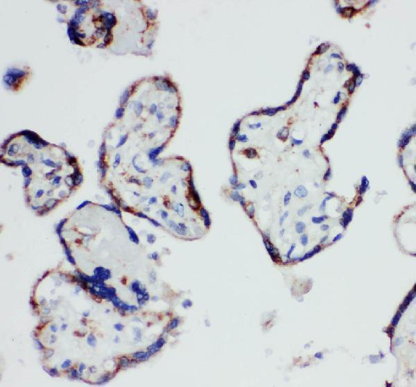

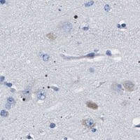


Comprehensive Human Protein Atlas IHC scoring per tissue (reliability: Enhanced — High consistency between antibody staining and RNA expression data.). Rows are taken directly from the HPA tissue chart — click any row's HPA link to view the source.
| Tissue | Cell type | Level | Evidence | Source |
|---|---|---|---|---|
| Placenta | Syncytiotrophoblasts - cell body | High | Protein (IHC) | HPA → |
| Tissue | Cell type | Level | Evidence | Source |
|---|---|---|---|---|
| Adipose tissue | Adipocytes | Not detected | Protein (IHC) | HPA → |
| Adrenal gland | Glandular cells | Not detected | Protein (IHC) | HPA → |
| Appendix | Glandular cells | Not detected | Protein (IHC) | HPA → |
| Bone marrow | Hematopoietic cells | Not detected | Protein (IHC) | HPA → |
| Breast | Adipocytes | Not detected | Protein (IHC) | HPA → |
Troubleshoot HSD17B1 staining in paraffin sections by checking retrieval, cytoplasmic localisation, cell identity and detection controls (datasheet PA1613-1; UniProt P14061; HPA tissue IHC).
Anti-HSD17B1 antibodies have paraffin-section IHC images from human placenta and rat liver and ovary (catalog IHC captions), plus fluorescence ICC data from U20S cells (A02198 IF caption).
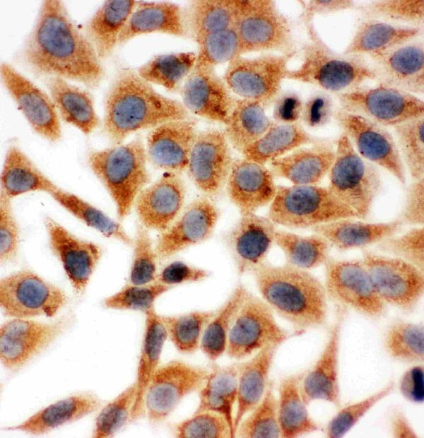
PA1613-1 has IHC images from rat liver, rat ovary, and human placenta paraffin sections, and an ICC image from Hela cells developed with DAB (catalog image captions). A02198 has an IHC image from a human placenta paraffin section and a fluorescence ICC image from U20S cells (catalog image captions).
Which to pick: For human placenta paraffin-section IHC, either SKU has an IHC image; for rat tissue IHC or human–rat coverage, choose PA1613-1 (PA1613-1 IHC captions; catalog reactivity: Human, Rat). For fluorescence IF/ICC, choose A02198 (catalog applications: IF, ICC; A02198 IF caption: fluorescent secondary and DAPI); PA1613-1’s ICC image uses DAB (PA1613-1 ICC caption). Both IHC captions specify paraffin sections, while the fixative is unreported (catalog IHC captions).