This website uses cookies to ensure you get the best experience on our website.


- Table of Contents
Plan paraffin-section HSPA1L IHC around strong staining in late spermatids of testis (HPA tissue IHC). Start with the catalog antibody at 2–5 μg/ml (datasheet A04286-3), and assess staining in light of the reported off-target binding (HPA tissue IHC).

Expected localisation, validated protocols, controls and antibodies — the at-a-glance facts below, then the full design guide.
| Expected localisation | Cytoplasmic and nuclear in spermatids (HPA tissue IHC) | |
| Staining pattern | Late spermatids show cytoplasmic and nuclear staining (HPA tissue IHC) | |
| Antigen retrieval | EDTA pH 8.0 HIER, heat-mediated (datasheet A04286-3) | |
| Positive control | Testis+3 more · see all | |
| Negative control | Adipose tissue+4 more · see all |
| Fixation | Keep fixation consistent across sections (standard IHC practice; not target-specific) | |
| Caveat | Off-target staining may resemble HSPA1L signal (HPA tissue IHC) | |
| Regulation | Spermatid expression; no regulator reported (UniProt) | |
| Isoform / epitope | One 1–641 chain; no isoforms annotated (UniProt) |
The catalog antibody’s IHC-P protocol (datasheet A04286-3) is accompanied by two published HSPA1L chromogenic IHC protocols (PMC13099889: Immunohistochemistry; PMC7979793: Morphological studies).
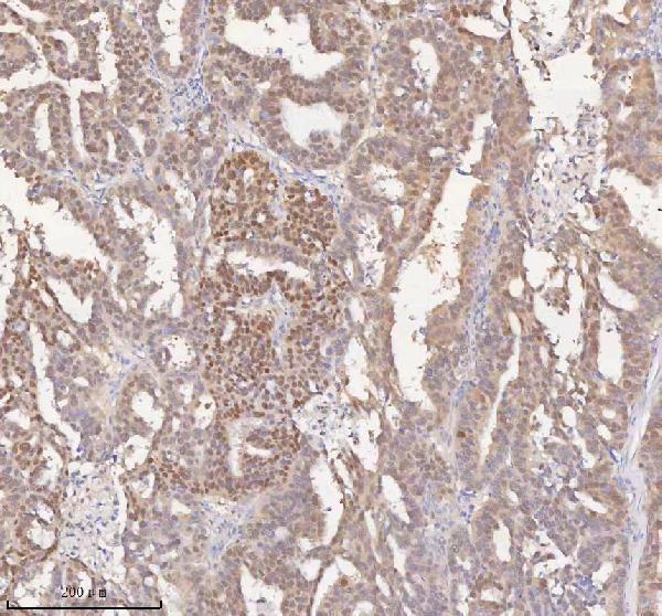
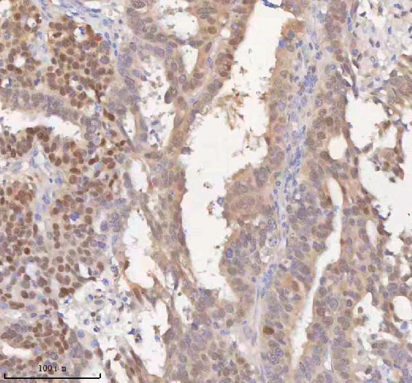
| Sample | Paraffin-embedded human ovarian cancer tissue; fixative not specified (datasheet A04286-3) |
| Fixation | Image fixative and duration unreported (datasheet A04286-3); verify before use. |
| Sectioning | 4–5 µm sections on charged slides (standard) |
| Deparaffinisation | Xylene, graded ethanol series to water (standard) |
| Antigen retrieval | Heat retrieval: EDTA pH 8.0 (datasheet A04286-3); 20 min, 95–100 °C (standard) |
| Peroxidase block | 3% H2O2, 10 min, room temperature (standard) |
| Blocking | 10% goat serum (datasheet A04286-3) |
| Primary antibody | Rabbit anti-HSPA1L, 2-5 μg/ml (datasheet A04286-3) |
| Primary incubation | Overnight at 4 °C (datasheet A04286-3) |
| Detection | HRP-conjugated secondary, DAB chromogen (datasheet A04286-3) |
| Counterstain | Hematoxylin, blue, dehydrate and mount (standard) |
| Expected result | HSPA1L-positive staining in elongated or late spermatids of testis (HPA tissue IHC: High). HPA tissue profile: Selective cytoplasmic and nuclear expression in spermatids in testis. No signal in the no-primary control. |
In paraffin-section IHC, expect selective cytoplasmic and nuclear staining in testicular spermatids, strongest in elongated or late spermatids (HPA: Enhanced tissue IHC; High in elongated or late spermatids). UniProt reports expression in spermatids but does not annotate a subcellular location (UniProt P34931). HSPA1L has no transmembrane segment, signal peptide, or propeptide (UniProt P34931 topology and processing). Interpret the pattern cautiously because HPA reports antibody recognition of proteins from more than one gene (HPA: reliability description).
| Strong cytoplasmic and nuclear staining in elongated or late spermatids, with other testicular cells less conspicuous (HPA: tissue IHC). | This matches the principal testis pattern; score the stained cell population as well as intensity (HPA: High in elongated or late spermatids; general IHC practice). |
| Predominantly membrane-restricted staining, without the expected spermatid cytoplasmic or nuclear pattern (HPA: tissue IHC). | Treat this as discordant with the reference pattern and investigate artefact or nonspecific staining (HPA: selective cytoplasmic and nuclear expression; general IHC practice). UniProt provides no membrane localisation (UniProt P34931). |
| Strong staining in an unexpected cell type while spermatids are weak or unstained (HPA: tissue IHC). | Consider cross-reactivity; also check endogenous chromogenic activity in the detection system (HPA: multiple-gene targeting and presumed off-target binding; general IHC practice). Medium staining in specified ciliated cells can be expected (HPA: bronchus, fallopian tube, nasopharynx). |
| Diffuse chromogen across many cells or tissue spaces, with little cell-specific contrast (general IHC practice). | This does not reproduce HPA's selective spermatid pattern (HPA: tissue IHC). Review blocking, washes, detection background, and counterstain before assigning a biological localisation (general IHC practice). |
| No detectable staining in elongated or late spermatids in a testis section (HPA: expected High staining). | First assess section quality and the IHC run with suitable controls (general IHC practice). An absent positive-control signal cannot establish absence of HSPA1L in the study specimen (HPA: High in elongated or late spermatids; general IHC practice). |
| Which tissue and cells give the clearest IHC reference? | Testis is RNA-enriched, and elongated or late spermatids stain High (HPA: RNA tissue specificity; tissue IHC). Adipocytes in adipose tissue and adrenal glandular cells are reported as not detected; use any comparator at the stated cell-type level (HPA: tissue IHC). |
| How much confidence does the reference pattern support? | HPA grades tissue IHC Enhanced, with medium agreement between staining and RNA data (HPA: reliability description). Its antibody caution includes recognition of proteins from multiple genes and disregarded presumed off-target staining; morphology alone cannot prove target identity (HPA: reliability description; general IHC practice). |
| Does topology predict a membrane or secreted pattern? | No transmembrane segment, signal peptide, propeptide, or annotated modified residues are reported (UniProt P34931 topology, processing, modified residues). These features do not establish a subcellular location; use the observed IHC pattern for slide interpretation (UniProt P34931; HPA: tissue IHC). |
| What does the separate IF/ICC evidence show? | Q: Where is IF/ICC signal reported? A: Mainly vesicles, flagellar centriole, and annulus; additionally perinuclear theca and calyx (HPA: subcellular ICC-IF). These approved locations carry HPA's multiple-gene antibody caution and do not replace the paraffin IHC reference pattern (HPA: subcellular caution; tissue IHC). |
| Is HSPA1L fixation sensitivity established here? | Target-specific fixation sensitivity is unreported in the supplied UniProt and HPA records (UniProt P34931; HPA: tissue IHC). Document the run's processing conditions when comparing sections; do not infer a HSPA1L fixation effect from staining intensity alone (general IHC practice). |
| Situation | Likely cause | Next action |
|---|---|---|
| Testis control lacks spermatid staining (HPA: expected High staining). | The run, detection reagents, tissue preservation, or antibody conditions may have failed (general IHC practice). | Confirm spermatids are present, inspect counterstain and controls, and review the antibody's IHC validation before interpreting study negatives (HPA: testis IHC; general IHC practice). |
| Signal is widespread and masks cell boundaries (general IHC practice). | Nonspecific reagent binding, inadequate washing, or excessive chromogen development can raise background (general IHC practice). | Compare a no-primary control; review blocking, washes, and detection development, then reassess selective spermatid staining (general IHC practice; HPA: tissue IHC). |
| Chromogen appears in unexpected cells or tissue components (HPA: selective spermatid pattern). | Cross-reactivity is plausible given HPA's multiple-gene and presumed off-target cautions; endogenous detection activity is another general IHC possibility (HPA: reliability description; general IHC practice). | Check a no-primary control and endogenous-activity blocking for the detection system; compare cell identity with HPA's tissue pattern (general IHC practice; HPA: tissue IHC). |
| Only cell borders stain in testis (HPA: cytoplasmic and nuclear pattern). | The compartment pattern conflicts with HPA tissue IHC and is not supported by UniProt topology (HPA: tissue IHC; UniProt P34931 topology). | Check section morphology and background controls; withhold a positive HSPA1L call unless the expected spermatid pattern is also present (general IHC practice; HPA: tissue IHC). |
| Ciliated cells stain outside testis (HPA: tissue IHC). | Medium staining is reported in bronchial and nasopharyngeal ciliated cell bodies and fallopian-tube ciliary rootlets (HPA: tissue IHC). | Check tissue and cell identity before calling this off-target; interpret intensity and compartment alongside the HPA antibody-specificity caution (HPA: tissue IHC; reliability description). |
| Study tissue is negative while the testis control is positive (HPA: testis IHC). | The study cell type may have little detectable staining under the assay conditions; HPA reports several specified cell populations as not detected (HPA: tissue IHC; general IHC practice). | Record the scored cell type and detection threshold, and report the result as not detected in that assay rather than claiming universal absence of protein (general IHC practice). |
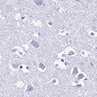


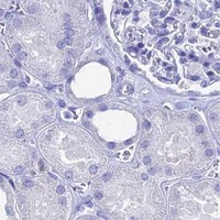


Comprehensive Human Protein Atlas IHC scoring per tissue (reliability: Enhanced — Medium consistency between antibody staining and RNA expression data. Caution, targets protein from more than one gene. Presumed off target binding observed and disregarded.). Rows are taken directly from the HPA tissue chart — click any row's HPA link to view the source.
| Tissue | Cell type | Level | Evidence | Source |
|---|---|---|---|---|
| Testis | Elongated or late spermatids | High | Protein (IHC) | HPA → |
| Bronchus | Ciliated cells (cell body) | Medium | Protein (IHC) | HPA → |
| Fallopian tube | Ciliated cells (ciliary rootlets) | Medium | Protein (IHC) | HPA → |
| Nasopharynx | Ciliated cells (cell body) | Medium | Protein (IHC) | HPA → |
| Tissue | Cell type | Level | Evidence | Source |
|---|---|---|---|---|
| Adipose tissue | Adipocytes | Not detected | Protein (IHC) | HPA → |
| Adrenal gland | Glandular cells | Not detected | Protein (IHC) | HPA → |
| Appendix | Glandular cells | Not detected | Protein (IHC) | HPA → |
| Bone marrow | Hematopoietic cells | Not detected | Protein (IHC) | HPA → |
| Breast | Adipocytes | Not detected | Protein (IHC) | HPA → |
Troubleshoot HSPA1L staining by checking retrieval, cellular context and antibody specificity before comparing signal across paraffin sections (datasheet A04286-3; HPA tissue IHC).
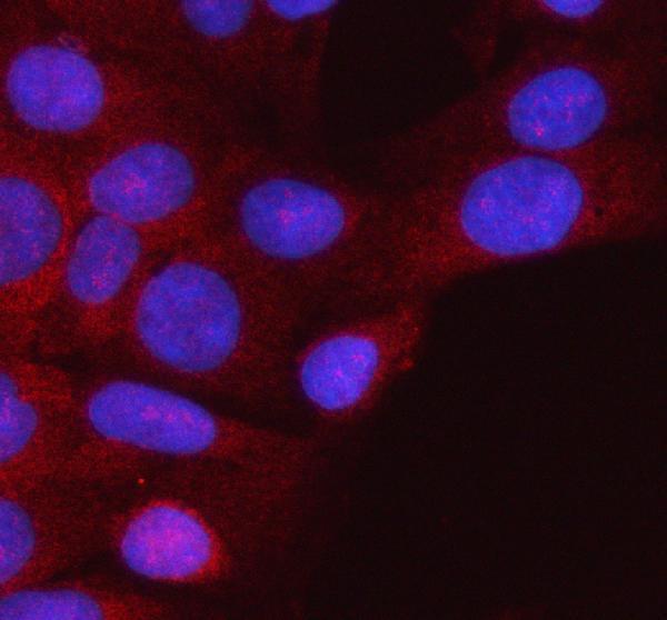
The IHC-validated antibody has paraffin-section images from human ovarian cancer and mouse and rat testis, plus IF data from HeLa cells (catalog image captions).
A04286-3 will render with an IHC image from a paraffin-embedded human ovarian cancer section; its other IHC captions show mouse and rat testis sections (catalog IHC captions). Its IF caption shows staining in HeLa cells (catalog IF caption).
Which to pick: Choose A04286-3 for paraffin-section IHC because its own caption documents that preparation; the fixative is unreported (A04286-3 IHC caption). Choose the same SKU for IF/ICC in human cells because IF and ICC are listed applications and its IF caption shows HeLa cells (catalog applications; A04286-3 IF caption). For cross-species IHC, its listed reactivity covers human, mouse and rat, with paraffin-section images in each species; the catalog lists no clone designation (catalog reactivity; A04286-3 IHC captions; catalog clone field).