This website uses cookies to ensure you get the best experience on our website.


- Table of Contents
Plan chromogenic IHC in paraffin sections around the cytoplasmic and nuclear tissue pattern (HPA tissue IHC). Start with the catalog antibody's 2–5 μg/ml IHC range (datasheet A03618-2), and assess ovarian follicle cells and cells in seminiferous ducts as high-staining references while accounting for uncertain HPA reliability (HPA tissue IHC).

Expected localisation, validated protocols, controls and antibodies — the at-a-glance facts below, then the full design guide.
| Expected localisation | Cytoplasmic and nuclear tissue staining (HPA tissue IHC) | |
| Staining pattern | Most tissues show cytoplasmic and nuclear staining (HPA tissue IHC) | |
| Antigen retrieval | EDTA pH 8.0 HIER, heat-mediated (datasheet A03618-2) | |
| Positive control | Ovary+4 more · see all | |
| Negative control | Adipose tissue+4 more · see all |
| Fixation | Keep fixation consistent (standard IHC practice; not target-specific); its effect on HSPA4 staining is unclear. | |
| Caveat | Staining and RNA show medium consistency; verify locally (HPA tissue IHC) | |
| Regulation | Stress response; induction unreported (UniProt) | |
| Isoform / epitope | 2 isoforms; epitope coverage is unreported (UniProt) |
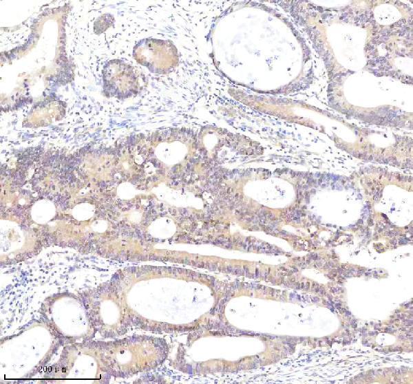
The catalog antibody protocol (datasheet A03618-2) is accompanied by one published chromogenic colorectal IHC protocol (PMC8214019).
| Sample | Paraffin-embedded human colon cancer tissue; fixative not specified (datasheet A03618-2) |
| Fixation | Image fixative and duration unreported (datasheet A03618-2); verify before use. |
| Sectioning | 4–5 µm sections on charged slides (standard) |
| Deparaffinisation | Xylene, graded ethanol series to water (standard) |
| Antigen retrieval | Heat retrieval: EDTA pH 8.0 (datasheet A03618-2); 20 min, 95–100 °C (standard) |
| Peroxidase block | 3% H2O2, 10 min, room temperature (standard) |
| Blocking | 10% goat serum (datasheet A03618-2) |
| Primary antibody | Rabbit anti-HSPA4, 2-5 μg/ml (datasheet A03618-2) |
| Primary incubation | Overnight at 4 °C (datasheet A03618-2) |
| Detection | HRP-conjugated secondary, DAB chromogen (datasheet A03618-2) |
| Counterstain | Hematoxylin, blue, dehydrate and mount (standard) |
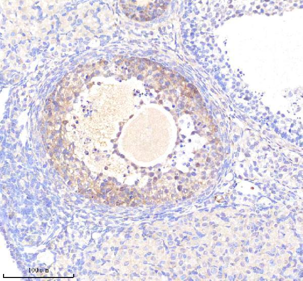
| Expected result | HSPA4-positive staining in follicle cells of ovary (HPA tissue IHC: High). HPA tissue profile: Cytoplasmic and nuclear expression in most tissues. No signal in the no-primary control. |
HSPA4 is a cytoplasmic protein without a transmembrane segment (UniProt P34932). Tissue IHC shows cytoplasmic and nuclear staining in most tissues, with high staining in ovarian follicle cells and cells in testicular seminiferous ducts (HPA tissue IHC). Treat those examples as useful reference patterns, not definitive controls: HPA rates tissue IHC reliability as Uncertain because antibody staining and RNA expression show only medium consistency (HPA tissue IHC).
| Strong cellular staining in ovarian follicle cells or cells in testicular seminiferous ducts, with cytoplasmic signal and possible nuclear signal (HPA tissue IHC). | This matches the reported high-staining cell populations and broad tissue compartment pattern (HPA tissue IHC). Assess staining in the named cells rather than averaging across the section. A matching appearance supports interpretation but does not establish antibody specificity, because tissue IHC reliability remains Uncertain (HPA tissue IHC). |
| Staining is confined to cell borders, extracellular material, or another location without convincing intracellular staining. | That distribution differs from the reported cytoplasmic and nuclear tissue pattern (HPA tissue IHC), the approved nucleoplasm and cytosol IF locations (HPA subcellular), and the lack of a transmembrane segment (UniProt P34932). Check morphology and controls before assigning it to HSPA4; compartment mismatch alone does not identify its cause. |
| Strong staining appears in adipocytes, cardiomyocytes, smooth muscle cells, or soft-tissue fibroblasts. | Those cell populations were reported as Not detected by tissue IHC (HPA tissue IHC). Recheck cell identity and examine a no-primary control for endogenous detection activity; persistent signal may reflect cross-reactivity. Because the tissue IHC assessment is Uncertain, a single discordant section cannot by itself resolve specificity (HPA tissue IHC). |
| Colour spreads across several cell types, stroma, and areas expected to have little cellular signal. | The reported pattern names specific cells and intracellular compartments, even though expression is widespread (HPA tissue IHC). Broad haze can obscure that distinction. Compare the no-primary control, inspect local tissue background, and reassess antibody concentration and washing as general IHC practice before scoring cells as positive. |
| No convincing staining appears in ovarian follicle cells or cells in testicular seminiferous ducts. | Both were reported as High in tissue IHC (HPA tissue IHC), so first check section quality, detection controls, and assay conditions as general IHC practice. Do not treat absence in one section as proof of biological absence: HPA labels its tissue IHC evidence Uncertain (HPA tissue IHC). |
| Compartment and topology | UniProt places HSPA4 in the cytoplasm and reports no transmembrane segment (UniProt P34932); HPA tissue IHC also reports nuclear staining, and ICC-IF approves nucleoplasm and cytosol (HPA tissue IHC; HPA subcellular). Interpret nuclear signal alongside these method-specific observations rather than requiring every positive cell to be cytoplasm-only. |
| Cell population and staining level | High staining is reported in ovarian follicle cells and cells in testicular seminiferous ducts; medium staining is reported in several glandular, respiratory epithelial, neuronal, and glial populations (HPA tissue IHC). Adipocytes and cardiomyocytes are reported as Not detected (HPA tissue IHC). Compare like cell populations when reviewing sections. |
| Strength of the IHC evidence | HPA rates the overall tissue IHC assessment Uncertain and describes medium consistency with RNA expression, pending external verification (HPA tissue IHC). Both listed antibodies have Uncertain IHC status, while HPA010023 has Approved ICC status (HPA antibodies). A familiar tissue pattern therefore remains a provisional specificity check, not validation of the catalog antibody. |
| Isoforms and processing | UniProt lists two HSPA4 isoforms and describes the annotated protein chain as residues 1–840, with no signal peptide or propeptide (UniProt P34932). The supplied records give no isoform-specific IHC pattern or antibody epitope, so section staining cannot be assigned to one isoform or explained as cleavage or shedding from these data. |
| Antigen retrieval and detection controls | No target-specific fixation or retrieval effect is supplied by UniProt or HPA (UniProt P34932; HPA tissue IHC). Retrieval optimisation, no-primary controls, and checks for endogenous detection activity are general IHC practice. Interpret changes from those steps as assay behaviour until a controlled comparison establishes their effect in the section being studied. |
| Situation | Likely cause | Next action |
|---|---|---|
| The expected positive cell population is faint or blank. | Possible assay under-detection should be considered before inferring absence; ovarian follicle cells and cells in testicular seminiferous ducts were reported as High, but tissue IHC reliability is Uncertain (HPA tissue IHC). | Confirm the intended cells are present, inspect section and detection controls, then optimise retrieval and antibody concentration within the assay as general IHC practice; record each condition rather than assuming a target-specific fixation effect. |
| Nearly every structure is dark, obscuring cell boundaries. | Excessive assay signal or background may overwhelm the cell-specific pattern reported by HPA (HPA tissue IHC); the appearance alone cannot distinguish antibody binding from endogenous detection activity. | Compare a no-primary control, inspect background outside cells, and adjust detection, washing, or antibody concentration one variable at a time as general IHC practice; score only interpretable cellular staining. |
| Strong staining appears where HPA reports no detection. | Adipocytes, cardiomyocytes, smooth muscle cells, and soft-tissue fibroblasts are listed as Not detected (HPA tissue IHC); cell misidentification, cross-reactivity, or endogenous activity could account for discordance. | Verify morphology and section identity, compare a no-primary control, and seek independent specificity evidence before calling that signal HSPA4; the HPA tissue IHC assessment itself is Uncertain (HPA tissue IHC). |
| The main signal follows membranes or extracellular spaces. | That location conflicts with the intracellular tissue and ICC-IF patterns (HPA tissue IHC; HPA subcellular) and with HSPA4 lacking a transmembrane segment (UniProt P34932). | Review the counterstained morphology and assay controls, then compare another validated section or antibody if available as general IHC practice; avoid interpreting an isolated border pattern as expected HSPA4 staining. |
| Cytoplasmic staining is present, but nuclear staining varies among cells. | UniProt annotates cytoplasm, while HPA reports cytoplasmic and nuclear tissue expression and approves nucleoplasm and cytosol by ICC-IF (UniProt P34932; HPA tissue IHC; HPA subcellular). | Score cytoplasmic and nuclear compartments separately and document which cells show each pattern; variation alone does not invalidate a section, but the Uncertain tissue IHC reliability limits a definitive localisation claim (HPA tissue IHC). |
| IF/ICC question: should nucleoplasmic signal be considered compatible with HSPA4? | HPA lists both nucleoplasm and cytosol as Approved ICC-IF locations, and reports cytoplasmic and nuclear expression in tissue IHC (HPA subcellular; HPA tissue IHC). | Yes: interpret nucleoplasmic signal alongside cytosolic signal and appropriate imaging controls (HPA subcellular; general IF practice). Use the separate IF/ICC guide for method decisions; this IHC interpretation does not establish an IF/ICC protocol. |


Comprehensive Human Protein Atlas IHC scoring per tissue (reliability: Uncertain — Medium consistency between antibody staining and RNA expression data. Pending external verification.). Rows are taken directly from the HPA tissue chart — click any row's HPA link to view the source.
| Tissue | Cell type | Level | Evidence | Source |
|---|---|---|---|---|
| Ovary | Follicle cells | High | Protein (IHC) | HPA → |
| Testis | Cells in seminiferous ducts | High | Protein (IHC) | HPA → |
| Adrenal gland | Glandular cells | Medium | Protein (IHC) | HPA → |
| Breast | Glandular cells | Medium | Protein (IHC) | HPA → |
| Bronchus | Respiratory epithelial cells | Medium | Protein (IHC) | HPA → |
| Tissue | Cell type | Level | Evidence | Source |
|---|---|---|---|---|
| Adipose tissue | Adipocytes | Not detected | Protein (IHC) | HPA → |
| Duodenum | Glandular cells | Not detected | Protein (IHC) | HPA → |
| Heart muscle | Cardiomyocytes | Not detected | Protein (IHC) | HPA → |
| Smooth muscle | Smooth muscle cells | Not detected | Protein (IHC) | HPA → |
| Soft tissue | Fibroblasts | Not detected | Protein (IHC) | HPA → |
Troubleshoot HSPA4 chromogenic IHC in paraffin sections by checking retrieval, compartment, controls and scoring before comparing specimens.
A03618-2 has paraffin-section IHC images from human colon cancer and mouse ovary (image captions). Both antibodies list IF/ICC applications (catalog); neither supplies an IF image (catalog).
A03618-2 lists IHC and IF/ICC applications and human, mouse, and rat reactivity (catalog); its IHC captions show human colon cancer and mouse ovary paraffin sections (image captions). M03618 lists IHC and IF/ICC applications and human, mouse, and rat reactivity (catalog), with no IHC or IF image supplied (catalog).
Which to pick: For tissue IHC, choose A03618-2: its captions document paraffin-section staining in human colon cancer and mouse ovary, but do not report the fixative (image captions). For human IF/ICC, A03618-2 has a stated 5 μg/ml condition (catalog); M03618 is a rabbit monoclonal alternative, clone IAB-8, with IF/ICC listed but no supplied image (catalog). Both list human, mouse, and rat reactivity (catalog), while the supplied IHC images for A03618-2 cover human and mouse only (image captions).