HSPB1 / Heat shock protein beta-1 · IHC design guide

Design Immunohistochemistry for HSPB1

Plan chromogenic HSPB1 IHC in paraffin sections using esophageal squamous epithelium as a high-staining reference (HPA tissue IHC). This guide covers expected localisation, fixation, controls and interpretation.

Evidence assembled Oct 2026 · For research use; verify linked source records and product datasheet before use
Immunohistochemistry protocol sheet for HSPB1 (IHC for HSPB1): expected localisation Cytoplasmic staining in squamous epithelium (HPA tissue IHC), antibody M00676-5, validated IHC image, and IHC protocol steps
Printable HSPB1 IHC protocol sheet — expected localisation Cytoplasmic staining in squamous epithelium (HPA tissue IHC), antibody M00676-5, controls and protocol steps. Open the full HSPB1 IHC guide →

HSPB1 Immunohistochemistry Experimental Design Guide

Expected localisation, validated protocols, controls and antibodies — the at-a-glance facts below, then the full design guide.

Must know before staining
Expected localisation Cytoplasmic staining in squamous epithelium (HPA tissue IHC)
Staining pattern Squamous epithelial cells show cytoplasmic staining (HPA tissue IHC)
Antigen retrieval Citrate pH 6 HIER, heat-mediated (datasheet M00676-5)
Positive control ⓘ Bronchus+4 more · see all
Negative control ⓘ Appendix+4 more · see all
Important caveats
Reasons your staining may differ from the expected pattern.
Fixation Keep fixation consistent across paraffin sections. (standard IHC practice; not target-specific)
Caveat Heat shock can shift HSPB1 into nuclei (UniProt)
Regulation Highest levels in heart and muscle tissues (UniProt)
Isoform / epitope No annotated isoforms; full-length 1–205 chain (UniProt)
Section 1

Recommended HSPB1 IHC & IF Protocols

The catalog antibody’s IHC-P protocol (datasheet: M00676-5) is accompanied by four published HSPB1 IHC protocols (PMC12972742; PMC4502388; PMC5370501; PMC4427334).

Recommended immunohistochemistry (IHC-P) protocol parameters
SampleParaffin-embedded human lung cancer tissues; fixative not specified (datasheet M00676-5)
FixationImage fixative and duration unreported (datasheet M00676-5); verify before use.
Sectioning4–5 µm sections on charged slides (standard)
DeparaffinisationXylene, graded ethanol series to water (standard)
Antigen retrievalHeat retrieval: Citrate pH 6, 20 min (datasheet M00676-5)
Peroxidase block3% H2O2, 10 min, room temperature (standard)
Blocking10% goat serum (datasheet M00676-5)
Primary antibodyMouse monoclonal (clone 3H3) anti-HSPB1, 0.5-1μg/ml (datasheet M00676-5)
Primary incubationOvernight at 4 °C (datasheet M00676-5)
DetectionStreptavidin-biotin complex (SABC), DAB chromogen (datasheet M00676-5)
CounterstainHematoxylin, blue, dehydrate and mount (standard)
Expected resultHSPB1-positive staining in respiratory epithelial cells of bronchus (HPA tissue IHC: High). HPA tissue profile: Cytoplasmic expression mainly in squamous epithelial cells. No signal in the no-primary control.
💡Decision noteStart with heat-mediated citrate retrieval at pH 6 (datasheet: M00676-5); a published HSPB1 protocol specifies pressure-cooker retrieval in citrate at pH 6.0 (PMC4502388).
Section 2

What Is the Expected HSPB1 Staining Pattern?

In paraffin-section IHC, expect HSPB1 mainly in the cytoplasm of squamous epithelial cells, with high staining in esophageal squamous epithelium and heart cardiomyocytes (HPA: tissue IHC, Enhanced reliability). UniProt places HSPB1 in the cytoplasm during interphase and also reports nuclear localization under heat stress (UniProt P04792: subcellular location). It has no transmembrane segment (UniProt P04792: topology).

What am I looking at on my slide?
Strong cytoplasmic staining in esophageal squamous epithelial cells or heart cardiomyocytes, with identifiable cell boundaries (HPA: High in both cell types).This matches reported positive cell types and the predominant tissue-IHC compartment (HPA: tissue IHC). Judge the named cells, since a tissue section also contains other cell populations whose staining may differ.
Predominantly nuclear staining in otherwise unstressed cells, with little cytoplasmic signal (UniProt P04792: cytoplasmic in interphase).Review specificity and staining conditions before accepting this as the expected IHC pattern (HPA: mainly cytoplasmic tissue IHC). Nuclear HSPB1 can occur during heat shock, so nuclear signal alone cannot establish an artefact (UniProt P04792: heat-shock translocation).
Staining appears in a cell population reported as not detected, such as appendix endocrine cells or lymph-node germinal-center cells (HPA: Not detected).Consider cross-reactivity or endogenous chromogenic activity, then inspect morphology and controls (standard IHC practice). The HPA calls apply to those cell types; they do not make every cell in either tissue a negative control (HPA: tissue IHC).
Uniform color covers cells and surrounding tissue without a readable cytoplasmic pattern.Diffuse background obscures the compartment and cannot be scored as HSPB1-positive staining (standard IHC practice). Compare a no-primary control and check blocking, washes, and detection chemistry before interpreting the section (standard IHC practice).
Esophageal squamous epithelial cells show no signal, despite being a reported high-staining population (HPA: High in esophagus).Treat the run as unresolved until controls and processing are checked (standard IHC practice). An absent signal could reflect assay performance; the HPA observation alone does not identify a cause or establish HSPB1-specific fixation sensitivity.
💡Expected HSPB1 appearanceCall a result positive when distinct cytoplasmic signal is visible in the relevant cells, especially high-staining esophageal squamous epithelium or cardiomyocytes (HPA: tissue IHC); uniform background or a nuclear-only pattern without context should not be scored as the expected result (HPA: mainly cytoplasmic; UniProt P04792: stress-associated nuclear translocation).
How each factor affects the staining
Compartment and cell state (UniProt P04792: subcellular location).Cytoplasmic localization is expected in interphase; spindle colocalization is reported in mitosis, and nuclear translocation during heat shock. Interpret unusual nuclear signal with the cell state in mind, while using the mainly cytoplasmic tissue-IHC profile as the routine reference (HPA: tissue IHC).
Choice of comparison cells (HPA: tissue IHC).Esophageal squamous epithelial cells and heart cardiomyocytes are reported High; appendix endocrine cells and lymph-node germinal-center cells are Not detected. Compare the specified populations rather than labeling an entire section positive or negative (HPA: cell-level observations).
Evidence behind the reference pattern (HPA: Enhanced tissue-IHC reliability).HPA reports high consistency between antibody staining and RNA expression, and lists IHC Enhanced status for HPA000497, CAB004439, and CAB047331 (HPA: tissue IHC; antibody validation). These are reference-pattern findings, not validation of an unspecified catalog antibody.
IF/ICC Q: should membrane or cytosolic fluorescence change the IHC call?A: HPA reports enhanced plasma-membrane and cytosol localization in ICC-IF (HPA: subcellular). Use that as IF/ICC context; the paraffin-section IHC reference remains mainly cytoplasmic expression in squamous epithelium (HPA: tissue IHC).
Why is my staining missing, weak or wrong?
SituationLikely causeNext action
Known-positive esophageal squamous cells are blank (HPA: High in esophagus).The section or staining run may have failed; this observation does not establish a target-specific fixation effect.Check section integrity, primary-antibody application, detection reagents, and a concurrently stained positive control (standard IHC practice). Repeat with documented retrieval and incubation settings if a run-level fault is found.
The whole slide shows weak, diffuse chromogen rather than distinct cytoplasm.Background from nonspecific binding or detection chemistry may hide the expected cell pattern (standard IHC practice; HPA: mainly cytoplasmic).Inspect the no-primary control, blocking step, washes, and chromogen development; adjust the general staining workflow before scoring (standard IHC practice).
A supposedly negative tissue shows scattered positive cells.The selected HPA negative call may describe only one cell type, such as appendix endocrine cells (HPA: Not detected), while other cells remain unevaluated by that call.Identify stained cells by morphology and compare the exact HPA cell population. Use a no-primary control to assess nonspecific detection (standard IHC practice).
Signal is predominantly nuclear across interphase cells.This differs from the mainly cytoplasmic tissue-IHC profile, although stress-related nuclear translocation is reported (HPA: tissue IHC; UniProt P04792: subcellular location).Confirm cell morphology and compare a known-positive section and no-primary control. Report the nuclear pattern separately rather than assigning it the routine cytoplasmic call (standard IHC practice).
Chromogen outlines membranes but cytoplasm is hard to see.HPA reports plasma-membrane localization in ICC-IF; that assay does not establish a membrane-dominant paraffin-section IHC pattern (HPA: subcellular; tissue IHC).Check focus, counterstain, and cellular boundaries, then compare with the cytoplasmic IHC reference and controls before scoring (HPA: tissue IHC; standard IHC practice).
A catalog antibody produces a pattern unlike the HPA reference.HPA's Enhanced assessment applies to its reported tissue pattern and listed antibodies; it does not validate an unspecified catalog antibody (HPA: antibody validation).Review that antibody's IHC-P validation and its documented conditions; compare positive cells, no-primary control, and the HPA cell-level pattern before interpreting discrepant staining (standard IHC practice; HPA: tissue IHC).

Sample controls for HSPB1 IHC & IF

🧪Run bronchus first; respiratory epithelial cells should stain for HSPB1 (HPA: High in bronchus respiratory epithelial cells). Use appendix endocrine cells as the negative comparator (HPA: Not detected in appendix endocrine cells); on the bronchus slide, internal negative cells should show counterstain without specific chromogen, although the supplied HPA row does not identify a reliably negative bronchial cell type (HPA: bronchus row).
Positive control tissue: Bronchus (Respiratory epithelial cells, HPA High)
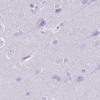
Negative control tissue: Appendix (HPA Not detected)
ICC-IF cell lines (HPA subcellular resource): HPA ICC-IF images show HSPB1 in A-431, U-251MG, U2OS, with annotated localisation: Plasma membrane (enhanced), Cytosol (enhanced) (HPA subcellular).
Technical controls: Include a no-primary, secondary-only control, a matched mouse IgG isotype control, and an HSPB1 knockout control or peptide block if the immunizing peptide is available (selected IHC caption: mouse primary; standard IHC practice). For bronchus, check endogenous peroxidase and biotin background with the caption’s SABC/DAB detection; check tissue autofluorescence if adapting the assay to IF (selected IHC caption: SABC/DAB; standard IHC/IF practice).
⚠️Feasibility: No matched source reports a target-specific fixation window or fixation effect, and the selected M00676-5 paraffin-section caption does not state the fixative (selected IHC caption). Heat-mediated citrate retrieval at pH 6 for 20 minutes is a documented starting condition, but retrieval dependency is unreported (selected IHC caption). Whether frozen sections or IF/ICC are easier is unreported; assess background from the SABC/DAB system in bronchus and autofluorescence if using IF (selected IHC caption: SABC/DAB; standard IHC/IF practice).

HPA tissue IHC evidence for HSPB1

Comprehensive Human Protein Atlas IHC scoring per tissue (reliability: Enhanced — High consistency between antibody staining and RNA expression data.). Rows are taken directly from the HPA tissue chart — click any row's HPA link to view the source.

Positive expression · recommended positive controls

TissueCell typeLevelEvidenceSource
Bronchus Respiratory epithelial cells High Protein (IHC) HPA →
Endometrium Glandular cells High Protein (IHC) HPA →
Epididymis Glandular cells High Protein (IHC) HPA →
Esophagus Squamous epithelial cells High Protein (IHC) HPA →
Fallopian tube Glandular cells High Protein (IHC) HPA →

Undetected expression · recommended negative controls

TissueCell typeLevelEvidenceSource
Appendix Endocrine cells Not detected Protein (IHC) HPA →
Caudate Glial cells Not detected Protein (IHC) HPA →
Cerebral cortex Endothelial cells Not detected Protein (IHC) HPA →
Hippocampus Glial cells Not detected Protein (IHC) HPA →
Lymph node Germinal center cells Not detected Protein (IHC) HPA →
Section 3

Advanced HSPB1 IHC Tips

Use the catalog antibody’s paraffin section evidence to troubleshoot HSPB1 staining, then assess signal by cell type, compartment and tissue quality.

What retrieval should I try first when HSPB1 staining is weak in paraffin sections?
Start with heat mediated retrieval in citrate buffer at pH 6 for 20 minutes (datasheet M00676-5). The selected paraffin section image also used 10% goat serum blocking and 1 μg/mL primary antibody overnight at 4°C, so check those steps before changing retrieval (datasheet M00676-5). Compare a reliably staining cell population on the same run, such as esophageal squamous epithelial cells, to separate a retrieval failure from low local expression (HPA: High in esophageal squamous epithelial cells; standard IHC practice). If staining remains weak, test a second retrieval condition on matched sections while keeping detection and exposure to chromogen consistent (standard IHC practice).
Could fixation explain weak or uneven HSPB1 staining?
The selected image identifies a paraffin embedded section but does not state its fixative, so target specific fixation sensitivity is unknown for this antibody (datasheet M00676-5 caption). Record the actual fixative and processing history for each specimen before comparing staining intensity across blocks (standard IHC practice). On matched sections, keep retrieval at citrate pH 6 for 20 minutes while checking whether patchy staining tracks processing differences or damaged tissue (datasheet M00676-5; standard IHC practice). Use morphology and an internal positive population to judge whether weak signal reflects preparation quality; do not assign a particular fixation effect to HSPB1 without a direct comparison (standard IHC practice).
Should HSPB1 appear in the cytoplasm, nucleus or along the cell membrane?
Expect predominantly cytoplasmic staining in interphase cells, with possible nuclear signal during heat shock and spindle association in mitotic cells (UniProt P04792 subcellular location). Tissue IHC chiefly reports cytoplasmic expression in squamous epithelial cells, providing a useful reference for routine section review (HPA: tissue IHC profile). HPA cell imaging also reports enhanced plasma membrane and cytosol localisation, while HSPB1 has no transmembrane segment; assess apparent membrane staining alongside cytoplasmic signal and cell boundaries (HPA: subcellular; UniProt P04792 topology; standard IHC practice). Score nuclear staining separately from cytoplasmic staining and document the tissue context before treating a compartment shift as a biological result (standard IHC practice).
Can HSPB1 modification or epitope accessibility change the IHC pattern?
The supplied record lists 0 isoforms and a single 1–205 amino acid chain, so these data do not support assigning distinct staining patterns to splice isoforms (UniProt P04792 processing and isoforms). HSPB1 has reported phosphoserine sites including residues 15, 26, 65 and 78, but the catalog antibody’s epitope is not specified here (UniProt P04792 modified residues; datasheet M00676-5). Do not interpret an intensity difference as phosphorylation specific without epitope information or a modification specific reagent (standard IHC practice). For weak signal, compare matched sections with consistent citrate pH 6 retrieval and primary incubation before drawing an epitope level conclusion (datasheet M00676-5; standard IHC practice).
How should I investigate HSPB1 localisation with IF after observing chromogenic IHC staining?
Use IF as a separate localisation experiment; the selected antibody evidence here is paraffin section chromogenic IHC, with no IF protocol specified for this SKU (datasheet M00676-5 caption). Multiplex HSPB1 with a validated marker for the expected cell type, such as squamous epithelial cells in esophagus, and check each channel alone before interpreting overlap (HPA: High in esophageal squamous epithelial cells; standard IF practice). Choose a fluorophore whose emission is readily separated from the specimen’s autofluorescence and include an unstained section to assess that background (standard IF practice). Because HSPB1 has no transmembrane segment and is reported in the cytosol, select and verify permeabilisation that permits access to intracellular epitopes in the IF preparation (UniProt P04792 topology; HPA: subcellular; standard IF practice).
How can I reduce diffuse brown background without losing genuine HSPB1 signal?
First inspect a section processed without primary antibody to identify detection or tissue background, then compare it with the complete stain (standard IHC practice). The selected image used 10% goat serum blocking, 1 μg/mL primary antibody, a biotinylated secondary and DAB detection; keep these documented conditions as the initial comparison point (datasheet M00676-5). Include appropriate endogenous peroxidase blocking for chromogenic detection and assess whether biotin based detection contributes background in the specimen (standard IHC practice; datasheet M00676-5 detection method). If background persists, titrate primary antibody and chromogen development on matched sections while retaining a positive cell population to monitor signal loss (standard IHC practice).
What is a defensible way to quantify HSPB1 across tissue sections? ⚠ ANSWER MARKED FOR VERIFICATION
Define the cell population and compartment before scoring; HSPB1 is chiefly cytoplasmic in interphase cells, and tissue IHC highlights squamous epithelial expression (UniProt P04792 subcellular location; HPA: tissue IHC profile). For chromogenic sections, report the percentage of positive cells and, when intensity is reproducible, an H-score calculated as the sum of each intensity category’s percentage multiplied by its category value (standard IHC practice). Normalise cell counts to the number of eligible cells, or positive area to viable tissue area in mm², while excluding folds, necrosis and section edges by prespecified rules (standard IHC practice). Keep retrieval, staining batch, imaging settings and scoring thresholds consistent, and report cytoplasmic and nuclear scores separately when both occur (datasheet M00676-5 retrieval; standard IHC practice).
How do I distinguish true HSPB1 staining from artefact in a tumor section?
Check that signal follows intact cells and a plausible compartment: HSPB1 is cytoplasmic in interphase, can enter nuclei during heat shock and can associate with mitotic spindles (UniProt P04792 subcellular location). Compare with an expected positive population; HPA reports high staining in bronchial respiratory epithelial cells and esophageal squamous epithelial cells, while some named cell populations are not detected (HPA: tissue IHC). Treat staining confined to section edges, necrotic regions or a no primary control as suspect, and evaluate endogenous peroxidase contribution to DAB signal (standard IHC practice). Interpret a nuclear or membrane predominant pattern cautiously, documenting morphology and control results before assigning it to HSPB1 biology (UniProt P04792 subcellular location and topology; HPA: subcellular; standard IHC practice).
Boster reagents

Best HSPB1 / Heat shock protein beta-1 IHC Antibodies

Three anti-HSPB1 antibodies have paraffin-section IHC images from human tissues (catalog IHC captions); M00676-2 also has a rat-tissue image, while M00676-5 and PB9237 have IF/ICC images (catalog image captions).

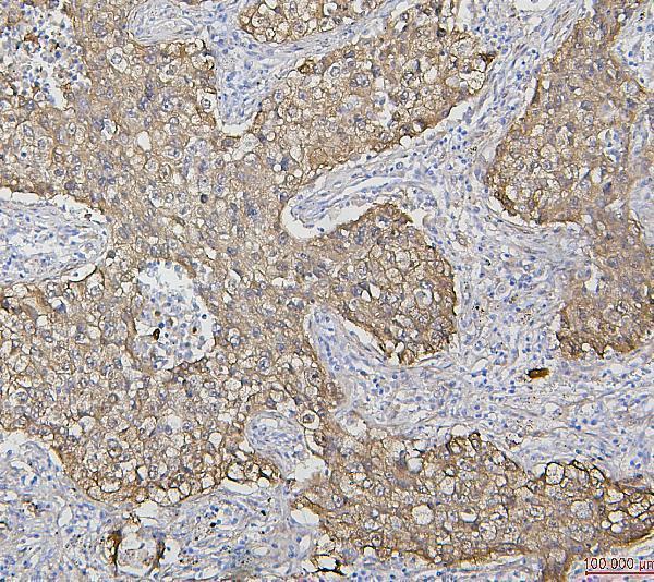
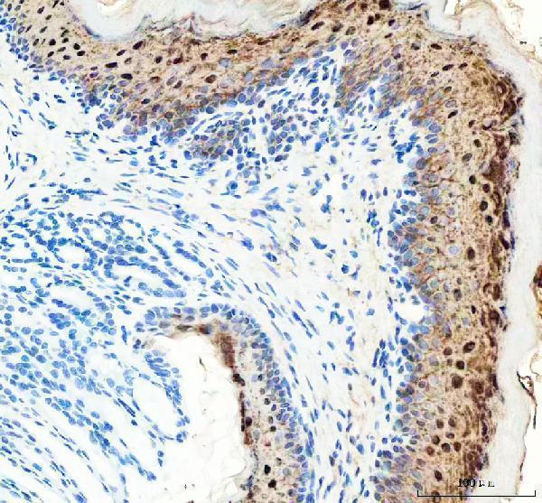
Real IHC data IHC analysis of HSP27 using anti-HSP27 antibody (M00676-5). HSP27 was detected in paraffin-embedded section of human lung cancer tissues. Heat mediated antigen retrieval was performed in citrate buffer (pH6, epitope retrieval solution) for 20 mins. The tissue section was blocked with 10% goat serum. The tissue section was then incubated with 1μg/ml mouse anti-HSP27 Antibody (M00676-5) overnight at 4°C. Biotinylated goat anti-mouse IgG was used as secondary antibody and incubated for 30 minutes at 37°C. The tissue section was developed using Strepavidin-Biotin-Complex (SABC)(Catalog # SA1021) with DAB as the chromogen.
Anti-Hsp27/HSPB1 Antibody ® (monoclonal, 3H3)
Cat # M00676-5
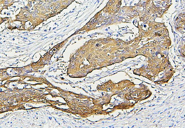
Real IHC data IHC analysis of HSP27 using anti-HSP27 antibody (PB9237). HSP27 was detected in paraffin-embedded section of human intestinal cancer tissue. Heat mediated antigen retrieval was performed in citrate buffer (pH6, epitope retrieval solution) for 20 mins. The tissue section was blocked with 10% goat serum. The tissue section was then incubated with 1μg/ml rabbit anti-HSP27 Antibody (PB9237) overnight at 4°C. Biotinylated goat anti-rabbit IgG was used as secondary antibody and incubated for 30 minutes at 37°C. The tissue section was developed using Strepavidin-Biotin-Complex (SABC)(Catalog # SA1022) with DAB as the chromogen.
Anti-Hsp27/HSPB1 Antibody ®
Cat # PB9237
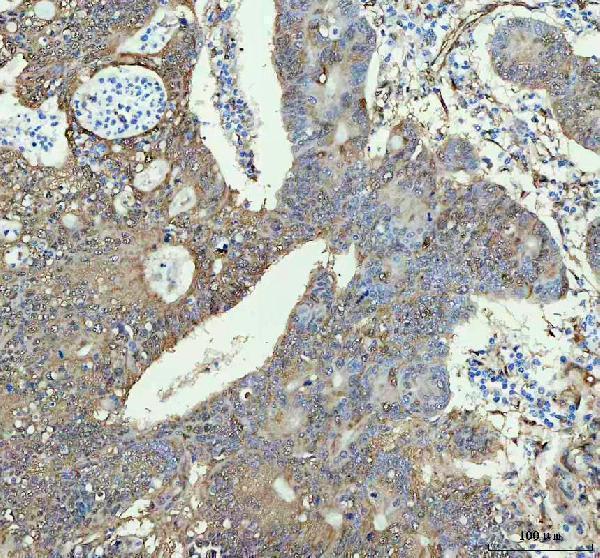
Real IHC data IHC analysis of HSPB1 using anti-HSPB1 antibody (M00676-2). HSPB1 was detected in a paraffin-embedded section of human colon cancer tissue. Heat mediated antigen retrieval was performed in EDTA buffer (pH 8.0, epitope retrieval solution). The tissue section was blocked with 10% goat serum. The tissue section was then incubated with 1:50 rabbit anti-HSPB1 Antibody (M00676-2) overnight at 4°C. Peroxidase Conjugated Goat Anti-rabbit IgG was used as secondary antibody and incubated for 30 minutes at 37°C. The tissue section was developed using HRP Conjugated Rabbit IgG Super Vision Assay Kit (Catalog # SV0002) with DAB as the chromogen.
Anti-Hsp27 HSPB1 Rabbit Monoclonal Antibody
Cat # M00676-2

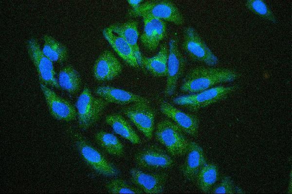
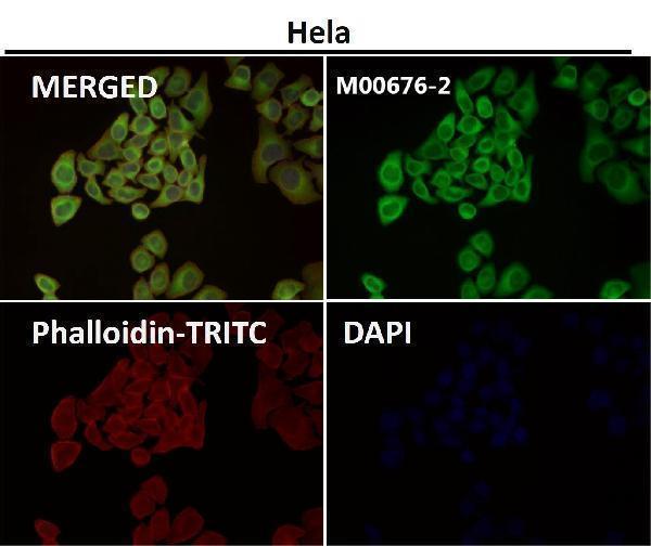
M00676-5 has a human lung cancer paraffin-section IHC image and an IF/ICC cell image (catalog image captions); PB9237 has a human intestinal cancer paraffin-section IHC image and an IF/ICC cell image (catalog image captions). M00676-2 has human colon cancer and rat stomach paraffin-section IHC images; its application list also includes IF/ICC (catalog IHC captions; catalog applications).

Which to pick: For human paraffin-section IHC, choose M00676-5 or PB9237 when citrate retrieval at pH 6 matches your workflow; each caption reports that retrieval and 1 μg/ml primary antibody (catalog IHC captions). For IF/ICC, M00676-5 and PB9237 each have a cell image at 2 μg/ml (catalog IF captions); for human, mouse or rat work, M00676-2 lists all three species and has human and rat IHC images with EDTA retrieval at pH 8 (catalog reactivity; catalog IHC captions). The paraffin-section captions do not report the fixative (catalog IHC captions).

Each figure is that product's own IHC / IF validation image from its datasheet.

References

  1. UniProt Consortium. UniProt entry P04792 (HSPB1_HUMAN, Heat shock protein beta-1).
  2. Human Protein Atlas. HSPB1 tissue IHC expression (reliability: Enhanced).
  3. Human Protein Atlas. HSPB1 subcellular location (ICC-IF): Localized to the plasma membrane and cytosol..
  4. Human Protein Atlas. HSPB1 antibody validation summary (5 antibodies).
  5. Cardiomyocyte-derived HSPB1 regulates TGF-β1 maturation and inhibits endothelial-to-mesenchymal transition in myocardial fibrosis. iScience 2026 — PMC12972742.
  6. Quantitative proteomic analysis shows differentially expressed HSPB1 in glioblastoma as a discriminating short from long survival factor and NOVA1 as a differentiation factor between low-grade astrocytoma and oligodendroglioma. BMC cancer 2015 — PMC4502388.
  7. Proteomic Characterization Reveals a Molecular Portrait of Nasopharyngeal Carcinoma Differentiation. Journal of Cancer 2017 — PMC5370501.
  8. Small Heat Shock Protein Beta-1 (HSPB1) Is Upregulated and Regulates Autophagy and Apoptosis of Renal Tubular Cells in Acute Kidney Injury. PloS one 2015 — PMC4427334.
  9. PubMed PMID:3714473 — UniProt-cited evidence.
  10. PubMed PMID:2243808 — UniProt-cited evidence.
  11. PubMed PMID:10777697 — UniProt-cited evidence.