This website uses cookies to ensure you get the best experience on our website.
- Table of Contents
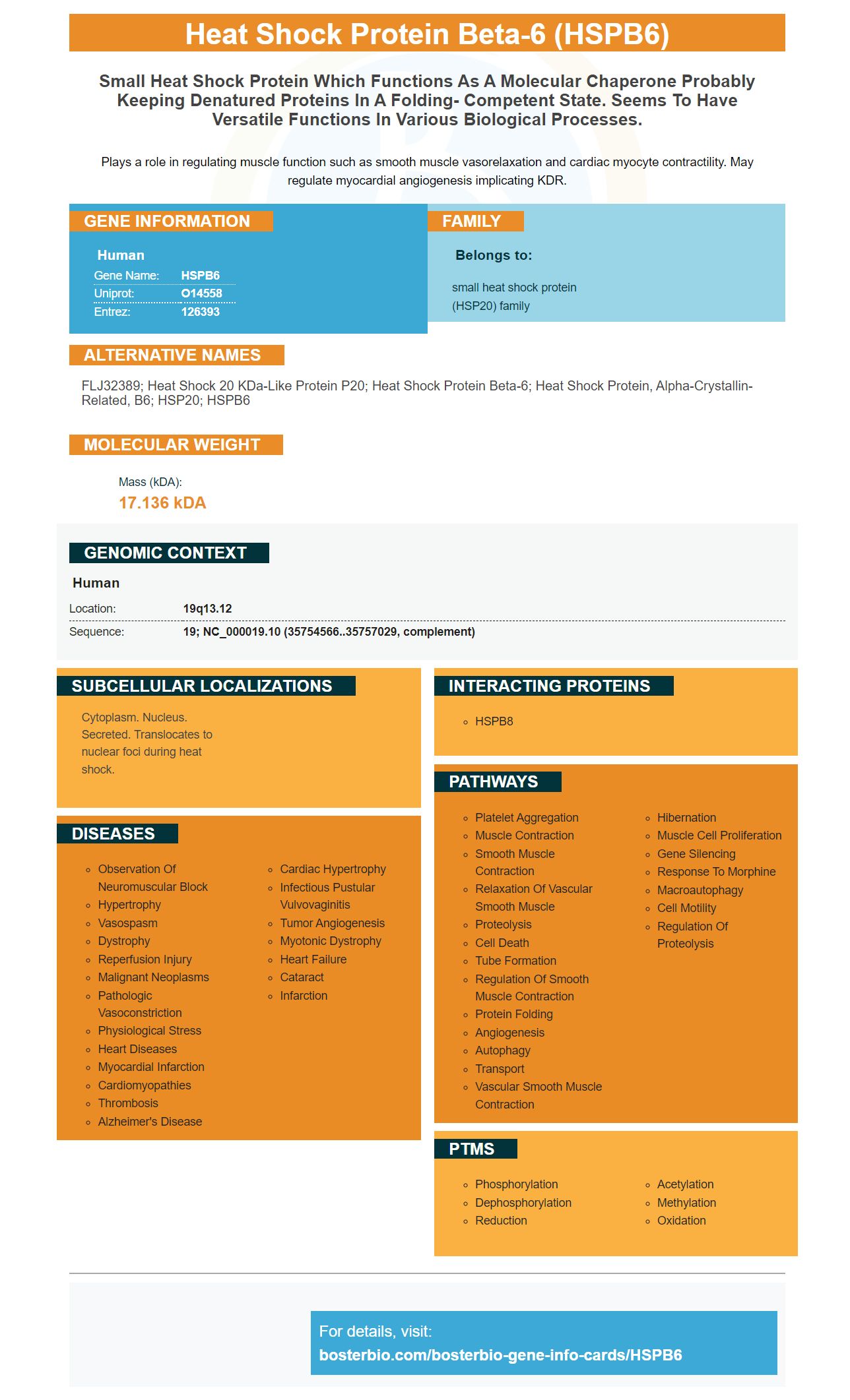
Facts about Heat shock protein beta-6.
Plays a role in regulating muscle function such as smooth muscle vasorelaxation and cardiac myocyte contractility. May regulate myocardial angiogenesis implicating KDR.
Human | |
---|---|
Gene Name: | HSPB6 |
Uniprot: | O14558 |
Entrez: | 126393 |
Belongs to: |
---|
small heat shock protein (HSP20) family |
FLJ32389; Heat shock 20 kDa-like protein p20; heat shock protein beta-6; heat shock protein, alpha-crystallin-related, B6; HSP20; HSPB6
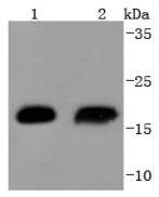
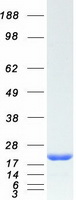
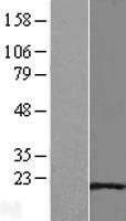
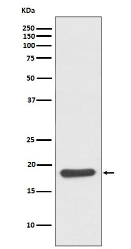
Mass (kDA):
17.136 kDA
Human | |
---|---|
Location: | 19q13.12 |
Sequence: | 19; NC_000019.10 (35754566..35757029, complement) |
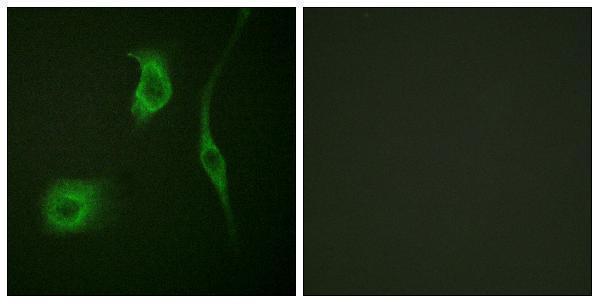
Cytoplasm. Nucleus. Secreted. Translocates to nuclear foci during heat shock.
This article will focus on the Boster Bio Anti–HSP20 HSPB6 marker in various applications including autoradiography films, Enhanced Chemiluminescence Detection, (ECL) and colorimetric detection. This marker has recently received a number of scientific publications, making it a useful tool for research labs. For more information, please contact us.
Boster Bio Anti-HSP20 HSP6 Marker can detect heat shock protein Beta-6 (HSP20), an 17 kDa protein, in various tissues. The protein is most abundantly expressed in vascular and airway muscle, colonic muscle, and uterine smooth muscle. Its special functions include platelet function, insulin resistance, and platelet function. Its validity is established by its use on known positive and negative samples.
Prepare a diluted solution containing 50 ul luminol and p-coumaric acids in PBS to perform ECL. Mix the solution with 3 ul H2O2 until it is well combined. Keep the solution at 4°C until you are ready to use it. Place the membrane on Saran Wrap.
The membranes were rinsed three times with TBST. After that, the secondary antibody was incubated at room temperature for 1 hour. This problem can easily be fixed by rinsing the membranes with MilliQ or twice-distilled water. However, membranes rinsed in water tend to dry faster than those with wash buffer. It is therefore important to quickly prepare the ECL detection agent.
Chemiluminescence is a more effective detection method than other methods. Chemiluminescence allows multiple exposures to optimize signal-to-noise ratio. It is also possible to remove and reprobe the detection agent, which allows for optimal detection of protein. Chemiluminescent detection is a popular choice for protein laboratories because of its high sensitivity.
Colorimetric Western-blotting is an economical method to identify the proteins of interest in samples. A secondary antibody conjugated in an enzyme reporter was used to create a visible signal. The resulting colored precipitate is used to identify the protein target directly on the membrane. The process is simple because it is color-dependent.
The signal exhibited a similar pattern for both ABTS and TMB-containing substrates. This pattern was observed for all tested HRP-antibody dilutions and substrate volumes. In addition, the signals from a stopped react were stable for longer periods of time than signals that were not stopped. The colorimetric signal generated from a stopped reaction was also generated on the exact substrate, regardless its concentration.
For this test, we used nine reagents: the Dragendorff reagent. To determine the differences between the time points, we used the color intensity analysis.
The sensitivity of the PAD was first tested by measuring the lowest concentration of allopurinol. The PAD was then subjected to various levels of paracetamol, a common treatment for gout, and caffeine. This test showed that both the green and yellow signals were produced by the PAD. This is because the reagents work in a similar way.
The HSPB6 protein is an essential part of several biomarkers. Boster has a wide range of high affinity primary antibodies. These have been cited over 25 years in peer-reviewed journal articles. This antibody can also used in immunohistochemistry as well as Western Blotting. This antibody can be used to determine the protein expression levels in a variety cells, tissues, and cell types by scientists from different fields.
PMID: 8195168 by Kato K., et al. Purification and characterization of a 20-kDa protein that is highly homologous to alpha B crystallin.
PMID: 19845507 by Fuchs M., et al. Identification of the key structural motifs involved in HspB8/HspB6- Bag3 interaction.