HSPB6 / Heat shock protein beta-6 · IHC design guide

Design Immunohistochemistry for HSPB6

Plan HSPB6 staining in paraffin sections using cytoplasmic staining in myocytes and adipocytes as a reference (HPA tissue IHC). Compare matched sections under consistent fixation and account for possible nuclear foci after heat shock (UniProt).

Evidence assembled Oct 2026 · For research use; verify linked source records and product datasheet before use
Immunohistochemistry protocol sheet for HSPB6 (IHC for HSPB6): expected localisation Cytoplasmic tissue staining (HPA tissue IHC), antibody A07981-1, validated IHC image, and IHC protocol steps
Printable HSPB6 IHC protocol sheet — expected localisation Cytoplasmic tissue staining (HPA tissue IHC), antibody A07981-1, controls and protocol steps. Open the full HSPB6 IHC guide →

HSPB6 Immunohistochemistry Experimental Design Guide

Expected localisation, validated protocols, controls and antibodies — the at-a-glance facts below, then the full design guide.

Must know before staining
Expected localisation Cytoplasmic tissue staining (HPA tissue IHC)
Staining pattern Cytoplasmic signal in myocytes, adrenal cells, stroma and adipocytes (HPA tissue IHC)
Antigen retrieval EDTA pH 8.0 HIER, heat-mediated (datasheet A07981-1)
Positive control ⓘ Adipose tissue+4 more · see all
Negative control ⓘ Appendix+4 more · see all
Important caveats
Reasons your staining may differ from the expected pattern.
Fixation Keep fixation conditions consistent across sections. (standard IHC practice; not target-specific)
Caveat Heat shock may shift signal to nuclear foci (UniProt)
Regulation PKA phosphorylates Ser16 (UniProt)
Isoform / epitope One chain, residues 1–160; no annotated isoforms (UniProt)
Section 1

Recommended HSPB6 IHC & IF Protocols

The catalog antibody protocol uses EDTA pH 8.0 retrieval (datasheet). The published IHC examples below cover rat myocardial injury, lumbar spine specimens, and multiple sclerosis tissue (PMC3459299; PMC11786202; PMC4688967; PMC6194336).

Recommended immunohistochemistry (IHC-P) protocol parameters
SampleParaffin-embedded human skeletal muscle tissue; fixative not specified (datasheet A07981-1)
FixationImage fixative and duration unreported (datasheet A07981-1); verify before use.
Sectioning4–5 µm sections on charged slides (standard)
DeparaffinisationXylene, graded ethanol series to water (standard)
Antigen retrievalHeat retrieval: EDTA pH 8.0 (datasheet A07981-1); 20 min, 95–100 °C (standard)
Peroxidase block3% H2O2, 10 min, room temperature (standard)
Blocking10% goat serum (datasheet A07981-1)
Primary antibodyRabbit anti-HSPB6, 0.5-1μg/ml (datasheet A07981-1)
Primary incubationOvernight at 4 °C (datasheet A07981-1)
DetectionStreptavidin-biotin complex (SABC), DAB chromogen (datasheet A07981-1)
CounterstainHematoxylin, blue, dehydrate and mount (standard)
Expected resultHSPB6-positive staining in adipocytes of adipose tissue (HPA tissue IHC: High). HPA tissue profile: Cytoplasmic expression in myocytes, adrenal gland, stromal cells and adipocytes. No signal in the no-primary control.
💡Decision noteStart with heat-mediated EDTA pH 8.0 for the catalog antibody (datasheet). Published protocols also report citrate pH 6.0 or EDTA pH 9 (PMC4688967; PMC6194336).
Section 2

What Is the Expected HSPB6 Staining Pattern?

HSPB6 should give predominantly cytoplasmic staining in myocytes, adipocytes and selected stromal or glandular cells (HPA: tissue IHC profile). Cytoplasmic localization is consistent with a protein lacking a transmembrane segment (UniProt O14558: topology). Nuclear signal can occur, particularly in foci during heat shock (UniProt O14558: subcellular location). Interpret intensity by cell type: HPA rates its tissue IHC profile Enhanced, while noting medium consistency with RNA expression (HPA: tissue IHC reliability).

What am I looking at on my slide?
Clear cytoplasmic staining in skeletal myocytes or cardiomyocytes, with restrained background.This fits the reported High staining in both cell types (HPA: skeletal muscle and heart muscle IHC). Score the target cells and their compartment, since a whole-section intensity score can obscure which cells carry the signal.
Strong signal confined to nuclei, or a membrane outline presented as the dominant pattern.Reassess compartment and controls before calling it HSPB6: cytoplasm is reported in tissue IHC, while nuclear foci are linked to heat shock (HPA: tissue IHC profile; UniProt O14558: subcellular location). A dominant membrane pattern lacks support from the reported topology (UniProt O14558: no transmembrane segment).
Strong staining in cells expected to be unstained, such as appendix glandular cells.Treat this as a specificity or detection concern: those glandular cells are Not detected in the HPA tissue survey (HPA: appendix IHC). Cross-reactivity or endogenous detection activity is possible; compare the stained cell type with a negative reagent control before assigning target expression (general IHC practice).
Uniform brown haze across tissue, including spaces between cells or areas without a coherent cell pattern.This is difficult to interpret as target staining because the reported pattern is cell-associated and mainly cytoplasmic (HPA: tissue IHC profile). Check background in a negative reagent control, then review blocking, washes and chromogen development (general IHC practice).
No staining in skeletal muscle myocytes or heart cardiomyocytes.A blank known-positive cell population conflicts with the HPA High ratings (HPA: skeletal muscle and heart muscle IHC). First check that the cells are present and preserved; then verify the IHC-validated antibody and detection run with controls (general IHC practice).
💡Expected HSPB6 appearanceCall a positive result when myocytes show convincing cytoplasmic signal at the reported High level (HPA: skeletal muscle and heart muscle IHC); diffuse section-wide haze or isolated staining in HPA Not detected appendix glandular cells is suspect (HPA: tissue IHC and appendix IHC).
How each factor affects the staining
Cell type and comparator tissueAdipocytes, adrenal glandular cells, endometrial stromal cells, decidual cells and Leydig cells are also rated High (HPA: tissue IHC). Appendix glandular and bone marrow hematopoietic cells are Not detected (HPA: tissue IHC). Compare like cell types rather than treating every cell in a positive organ as positive.
IHC evidence strengthThe tissue profile is rated Enhanced but has medium consistency with RNA expression; staining also occurs in cells or structures not fully annotated (HPA: tissue IHC reliability). HPA044153, HPA054811 and CAB001974 each have Enhanced IHC validation (HPA: antibody validation). Use these ratings as support, not a substitute for slide-level controls.
Compartment and stress stateUniProt places HSPB6 in cytoplasm and nucleus and reports movement to nuclear foci during heat shock (UniProt O14558: subcellular location). A nuclear focus therefore needs context; routine tissue IHC is described chiefly as cytoplasmic (HPA: tissue IHC profile). No transmembrane segment is reported (UniProt O14558: topology).
Protein form and epitope claimsUniProt reports one chain spanning residues 1–160, no signal peptide or propeptide, and no listed isoforms (UniProt O14558: processing and isoforms). It also lists phosphoserine at residue 16 and deamidation at residue 66 (UniProt O14558: modified residues). These facts do not establish which epitope an antibody recognizes or predict a staining change.
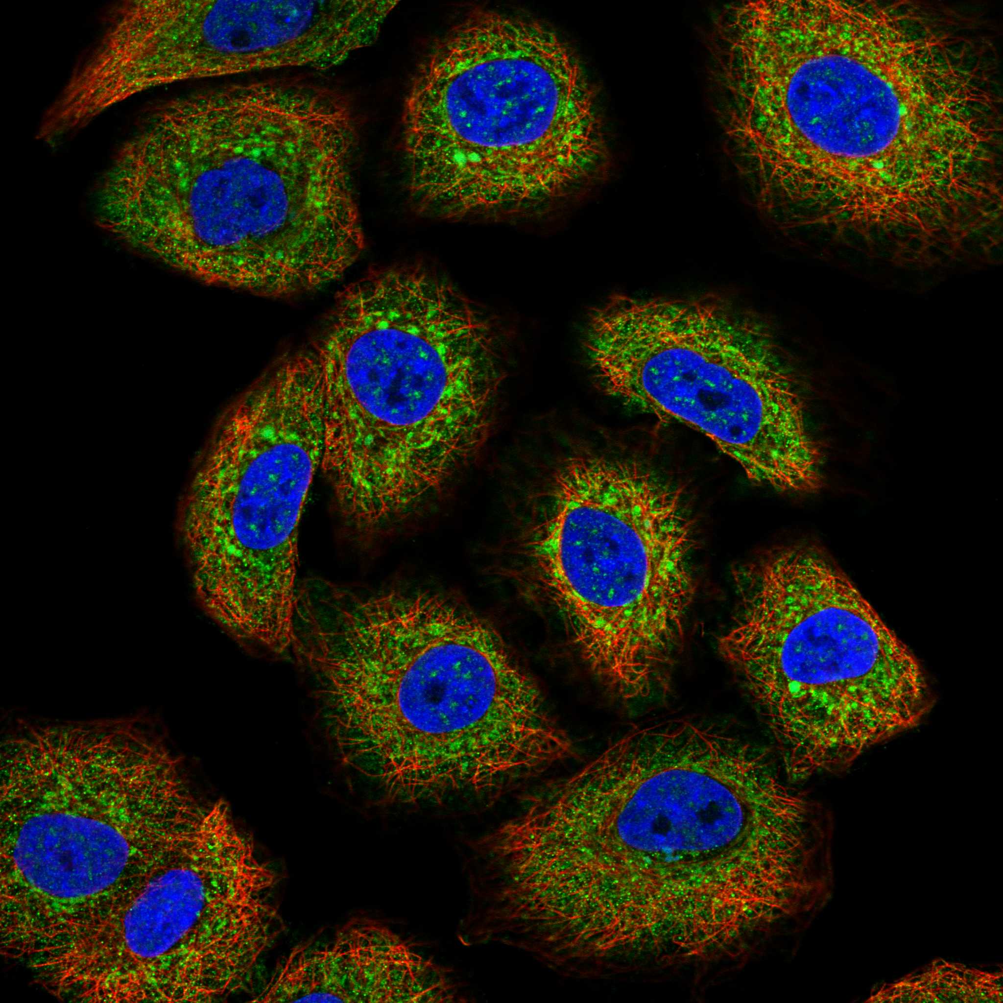
IF/ICC Q&A: what pattern is supported?In ICC-IF, HPA reports mainly cytosol and mitochondria, with additional nuclear speckles; the respective location assessments are supported or approved (HPA: subcellular). HPA044153 has Supported ICC validation (HPA: antibody validation). Interpret this as ICC-IF evidence, without treating mitochondrial or speckle staining as a required paraffin IHC pattern.
Why is my staining missing, weak or wrong?
SituationLikely causeNext action
Known-positive myocytes are blank.The run or detection chain may have failed; skeletal myocytes and cardiomyocytes are rated High (HPA: tissue IHC).Verify tissue identity, the IHC-validated antibody, and positive control development before interpreting the sample as negative (general IHC practice).
Myocyte staining is faint or patchy.The slide may lack a stable comparator; HPA reports High staining in skeletal myocytes and cardiomyocytes (HPA: tissue IHC).Compare a known-positive section processed in the same run; review section quality, retrieval settings and detection performance without assuming HSPB6-specific retrieval sensitivity (general IHC practice).
Appendix glandular cells stain strongly.The observed cell type conflicts with the HPA Not detected rating; cross-reactivity or endogenous detection activity is possible (HPA: appendix IHC; general IHC practice).Check a negative reagent control and the staining compartment; repeat specificity assessment with an independently validated IHC antibody if available (general IHC practice).
Brown signal appears across most of the section without cell boundaries.Diffuse background can arise from detection chemistry or inadequate washing (general IHC practice); it does not match the reported cell-associated cytoplasmic profile (HPA: tissue IHC).Inspect the negative reagent control, washing and chromogen development, then score only reproducible cell-associated signal (general IHC practice).
Only nuclei or membrane outlines are prominent.HSPB6 may enter nuclear foci during heat shock, but routine tissue IHC is chiefly cytoplasmic; no transmembrane segment is reported (UniProt O14558: subcellular location and topology; HPA: tissue IHC).Record the compartment, check controls and reassess antibody specificity before calling the pattern positive (general IHC practice).
Cells within one tissue show different intensities.HPA rates specific cell populations, not every cell in an organ; its tissue IHC profile also notes incompletely annotated stained structures (HPA: tissue IHC).Score named cell populations separately and compare their compartments with the HPA entries; keep unannotated structures descriptive rather than assigning them a target-positive identity.

Sample controls for HSPB6 IHC & IF

🧪Run heart muscle first; cardiomyocytes should stain strongly (HPA: High in cardiomyocytes). Run appendix glandular cells as a negative comparator (HPA: Not detected in appendix glandular cells); on the heart slide, treat non-cardiomyocyte cells as candidate internal negatives only if their staining stays at background, since their HSPB6 status is not specified by the supplied HPA row.
Positive control tissue: Adipose tissue (Adipocytes, HPA High)
Negative control tissue: Appendix (HPA Not detected)
ICC-IF cell lines (HPA subcellular resource): HPA ICC-IF images show HSPB6 in A-549, BJ [Human fibroblast], U2OS, with annotated localisation: Mitochondria (approved), Cytosol (supported) (HPA subcellular).
Technical controls: Include a no-primary (secondary-only) slide, a matched rabbit IgG isotype control, and a biological specificity control such as HSPB6 knockout material or peptide competition when available (selected-SKU caption: rabbit primary antibody). Quench endogenous peroxidase and check for endogenous biotin in heart sections because the documented detection uses biotinylated secondary antibody, streptavidin–biotin complex, and DAB (selected-SKU tissue-IHC caption).
⚠️Feasibility: Paraffin-section IHC is documented with heat retrieval in EDTA at pH 8.0, but whether HSPB6 staining depends on that retrieval condition is unreported (selected-SKU tissue-IHC caption). A target-specific fixation window and fixation effect are unreported, and the exact selected-SKU caption does not report a fixative (selected-SKU tissue-IHC caption). The supplied evidence does not establish that frozen sections or tissue IF are easier; HPA documents ICC-IF images in A-549, BJ, and U2OS cells (HPA subcellular), while the documented chromogenic heart-section workflow warrants checks for endogenous peroxidase and biotin (selected-SKU tissue-IHC caption).

HPA tissue IHC evidence for HSPB6

Comprehensive Human Protein Atlas IHC scoring per tissue (reliability: Enhanced — Medium consistency between antibody staining and RNA expression data. Antibody staining in cells/structures not annotated, view images.). Rows are taken directly from the HPA tissue chart — click any row's HPA link to view the source.

Positive expression · recommended positive controls

TissueCell typeLevelEvidenceSource
Adipose tissue Adipocytes High Protein (IHC) HPA →
Adrenal gland Glandular cells High Protein (IHC) HPA →
Endometrium Cells in endometrial stroma High Protein (IHC) HPA →
Heart muscle Cardiomyocytes High Protein (IHC) HPA →
Placenta Decidual cells High Protein (IHC) HPA →

Undetected expression · recommended negative controls

TissueCell typeLevelEvidenceSource
Appendix Glandular cells Not detected Protein (IHC) HPA →
Bone marrow Hematopoietic cells Not detected Protein (IHC) HPA →
Bronchus Respiratory epithelial cells Not detected Protein (IHC) HPA →
Caudate Glial cells Not detected Protein (IHC) HPA →
Cerebellum Cells in granular layer Not detected Protein (IHC) HPA →
Section 3

Advanced HSPB6 IHC Tips

Troubleshoot HSPB6 staining in paraffin sections by checking retrieval, cell type, compartment and detection controls before scoring chromogenic signal.

Which retrieval conditions should I use when HSPB6 staining is weak?
Use heat-mediated antigen retrieval in EDTA at pH 8.0 for this paraffin-section IHC assay (datasheet A07981-1). The selected skeletal-muscle image used that retrieval before primary antibody incubation at 1 μg/ml overnight at 4°C (caption A07981-1). If staining remains weak, compare a modestly longer heating period on matched sections while keeping buffer and detection conditions constant (standard IHC practice). Include skeletal-muscle myocytes as a positive cell population and assess their cytoplasmic staining before changing antibody concentration (HPA tissue IHC). Record retrieval temperature and duration for each run so apparent gains can be checked against tissue damage and background (standard IHC practice).
Can fixation explain variable HSPB6 staining across paraffin sections?
Target-specific sensitivity to fixation is unknown: the selected paraffin-section caption does not state a fixative (caption A07981-1). Record the actual fixative, fixation duration and processing history for each specimen before attributing an intensity difference to HSPB6 abundance (standard IHC practice). Compare sections processed together, using the same EDTA pH 8.0 retrieval and 1 μg/ml primary concentration used in the selected image (caption A07981-1). Check whether morphology and a positive myocyte population remain interpretable after retrieval, since damaged sections can distort cell-level scoring (standard IHC practice; HPA tissue IHC). Do not infer a fixation effect from HSPB6 localisation or phosphorylation annotations (UniProt O14558).
How should I assess cytoplasmic, mitochondrial or nuclear HSPB6 staining?
Start by identifying the stained cell type: HPA reports cytoplasmic expression in myocytes, adrenal gland, stromal cells and adipocytes (HPA tissue IHC). Cytosol and mitochondria are main subcellular locations in its IF profile, with nuclear speckles additionally reported (HPA subcellular). UniProt also lists cytoplasm, nucleus and secretion, and describes translocation to nuclear foci during heat shock (UniProt O14558). In chromogenic sections, assess whether apparent nuclear or punctate signal lies within intact cells, using a counterstain and adjacent morphology before assigning a compartment (standard IHC practice). Compare the suspected pattern with a secondary-only control; nuclear staining alone does not establish heat shock or secretion (standard IHC practice; UniProt O14558).
Could HSPB6 processing or modification alter epitope detection?
UniProt records 0 isoforms and one HSPB6 chain spanning residues 1–160, so the supplied record offers no alternative isoform to explain discordant staining (UniProt O14558). Its small heat-shock protein domain spans residues 55–160, while documented modifications include PKA-dependent phosphoserine at 16 and deamidated glutamine at 66 (UniProt O14558). The antibody epitope is not specified in the supplied caption, so modification-dependent recognition cannot be assigned to this reagent (caption A07981-1). HSPB6 forms flexible oligomers and heterooligomers, making epitope accessibility a testable possibility rather than an established cause of weak IHC (UniProt O14558). Compare matched retrieval conditions and an independent validated antibody if an epitope effect remains plausible (standard IHC practice).
How can I check an HSPB6 IHC pattern by multiplex immunofluorescence?
Pair HSPB6 with a validated marker for the expected cell type, such as a myocyte marker when examining skeletal muscle, and compare cell-level overlap (HPA tissue IHC; standard IF practice). Choose spectrally separated fluorophores and evaluate unstained tissue autofluorescence before assigning weak signal, especially when comparing tissue regions (standard IF practice). Because HSPB6 lacks a transmembrane segment and is reported in cytosol, mitochondria and nuclear speckles, assess permeabilisation appropriate to intracellular epitopes (UniProt O14558; HPA subcellular; standard IF practice). Include single-label and secondary-only controls to detect bleed-through and nonspecific fluorescence (standard IF practice). The paraffin-section caption supports this catalog antibody's chromogenic IHC workflow, so validate its IF performance separately rather than assuming transferability (caption A07981-1; standard IF practice).
What should I change when DAB staining is diffuse or widespread?
First compare stained sections with a secondary-only control to distinguish primary-dependent signal from detection background (standard IHC practice). The selected image used 10% goat-serum blocking, a biotinylated secondary antibody, a streptavidin-biotin complex and DAB (caption A07981-1). For that detection scheme, check endogenous peroxidase and endogenous biotin with appropriate blocking or control sections before increasing antibody dilution (standard IHC practice). Wash consistently and inspect section edges, folds and damaged areas, where deposited chromogen can mislead visual interpretation (standard IHC practice). Retain cell-type context: strong staining in adipocytes or myocytes can be plausible, whereas diffuse staining across unrelated cells requires control-based review (HPA tissue IHC; standard IHC practice).
How should I score HSPB6 staining across heterogeneous tissue sections? ⚠ ANSWER MARKED FOR VERIFICATION
Define the target compartment and cell population before scoring; HPA reports cytoplasmic staining in myocytes, adipocytes, adrenal gland and stromal cells (HPA tissue IHC). For each specified population, report percent positive cells and intensity on a 0–3 scale, or calculate an H-score from 0–300 (standard IHC practice). If cell abundance varies, report positive-cell density per mm² alongside the fraction of positive cells, and define the sampled tissue area (standard IHC practice). Normalise comparisons to the number of eligible target cells or sampled area, using matched retrieval, detection and counterstaining conditions (standard IHC practice). Keep cytoplasmic and nuclear scores separate when both patterns occur (HPA tissue IHC; UniProt O14558; standard IHC practice).
How do I distinguish true HSPB6 signal from staining artefact?
Treat coherent cytoplasmic staining in myocytes or adipocytes as biologically plausible, because these populations stain in HPA tissue IHC (HPA tissue IHC). Review the cell identity before calling staining in an unexpected compartment positive; cytosol and mitochondria are main IF locations, while nuclear speckles and stress-associated nuclear foci are also reported (HPA subcellular; UniProt O14558). Edge enhancement, folds, necrotic areas and diffuse signal in a secondary-only control favour artefact over cell-specific antigen detection (standard IHC practice). For DAB detection, assess endogenous peroxidase and, with the selected biotin-based method, endogenous biotin as possible sources of false signal (caption A07981-1; standard IHC practice). Interpret unexpected staining cautiously because HPA rates tissue evidence Enhanced with medium staining-to-RNA consistency (HPA tissue IHC).
Boster reagents

Best HSPB6 / Heat shock protein beta-6 IHC Antibodies

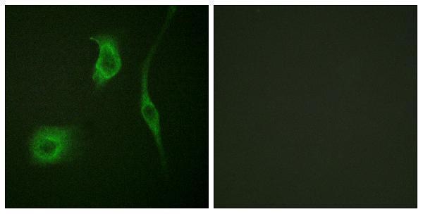
IHC images show HSPB6 staining in human skeletal muscle and breast carcinoma, rat cardiac muscle, and mouse lung (A07981-1 and A07981S16 image captions); IF images show A431 and HeLa cells (catalog IF image captions).

Real IHC data IHC analysis of Hsp20 using anti-Hsp20 antibody (A07981-1). Hsp20 was detected in paraffin-embedded section of human skeletal muscle tissue. Heat mediated antigen retrieval was performed in EDTA buffer (pH8.0, epitope retrieval solution). The tissue section was blocked with 10% goat serum. The tissue section was then incubated with 1μg/ml rabbit anti-Hsp20 Antibody (A07981-1) overnight at 4°C. Biotinylated goat anti-rabbit IgG was used as secondary antibody and incubated for 30 minutes at 37°C. The tissue section was developed using Strepavidin-Biotin-Complex (SABC) (Catalog # SA1022) with DAB as the chromogen.
Anti-Hsp20/HSPB6 Antibody ®
Cat # A07981-1
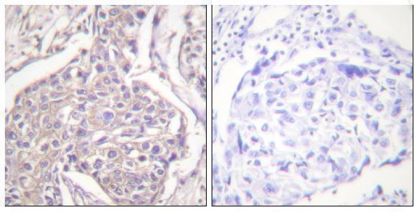
Real IHC data Immunohistochemistry analysis of paraffin-embedded human breast carcinoma, using HSP20 (Phospho-Ser16) Antibody. The picture on the right is blocked with the phospho peptide.
Anti-Phospho-HSP20 (S16) HSPB6 Antibody
Cat # A07981S16

A07981-1 has paraffin-section IHC images from human skeletal muscle and mammary cancer, rat cardiac muscle, and mouse lung, plus IF data from A431 cells (A07981-1 image captions). A07981S16 has paraffin-section IHC data from human breast carcinoma and IF data from HeLa cells, both with phospho-peptide blocking images (A07981S16 image captions).

Which to pick: For tissue IHC, choose A07981-1 for the imaged human, rat, and mouse paraffin-section examples; choose A07981S16 when the target is HSPB6 phosphorylated at Ser16 (A07981-1 IHC image captions; A07981S16 title and IHC image caption). For IF/ICC, A07981-1 lists both applications and has an A431 IF image; A07981S16 lists IF and has a HeLa IF image (catalog applications and IF image captions). Both list human, mouse, and rat reactivity, but the cited A07981S16 IHC image is human only; neither IHC caption reports the fixative (catalog reactivity; IHC image captions).

Each figure is that product's own IHC / IF validation image from its datasheet.

References

  1. UniProt Consortium. UniProt entry O14558 (HSPB6_HUMAN, Heat shock protein beta-6).
  2. Human Protein Atlas. HSPB6 tissue IHC expression (reliability: Enhanced).
  3. Human Protein Atlas. HSPB6 subcellular location (ICC-IF): Mainly localized to the cytosol and mitochondria. In addition localized to the nuclear speckles..
  4. Human Protein Atlas. HSPB6 antibody validation summary (3 antibodies).
  5. Effects of buyang huanwu decoction on ventricular remodeling and differential protein profile in a rat model of myocardial infarction. Evidence-based complementary and alternative medicine : eCAM 2012 — PMC3459299.
  6. Unveiling MiR-3085-3p as a modulator of cartilage degeneration in facet joint osteoarthritis: A novel therapeutic target. Journal of orthopaedic translation 2025 — PMC11786202.
  7. Small heat shock proteins are induced during multiple sclerosis lesion development in white but not grey matter. Acta neuropathologica communications 2015 — PMC4688967.
  8. Heat shock proteins are differentially expressed in brain and spinal cord: implications for multiple sclerosis. Clinical and experimental immunology 2018 — PMC6194336.
  9. PubMed PMID:14702039 — UniProt-cited evidence.
  10. PubMed PMID:15057824 — UniProt-cited evidence.
  11. PubMed PMID:15489334 — UniProt-cited evidence.