HTT / Huntingtin · IHC design guide

Design Immunohistochemistry for HTT

Plan HTT staining in paraffin sections using the cytoplasmic tissue pattern and high staining in cortical neurons and cerebellar Purkinje cells (HPA tissue IHC). The catalog antibody’s IHC-P example uses 2 μg/ml primary antibody with chromogenic detection (datasheet A00134-2).

Evidence assembled Oct 2026 · For research use; verify linked source records and product datasheet before use
Immunohistochemistry protocol sheet for HTT (IHC for HTT): expected localisation Cytoplasmic in most tissues (HPA tissue IHC), antibody A00134-2, validated IHC image, and IHC protocol steps
Printable HTT IHC protocol sheet — expected localisation Cytoplasmic in most tissues (HPA tissue IHC), antibody A00134-2, controls and protocol steps. Open the full HTT IHC guide →

HTT Immunohistochemistry Experimental Design Guide

Expected localisation, validated protocols, controls and antibodies — the at-a-glance facts below, then the full design guide.

Must know before staining
Expected localisation Cytoplasmic in most tissues (HPA tissue IHC)
Staining pattern Cytoplasmic staining in cells of most tissues (HPA tissue IHC)
Antigen retrieval EDTA pH 8.0 HIER, heat-mediated (datasheet A00134-2)
Positive control ⓘ Adrenal gland+4 more · see all
Negative control ⓘ Heart muscle
Important caveats
Reasons your staining may differ from the expected pattern.
Fixation Keep fixation conditions consistent across sections (standard IHC practice; not target-specific)
Caveat Staining shows medium consistency with RNA levels (HPA tissue IHC)
Regulation Widely expressed; highest in brain (UniProt)
Isoform / epitope No isoforms annotated; check epitope against the 551–584 fragment (UniProt)
Section 1

Recommended HTT IHC & IF Protocols

The catalog antibody uses EDTA pH 8.0 retrieval (datasheet: A00134-2). Published HTT staining protocols below cover free-floating brain sections and zQ175 brain sections (PMC6526682; PMC9901570).

Recommended immunohistochemistry (IHC-P) protocol parameters
SampleParaffin-embedded human breast cancer tissue; fixative not specified (datasheet A00134-2)
FixationImage fixative and duration unreported (datasheet A00134-2); verify before use.
Sectioning4–5 µm sections on charged slides (standard)
DeparaffinisationXylene, graded ethanol series to water (standard)
Antigen retrievalHeat retrieval: EDTA pH 8.0 (datasheet A00134-2); 20 min, 95–100 °C (standard)
Peroxidase block3% H2O2, 10 min, room temperature (standard)
Blocking10% goat serum (datasheet A00134-2)
Primary antibodyRabbit anti-HTT, 2-5 μg/ml (datasheet A00134-2)
Primary incubationOvernight at 4 °C (datasheet A00134-2)
DetectionHRP-conjugated secondary, DAB chromogen (datasheet A00134-2)
CounterstainHematoxylin, blue, dehydrate and mount (standard)
Expected resultHTT-positive staining in glandular cells of adrenal gland (HPA tissue IHC: High). HPA tissue profile: Cytoplasmic expression in most tissues. No signal in the no-primary control.
💡Decision noteStart with heat-mediated EDTA pH 8.0 for the catalog antibody (datasheet: A00134-2). Formic acid exposed polyglutamine staining with 4H7H7 in zQ175 sections (PMC9901570).
Section 2

What Is the Expected HTT Staining Pattern?

In paraffin tissue IHC, expect mainly cytoplasmic HTT staining across many tissues (HPA: cytoplasmic expression in most tissues; IHC reliability Approved, with medium consistency against RNA). Strong examples include cerebellar Purkinje cells and cerebral cortical neurons (HPA: High in both). HTT also localizes to the nucleus and intracellular vesicles (UniProt P42858: subcellular location), and has no transmembrane segment (UniProt P42858: topology).

What am I looking at on my slide?
Cytoplasmic staining in cerebellar Purkinje cells or cerebral cortical neurons.This matches the tissue IHC distribution; both cell populations are scored High (HPA: cerebellum and cerebral cortex). Judge the pattern within identifiable cells and compare it with the section's background (general IHC practice). HPA's Approved rating carries a medium-consistency caveat against RNA data (HPA: tissue IHC reliability).
Signal confined to cell surfaces, extracellular spaces, or a tissue-wide deposit.These locations do not match the mainly cytoplasmic tissue pattern (HPA: tissue IHC) or the recorded intracellular locations (UniProt P42858: subcellular location). Treat an isolated pattern there as a possible staining artefact; check morphology and detection controls before assigning HTT positivity (general IHC practice).
Strong staining in cardiomyocytes, especially if other unexpected cell types also stain.Cardiomyocytes are reported as Not detected (HPA: heart muscle). Unexpected color may reflect cross-reactivity or endogenous detection activity (general IHC practice); that possibility is a troubleshooting lead, not proof of the cause. Compare a reagent-omission control and an HPA High cell population processed in the same run (general IHC practice; HPA: tissue IHC).
Diffuse color over cells, empty spaces, and the surrounding section.Background that does not follow cell boundaries cannot establish the reported cytoplasmic pattern (HPA: tissue IHC; general IHC practice). Inspect a primary-antibody omission control and the counterstained morphology, then review blocking, washes, and chromogen development for this run (general IHC practice).
No discernible signal in cerebellar Purkinje cells or cerebral cortical neurons.Both are reported High, so a blank result in a suitable positive-control section calls for a workflow check (HPA: cerebellum and cerebral cortex; general IHC practice). It does not by itself show that HTT is absent. Check section quality, the documented IHC-P workflow, and detection reagents (general IHC practice).
💡Expected HTT appearanceCall a result positive when identifiable Purkinje cells or cortical neurons show clear cytoplasmic staining consistent with their High HPA scores (HPA: tissue IHC); widespread extracellular color or isolated cardiomyocyte staining is a warning pattern (HPA: heart muscle; general IHC practice).
How each factor affects the staining
Tissue and cell choicePurkinje cells and cortical neurons are High; cardiomyocytes are Not detected (HPA: tissue IHC). Use these contrasts to read a run, while allowing for HPA's medium consistency between staining and RNA expression (HPA: reliability description).
Compartment and applicationTissue IHC is mainly cytoplasmic (HPA: tissue IHC). IF/ICC answer: supported images place HTT mainly in the nucleoplasm, with additional cytosol (HPA: subcellular ICC-IF). Read each application in its own context; UniProt also records cytoplasmic and nuclear localization (UniProt P42858).
Antibody validationHPA026114, HPA051524, and CAB002756 each have Approved IHC status (HPA: antibody validation). That status supports comparison with HPA tissue observations; it does not identify the cause of an unexpected stain in an individual section (general IHC practice).
Intracellular distributionHTT has no transmembrane segment (UniProt P42858: topology), and UniProt records early-endosome and autophagosome localization alongside cytoplasm and nucleus (UniProt P42858: subcellular location). A solely cell-surface pattern therefore needs scrutiny; it is not the expected tissue IHC readout (HPA: tissue IHC).
Why is my staining missing, weak or wrong?
SituationLikely causeNext action
A High-control region is blank.Purkinje cells and cortical neurons should provide visible High-scored examples (HPA: tissue IHC); the failed step is undetermined.Confirm that the expected cells are present, then review the IHC-P antibody instructions and check primary antibody, detection reagents, and chromogen with appropriate controls (general IHC practice).
The whole section develops a diffuse haze.Nonspecific binding, residual detection reagent, or overdevelopment can obscure cell-level interpretation (general IHC practice).Compare the primary-antibody omission control; review blocking, wash steps, and development time before scoring cytoplasmic cells (general IHC practice; HPA: cytoplasmic tissue pattern).
Cardiomyocytes stain strongly.They are scored Not detected (HPA: heart muscle); cross-reactivity or endogenous detection activity is possible (general IHC practice).Compare omission and positive-control sections from the same run, then assess whether color follows cardiomyocyte boundaries or nonspecific deposits (general IHC practice).
Only extracellular or cell-surface color appears.That distribution conflicts with the mainly cytoplasmic tissue profile (HPA: tissue IHC) and intracellular UniProt locations (UniProt P42858).Check section morphology and omission controls before calling cells positive; inspect the detection and wash steps for deposits (general IHC practice).
A nuclear signal appears alongside cytoplasmic staining.Nuclear localization is recorded (UniProt P42858); supported ICC-IF images show mainly nucleoplasmic signal (HPA: subcellular).Score the tissue IHC cytoplasmic pattern separately (HPA: tissue IHC). Check cell boundaries and controls before deciding whether the nuclear component is interpretable (general IHC practice).
Different tissue regions stain with different strength.HPA reports High cells in several tissues and Not detected cardiomyocytes, with low tissue RNA specificity and medium IHC-to-RNA consistency (HPA: tissue IHC).Identify the stained cell types, compare equivalent regions and controls, and avoid treating regional intensity alone as proof of specificity (general IHC practice).

Sample controls for HTT IHC & IF

🧪Run breast first: glandular cells should stain (HPA: High in breast glandular cells; catalog caption: breast cancer section). Run heart muscle as the negative tissue (HPA: cardiomyocytes Not detected); on the breast slide, use any unstained nonglandular cells to assess background, without assuming they are HTT-negative (HPA: breast row specifies glandular cells only).
Positive control tissue: Adrenal gland (Glandular cells, HPA High)
Negative control tissue: Heart muscle (HPA Not detected)
ICC-IF cell lines (HPA subcellular resource): HPA ICC-IF images show HTT in A-431, U-251MG, U2OS, with annotated localisation: Nucleoplasm (supported) (HPA subcellular).
Technical controls: Include a no-primary (secondary-only) control, a rabbit isotype control matched to the primary antibody, and HTT-knockout material as a biological negative where available (catalog caption: rabbit primary; standard IHC practice). Block endogenous peroxidase and inspect background before interpreting DAB staining in the breast section (catalog caption: HRP/DAB detection; standard IHC practice).
⚠️Feasibility: A target-specific fixation window or fixation effect is unreported, and the selected A00134-2 paraffin-section caption does not state a fixative (catalog caption: fixative not stated). The demonstrated breast-section procedure used heat retrieval in EDTA at pH 8.0; whether HTT staining depends on that retrieval condition is unreported (catalog caption: EDTA retrieval). The supplied evidence does not establish that frozen sections or IF are easier, and it reports no breast-specific artefact; assess background in the illustrated breast cancer tissue (catalog caption: paraffin-embedded breast cancer section; standard IHC practice).

HPA tissue IHC evidence for HTT

Comprehensive Human Protein Atlas IHC scoring per tissue (reliability: Approved — Medium consistency between antibody staining and RNA expression data.). Rows are taken directly from the HPA tissue chart — click any row's HPA link to view the source.

Positive expression · recommended positive controls

TissueCell typeLevelEvidenceSource
Adrenal gland Glandular cells High Protein (IHC) HPA →
Breast Glandular cells High Protein (IHC) HPA →
Bronchus Respiratory epithelial cells High Protein (IHC) HPA →
Cerebellum Purkinje cells High Protein (IHC) HPA →
Cerebral cortex Neuronal cells High Protein (IHC) HPA →

Undetected expression · recommended negative controls

TissueCell typeLevelEvidenceSource
Heart muscle Cardiomyocytes Not detected Protein (IHC) HPA →
Section 3

Advanced HTT IHC Tips

Troubleshoot chromogenic HTT staining in paraffin sections by checking retrieval, controls, cellular localisation, and scoring before interpreting biological differences.

What retrieval should I use when HTT staining is weak in paraffin sections?
Use heat-mediated antigen retrieval in EDTA at pH 8.0 for this paraffin-section assay (datasheet A00134-2). The selected tissue image used this retrieval before 2 μg/ml primary antibody overnight at 4°C, so reproduce those conditions as the starting comparison (caption A00134-2). If signal remains weak, compare retrieval times on adjacent sections while keeping primary concentration and DAB development time constant (standard IHC practice). Test another buffer only as a fallback, with a no-primary section and a positive section alongside it, to distinguish improved epitope access from increased background (standard IHC practice).
Could fixation explain inconsistent HTT staining between paraffin specimens?
The selected image documents a paraffin-embedded human breast cancer section, but its fixative is unreported (caption A00134-2). Target-specific fixation sensitivity is therefore unknown; the image provides no basis for predicting how HTT staining responds to a particular fixative or fixation duration (caption A00134-2). Record each specimen’s fixative and processing history, then compare similarly processed sections using the same pH 8.0 EDTA retrieval and antibody conditions (datasheet A00134-2; standard IHC practice). If processing histories differ, include matched positive controls where available and assess tissue preservation before assigning a staining difference to HTT abundance (standard IHC practice).
Should I expect HTT staining in cytoplasm, nuclei, or both?
Score cytoplasmic and nuclear staining separately: tissue IHC reports cytoplasmic expression in most tissues, while subcellular imaging supports nucleoplasm and cytosol localisation (HPA tissue IHC; HPA subcellular). HTT also localises to early endosomes and autophagosomes and can shuttle between cytoplasm and nucleus (UniProt P42858). In brain sections, examine the relevant neuronal cells and neuropil context because HTT is reported in nerve fibers, varicosities, and nerve endings (UniProt P42858). A nuclear signal alone is therefore not automatically artefactual, but an unexpected compartment should prompt review of morphology, counterstain, and no-primary controls (HPA subcellular; standard IHC practice).
How can epitope choice change what HTT IHC detects?
Confirm the catalog antibody’s epitope before interpreting staining: the supplied tissue-image caption identifies A00134-2 but does not map its binding site (caption A00134-2). The record annotates 0 isoforms and a 1–3142 Huntingtin chain, plus a 551–584 myristoylated fragment (UniProt P42858). Consequently, a fragment-specific interpretation requires evidence that the antibody recognises an epitope retained in that fragment (UniProt P42858; standard IHC practice). HTT also has annotated acetylated lysines and a phosphoserine, so document epitope position before proposing modification-sensitive staining; section staining alone cannot establish which molecular form produced the signal (UniProt P42858; standard IHC practice).
How should I plan companion HTT immunofluorescence without misreading the IHC result?
For a companion IF/ICC experiment, verify the chosen primary antibody’s IF suitability independently; the supplied A00134-2 tissue example documents chromogenic IHC in a paraffin section (caption A00134-2). Multiplex HTT with a marker identifying the expected cell population, such as neuronal cells in cerebral cortex or Purkinje cells in cerebellum (HPA tissue IHC; standard IF practice). Choose spectrally separated fluorophores, and consider a far-red HTT channel if tissue autofluorescence overlaps shorter wavelengths (standard IF practice). Because HTT is intracellular and has no transmembrane segment, optimise permeabilisation for access to cytosolic or nuclear epitopes, checking morphology and controls alongside signal (UniProt P42858; standard IF practice).
What should I check when HTT DAB staining appears diffuse?
Start with the documented 10% goat-serum block, 2 μg/ml primary antibody overnight at 4°C, and peroxidase-linked secondary incubation for 30 minutes at 37°C (caption A00134-2). Review a no-primary control for nonspecific secondary staining and endogenous peroxidase activity, and include a peroxidase-blocking step as part of the general chromogenic workflow (standard IHC practice). Shorten DAB development or adjust primary concentration systematically if diffuse colour obscures cellular boundaries (standard IHC practice). Compare these changes on adjacent sections, since HTT has reported cytoplasmic expression in most tissues and genuine widespread staining is possible (HPA tissue IHC).
How can I quantify HTT IHC across specimens with different cellularity? ⚠ ANSWER MARKED FOR VERIFICATION
Define the cell population and compartment before scoring, since HTT tissue staining is commonly cytoplasmic while nuclear localisation is also supported (HPA tissue IHC; HPA subcellular). For cellular staining, record the percentage of positive viable cells and intensity by compartment; an H-score sums intensity-weighted percentages on a 0–300 scale (standard IHC practice). If measuring puncta or stained area, report density per mm² of viable annotated tissue and state how thresholds were set (standard image-analysis practice). Normalise comparisons to the same eligible cell type or viable tissue area, using consistent retrieval, DAB development, illumination, and scoring rules across specimens (standard IHC practice).
How do I distinguish true HTT staining from tissue artefact?
Look for reproducible staining within intact cells in plausible compartments: HTT has broad cytoplasmic tissue expression and supported nucleoplasmic and cytosolic localisation (HPA tissue IHC; HPA subcellular). Compare cell identity with the section’s anatomy; neuronal cells in cerebral cortex and Purkinje cells in cerebellum show high reported staining, whereas cardiomyocytes were not detected in the supplied HPA profile (HPA tissue IHC). Treat staining confined to section edges, folds, or necrotic areas cautiously and compare it with well-preserved interior tissue (standard IHC practice). A no-primary control helps reveal secondary-antibody or endogenous-enzyme colour, while consistent localisation across replicate sections strengthens interpretation (standard IHC practice).
Boster reagents

Best HTT / Huntingtin IHC Antibodies

Human paraffin-section IHC is documented for A00134-2 (A00134-2 image captions). M00134 lists human, mouse, and rat IHC/ICC/IF, with no supplied IHC or IF image (M00134 catalog).

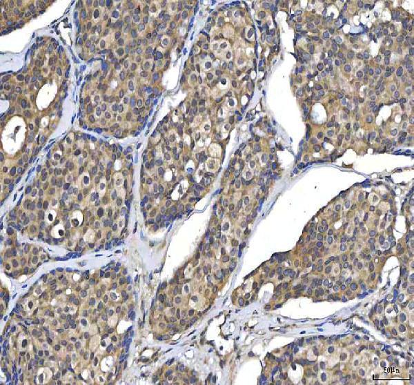
Real IHC data IHC analysis of Huntingtin/HTT using anti-Huntingtin/HTT antibody (A00134-2). Huntingtin/HTT was detected in a paraffin-embedded section of human breast cancer tissue. Heat mediated antigen retrieval was performed in EDTA buffer (pH 8.0, epitope retrieval solution). The tissue section was blocked with 10% goat serum. The tissue section was then incubated with 2 μg/ml rabbit anti-Huntingtin/HTT Antibody (A00134-2) overnight at 4°C. Peroxidase Conjugated Goat Anti-rabbit IgG was used as secondary antibody and incubated for 30 minutes at 37°C. The tissue section was developed using HRP Conjugated Rabbit IgG Super Vision Assay Kit (Catalog # SV0002) with DAB as the chromogen.
Anti-Huntingtin/HTT Antibody
Cat # A00134-2

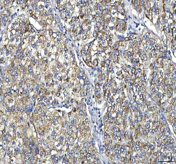
A00134-2 has IHC images from paraffin-embedded human breast cancer, cervix squamous cell carcinoma, colon adenocarcinoma, and endometrioid adenocarcinoma (A00134-2 image captions). M00134 is listed for IHC and ICC/IF in human, mouse, and rat, but has no supplied IHC or IF image (M00134 catalog).

Which to pick: For human paraffin-section IHC, choose A00134-2: its images document EDTA retrieval at pH 8.0 and a 2 μg/ml primary antibody incubation overnight at 4°C; the fixative is unreported (A00134-2 image captions). For IF/ICC, M00134 is the listed option because it is a rabbit monoclonal, clone ADGC-8, with ICC/IF among its applications; no IF image is supplied (M00134 catalog). For mouse or rat IHC, M00134 is the only listed reactive SKU, but its IHC image field is empty, so verify staining in the intended specimen (catalog reactivity and image alts).

Each figure is that product's own IHC / IF validation image from its datasheet.

References

  1. UniProt Consortium. UniProt entry P42858 (HD_HUMAN, Huntingtin).
  2. Human Protein Atlas. HTT tissue IHC expression (reliability: Approved).
  3. Human Protein Atlas. HTT subcellular location (ICC-IF): Mainly localized to the nucleoplasm. In addition localized to the cytosol..
  4. Human Protein Atlas. HTT antibody validation summary (3 antibodies).
  5. Case Report: Membranous/cytoplasmic Ki-67 staining and PAX8-GLIS3 fusion: defining the clinicopathological spectrum of hyalinizing trabecular tumor to optimize patient management. Frontiers in medicine 2026 — PMC12950578.
  6. Thalamic adenylyl cyclase 1 is required for barrel formation in the somatosensory cortex. Neuroscience 2015 — PMC5994750.
  7. Endogenous mouse huntingtin is highly abundant in cranial nerve nuclei, co-aggregates to Abeta plaques and is induced in reactive astrocytes in a transgenic mouse model of Alzheimer's disease. Acta neuropathologica communications 2019 — PMC6526682.
  8. Early detection of exon 1 huntingtin aggregation in zQ175 brains by molecular and histological approaches. Brain communications 2023 — PMC9901570.
  9. PubMed PMID:8458085 — UniProt-cited evidence.
  10. PubMed PMID:11013077 — UniProt-cited evidence.
  11. PubMed PMID:15815621 — UniProt-cited evidence.