This website uses cookies to ensure you get the best experience on our website.


- Table of Contents
Plan chromogenic HVCN1 IHC in paraffin sections around the cytoplasmic staining seen in immune and seminiferous duct cells (HPA tissue IHC). This guide covers fixation, epitope mapping, and interpretation of cell-specific signal.

Expected localisation, validated protocols, controls and antibodies — the at-a-glance facts below, then the full design guide.
| Expected localisation | Cytoplasmic staining in immune and seminiferous duct cells (HPA tissue IHC) | |
| Staining pattern | Cytoplasmic signal in immune and seminiferous duct cells (HPA tissue IHC) | |
| Antigen retrieval | Citrate pH 6.0 HIER, 95–98 °C, 20 min (rule: cytoplasmic / membrane antigen) | |
| Positive control | Lymph node+4 more · see all | |
| Negative control | Adipose tissue+4 more · see all |
| Fixation | Keep fixation consistent across paraffin sections. (standard IHC practice; not target-specific) | |
| Caveat | Immune cells may contribute signal within other tissues (HPA tissue IHC) | |
| Regulation | Staining varies with immune-cell abundance (HPA tissue IHC) | |
| Isoform / epitope | 4 isoforms; check whether the cytoplasmic or extracellular epitope is shared (UniProt) |
The catalog antibody’s IHC-P protocol (datasheet) appears alongside published paraffin-section methods for human tonsil (PMC3030552) and Crohn’s disease tissue (PMC8085323).
| Sample | Tissue sections; selected-image fixative not specified (standard IHC workflow) |
| Fixation | Image fixative and duration unreported (datasheet A05922); verify before use. |
| Sectioning | 4–5 µm sections on charged slides (standard) |
| Deparaffinisation | Xylene, graded ethanol series to water (standard) |
| Antigen retrieval | Heat-induced epitope retrieval in citrate buffer, pH 6.0, 20 min at 95–98 °C (standard rule: cytoplasmic / membrane antigen) |
| Peroxidase block | 3% H2O2, 10 min, room temperature (standard) |
| Blocking | 10% normal serum of the secondary host, 30 min, room temperature (standard) |
| Primary antibody | Rabbit anti-HVCN1, 5 μg/mL (datasheet A05922) |
| Primary incubation | Overnight at 4 °C (standard) |
| Detection | HRP-polymer secondary, DAB chromogen 5–10 min (standard) |
| Counterstain | Hematoxylin, blue, dehydrate and mount (standard) |
| Expected result | HVCN1-positive staining in non-germinal center cells of lymph node (HPA tissue IHC: High). HPA tissue profile: Cytoplasmic expression in cells in seminiferous duscts and immune cells. No signal in the no-primary control. |
HVCN1 is a four-pass membrane protein found at cell, apical and phagosomal membranes (UniProt Q96D96 topology and localization). In paraffin-section IHC, HPA describes mainly cytoplasmic staining in immune cells and cells in seminiferous ducts, with high staining in non-germinal center cells of lymph node and tonsil (HPA tissue IHC; reliability: Enhanced). Interpret the visible pattern by cell type as well as compartment.
| Distinct staining in lymph-node or tonsil non-germinal center cells, with cytoplasmic or peripheral emphasis. | This fits HPA's high cell-specific IHC signal and reported cytoplasmic pattern (HPA tissue IHC). Peripheral emphasis is compatible with membrane localization (UniProt Q96D96); chromogenic staining need not resolve an individual membrane. |
| Predominantly nuclear staining, especially when expected positive cells show little cytoplasmic signal. | An isolated nuclear pattern conflicts with the reported membrane localization and HPA cytoplasmic IHC profile (UniProt Q96D96; HPA tissue IHC). Treat it as suspect and check morphology, counterstain and a control section before scoring. |
| Strong staining in a cell population listed as undetected, or uniform staining across unrelated cell types. | That distribution does not match the cell-resolved HPA pattern (HPA tissue IHC). Cross-reactivity or endogenous chromogen activity are possibilities, especially if the no-primary control also develops color (general IHC practice). |
| Diffuse color obscures cell borders and makes positive cells hard to distinguish. | This is background rather than a reliably scoreable HVCN1 pattern. Excess detection signal or inadequate blocking or washing can contribute (general IHC practice); compare a no-primary control and intact tissue structures. |
| No staining in lymph-node or tonsil non-germinal center cells. | Absence in these high reference populations raises a sensitivity or workflow question (HPA tissue IHC). Review antibody use, retrieval, detection and section quality as general IHC checks; the payload gives no HVCN1-specific retrieval condition. |
| Cell population selected for review | HPA reports high staining in lymph-node and tonsil non-germinal center cells, but medium staining in appendix germinal center cells and testis elongated or late spermatids (HPA tissue IHC). Compare like cells when judging intensity. |
| Membrane topology and visible compartment | Four transmembrane segments and cytoplasmic regions support a membrane protein (UniProt Q96D96 topology). HPA nevertheless summarizes tissue IHC as cytoplasmic; avoid requiring a sharply outlined membrane in chromogenic sections (HPA tissue IHC). |
| Interpretation of low or absent staining | HPA lists lung macrophages as low and bronchial respiratory epithelial cells as not detected by tissue IHC (HPA tissue IHC). UniProt reports respiratory epithelial expression (UniProt Q96D96); those statements do not guarantee visible bronchial IHC. |
| Antibody evidence | HPA039329 has an Enhanced IHC validation status, and the tissue profile has Enhanced reliability through staining/RNA consistency (HPA antibodies; HPA tissue IHC). These ratings do not validate every cell-level signal. |
| Isoforms and epitope coverage | UniProt lists four isoforms (UniProt Q96D96). The supplied evidence does not map the IHC antibody's epitope or show isoform-specific staining, so a weak or missing signal cannot be assigned to one isoform. |
| Can the IHC pattern be applied to IF/ICC? | HPA's subcellular summary says membrane, but supplies no ICC/IF cell-line images; HPA039329 has no listed ICC validation (HPA subcellular; HPA antibodies). Use the separate IF/ICC guide for assay design. |
| Situation | Likely cause | Next action |
|---|---|---|
| Expected lymph-node or tonsil cells are unstained. | These cells are high by HPA IHC, so a missed signal is plausible; the record does not identify a target-specific fixation or retrieval effect (HPA tissue IHC). | Check section integrity and detection controls, then optimize retrieval and primary-antibody concentration as general paraffin-IHC steps. Do not infer a specific HVCN1 retrieval condition from HPA. |
| Only a nuclear deposit is visible. | Nuclear-only color conflicts with membrane localization and the reported cytoplasmic tissue profile (UniProt Q96D96; HPA tissue IHC). Counterstain or nonspecific signal may complicate reading (general IHC practice). | Inspect a no-primary control, verify focus and counterstain, and reassess whether color follows cell boundaries or cytoplasm before assigning HVCN1 positivity. |
| Color appears in nearly every cell or covers the section. | A uniform distribution is unlike HPA's cell-selective IHC profile (HPA tissue IHC). Detection background, endogenous enzyme activity or excess antibody are possible (general IHC practice). | Compare no-primary and tissue controls; review endogenous-enzyme blocking for enzyme-based detection, primary concentration and washing as general IHC checks. |
| Testis staining is weaker than lymph-node staining. | HPA rates elongated or late spermatids medium and lymph-node non-germinal center cells high (HPA tissue IHC). Equal intensity is therefore an unsuitable acceptance rule. | Score the identified cell populations separately and compare each with its HPA reference level; avoid treating the whole testis section as uniformly positive. |
| Bronchial respiratory epithelium is unstained despite reported expression. | UniProt records respiratory epithelial expression, while HPA records bronchial respiratory epithelial IHC as not detected (UniProt Q96D96; HPA tissue IHC). | Use a HPA high lymph-node or tonsil cell population to check assay performance. Do not use bronchial epithelial staining as the sole positive-control criterion. |
| A supposedly negative tissue contains a few stained cells. | HPA's negative calls identify particular cell types, such as adipocytes in adipose tissue; they are not blanket claims for every cell in a section (HPA tissue IHC). | Identify the stained cells morphologically, compare the specified HPA cell type, and inspect a no-primary control before calling the signal cross-reactive. |


Comprehensive Human Protein Atlas IHC scoring per tissue (reliability: Enhanced — High consistency between antibody staining and RNA expression data. Antibody staining in cells/structures not annotated, view images.). Rows are taken directly from the HPA tissue chart — click any row's HPA link to view the source.
| Tissue | Cell type | Level | Evidence | Source |
|---|---|---|---|---|
| Lymph node | Non-germinal center cells | High | Protein (IHC) | HPA → |
| Tonsil | Non-germinal center cells | High | Protein (IHC) | HPA → |
| Appendix | Germinal center cells | Medium | Protein (IHC) | HPA → |
| Rectum | Enterocytes - Microvilli | Medium | Protein (IHC) | HPA → |
| Small intestine | Enterocytes - Microvilli | Medium | Protein (IHC) | HPA → |
| Tissue | Cell type | Level | Evidence | Source |
|---|---|---|---|---|
| Adipose tissue | Adipocytes | Not detected | Protein (IHC) | HPA → |
| Adrenal gland | Glandular cells | Not detected | Protein (IHC) | HPA → |
| Breast | Adipocytes | Not detected | Protein (IHC) | HPA → |
| Bronchus | Respiratory epithelial cells | Not detected | Protein (IHC) | HPA → |
| Cerebellum | Cells in granular layer | Not detected | Protein (IHC) | HPA → |
Troubleshoot chromogenic HVCN1 staining in paraffin sections by checking retrieval, cell identity, subcellular pattern, controls, and scoring.
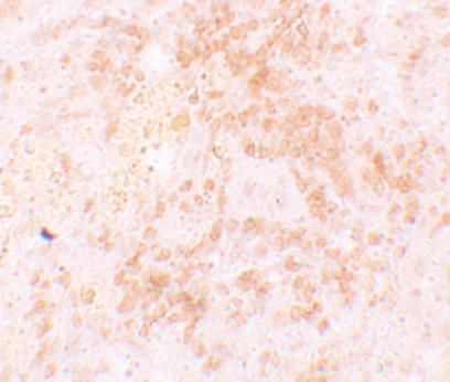
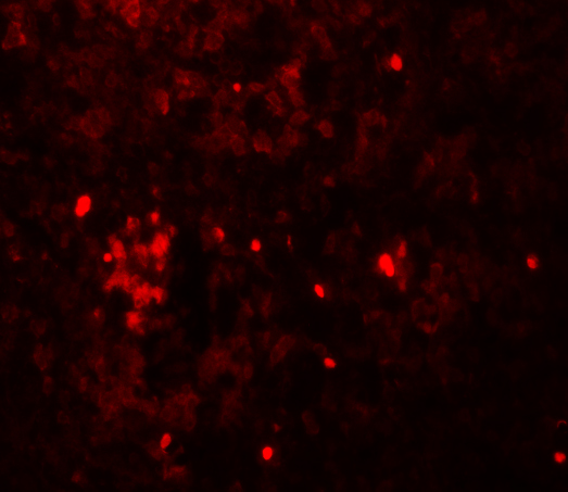
The catalog antibody has IHC data from rat spleen tissue and IF data from rat spleen cells (IHC and IF figure captions); it lists Human, Mouse and Rat reactivity (catalog: A05922).
A05922 is the SKU with a rendered IHC card: its figure shows rat spleen tissue stained at 5 μg/mL (IHC figure caption). The same SKU lists IHC-P and IF applications, with an IF figure from rat spleen cells at 20 μg/mL (catalog: A05922; IF figure caption).
Which to pick: For paraffin-section IHC, choose A05922 (catalog: IHC-P; IHC figure caption: rat spleen tissue); the figure caption does not report a fixative. For IF, choose A05922 (catalog: IF; IF figure caption: rat spleen cells); the payload does not establish separate ICC validation. For cross-species planning, A05922 lists Human, Mouse and Rat reactivity, while A05922-1 is a human-reactive polyclonal IHC option without a supplied IHC figure (catalog: A05922 and A05922-1).