This website uses cookies to ensure you get the best experience on our website.
- Table of Contents
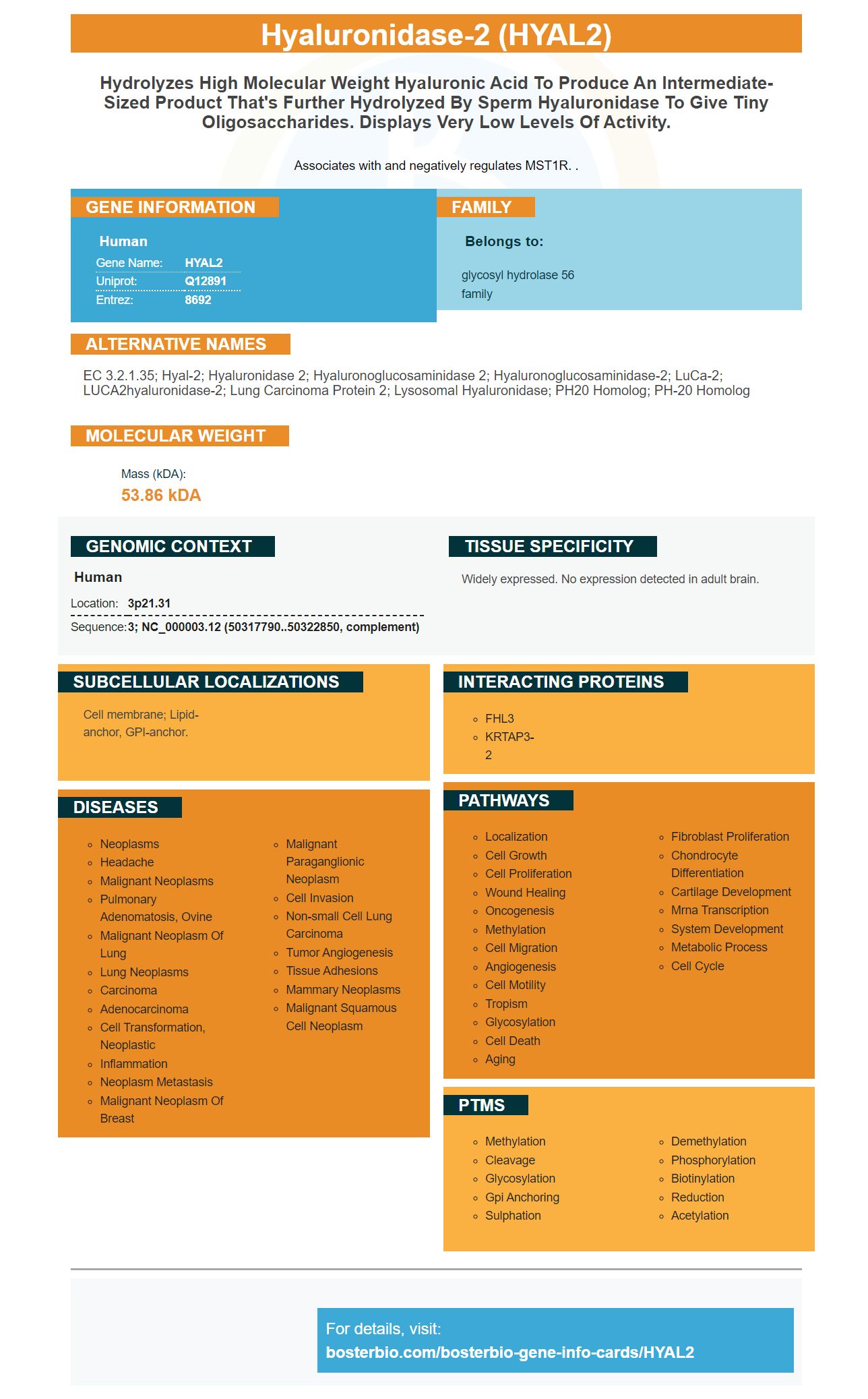
Facts about Hyaluronidase-2.
Associates with and negatively regulates MST1R. .
Human | |
---|---|
Gene Name: | HYAL2 |
Uniprot: | Q12891 |
Entrez: | 8692 |
Belongs to: |
---|
glycosyl hydrolase 56 family |
EC 3.2.1.35; Hyal-2; hyaluronidase 2; hyaluronoglucosaminidase 2; hyaluronoglucosaminidase-2; LuCa-2; LUCA2hyaluronidase-2; Lung carcinoma protein 2; lysosomal hyaluronidase; PH20 homolog; PH-20 homolog
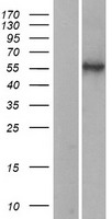
Mass (kDA):
53.86 kDA
Human | |
---|---|
Location: | 3p21.31 |
Sequence: | 3; NC_000003.12 (50317790..50322850, complement) |
Widely expressed. No expression detected in adult brain.
Cell membrane; Lipid-anchor, GPI-anchor.
This article focuses on the HYAL2 marker. Learn more about the HYAL2 Marker, Biotin-conjugated goat anti-rabbit and Horseradish peroxidase-conjugated goat anti-mouse, and a few other related topics. Read on to find out which HYAL2 marker is right for your research.
In a study by Miller et al. Miller et. al. (2002) found no evidence of HYAL2 mRNA and protein in adult brain tissue. HYAL2 cDNA however was expressed in C6 glioma cellular cells. Western blot demonstrated that these cells had a level of 3.8 hyaluronidase activities. However, they were negative for hyaluronidase activation in the presence of a placebo.
Although the HYAL2 mark is a candidate marker for targeting tumour cells it has been shown to have only minor effects on the environment. However, HYAL2 can inhibit tumour growth in vivo. Similarly, it also inhibits tumour growth in vitro. HYAL1 & HYAL2 might regulate the microenvironment of tumor cells and their intercellular interactions.
HYAL2 is a cell surface receptor for the JSRV virus. This virus causes pulmonary cancer in sheep. It is found within type II aveolar Epithelial Cells. These cells contain Hyal2 on the cell surface and it inhibits Mst1r. Antiserum directed against HYAL2 mRNA or RNA crossreacts also with 30% human pulmonary adenocarcinomas.
Human HYAL2 is expressed in all tissues, except the adult brain. The developmentally regulated expression of HYAL2 proteins is possible. The HYAL2 gene is expressed in the brain tissue of newborn mice but disappears shortly after birth. This gene is also expressed by mice's adult brains. These findings suggest that the expression of HYAL2 in human cells may be developmentally regulated.
The HYAL2 gene coding for a polypeptide containing two Hyaluronidase domains is a common marker of chondroitinase in cartilage. The enzyme is composed two chains, one single and one double chain. The former contains more Hyaluronidase activities. HYAL1 & HYAL2 prototypically act as acid-active enzymes. HYAL4 meanwhile, is a chondroitinase which has no hyaluronan catalytic activities.
This goat anti-rabbit secondary antibody has been purified of antiserum by removing goat sera. It is then combined with human serum proteins via a solid phase method. The biotin conjugation of this goat anti-rabbit secondary antibody makes it an excellent choice for applications requiring indirect sensitive immunodetection and quantification of low-abundance targets. Biotin-conjugated goat anti-rabbit secondary antibodies can also be used with reporter-labeled signal amplification systems to detect protein expression in biological samples.
This biotin conjugated goat antirabbit protein has a very specific reactivity towards Rabbit. This antibody can be used in western blotting and ELISA. It is compatible with many chemiluminescence substrates, and can be used to develop ELISA kits. Its biotin analog, 6-aminocaproic Acid, is it.
A biotin-conjugated goat monoclonal antibody reacts to rabbit IgG. It also reacts well with all the light chains in all rabbit immunoglobulins. It has minimal cross-reactivity with other rabbit serum proteins, bovine serum, and human serum. It contains one percent BSA (w/v), and sodium azide to preserve it.
Anti-Rabbit IgG biotin conjugate is suitable for western-blotting, immunoelectrophoresis, and IHC. Signal detection requires that the dilution be optimized. It can be distinguished by a single precipitin-arc when used in combination with a suitable substrate. It has been tested against rabbit sera by immunoelectrophoresis and immunoblotting and has been validated by ELISA and Western-blotting methods.
To detect IgG antibodies in mice, HRP-conjugated goat-anti-mice markers can also be used. This goat antiserum has been produced by immunoaffinity Chromatography and has been conjugated to horseradish peroxidase. The goat anti-mouse marker can be used in immunoaffinity chromatography, Western blotting, and ELISA.
The widely used enzyme HRP is used for secondary antibody labeling. It is widely used in immunohistochemistry, ELISA, western blot, and dot blot. Hydrogen peroxide helps make HRP visible. There are three types if substrates that work with HRP: fluorogenic, chromogenic and chemiluminescent. The level of peroxidase activity is indicated by the intensity of the signal.
After transferring the markers to the NC membranes, they were stained with secondary antibodies. Thermo Scientific, Invitrogen, Jackson Immuno-Research Laboratories supplied the secondary antibodies. The detection of M&R LE proteins markers was done as described previously. This method can be used for determining if the marker was auto-detected.
M&R LE protein markers were created by fusing a rabbit and mouse linear epitope. The leader peptide was fused together with an E. coli extra membrane protein, a transcription molecule, and a signal peptide. Secondary antibodies can be used to visualize M&R LE protein markers under denaturing conditions. They provide a reliable tool for protein analysis, and are convenient for proteomic studies.
An anti-rat secondary antibodies that targets the HYAL2 marker, is useful for immunofluorescence sperm staining. IP studies revealed that HYAL2 immunoprecipitated sperm lysates with high efficiency. This antibody is also highly specific and detects HYAL2 in sperm. We developed a protocol that uses FITC-PNA-conjugated secondary antibodies and a fluorescent substrate.
Primary antibodies are made by purifying whole IgG fragments or the F (ab')2 segment and then conjugated using agarose. Affinity purification eliminates non-specific antibodies, resulting in high specificity and low background signals. The antibodies are affinity purified before labeling and further processed by adsorption on an affinity column containing immobilized serum proteins or IgG of the selected species. This reduces cross-reactivity and makes them Highly Cross-Adsorbed antibodies (HCA).
The anti-rat antibody 15115-1 AP targets the HYAL2 gene. It can be used to perform WB, IHC or FC experiments. This antibody exhibits good reactivity with mouse and human samples. However, in addition to targeting HYAL2, it may also be useful for immunohistochemistry. Anti-rat antibodies targeting HYAL2 will provide a better understanding about the location of fusion proteins in rats.
A hyaluronidase, HYAL2, has an unusual activity. It cleaves high-molecular-weight hyaluronic acid to 20-kDa species, and it may have different biological functions such as stimulating angiogenesis and gene regulation. These findings raise the possibility that HYAL2 is a key player in cancer research.
PMID: 9712871 by Lepperdinger G., et al. HYAL2, a human gene expressed in many cells, encodes a lysosomal hyaluronidase with a novel type of specificity.
PMID: 11731268 by Lepperdinger G., et al. Hyal2 -- less active, but more versatile?