IAPP / Islet amyloid polypeptide · IHC design guide

Design Immunohistochemistry for IAPP

Plan paraffin-section IHC for IAPP using pancreatic endocrine cells as a high-expression reference (HPA tissue IHC). Assess the distinct cytoplasmic staining pattern while accounting for the protein’s secretion (HPA tissue IHC; UniProt).

Evidence assembled Oct 2026 · For research use; verify linked source records and product datasheet before use
Immunohistochemistry protocol sheet for IAPP (IHC for IAPP): expected localisation Cytoplasmic in pancreatic islets (HPA tissue IHC), antibody A00414-1, validated IHC image, and IHC protocol steps
Printable IAPP IHC protocol sheet — expected localisation Cytoplasmic in pancreatic islets (HPA tissue IHC), antibody A00414-1, controls and protocol steps. Open the full IAPP IHC guide →

IAPP Immunohistochemistry Experimental Design Guide

Expected localisation, validated protocols, controls and antibodies — the at-a-glance facts below, then the full design guide.

Must know before staining
Expected localisation Cytoplasmic in pancreatic islets (HPA tissue IHC)
Staining pattern Pancreatic endocrine cells show distinct cytoplasmic staining (HPA tissue IHC)
Antigen retrieval Citrate pH 6.0 HIER, 95–98 °C, 20 min (rule: cytoplasmic / membrane antigen)
Positive control ⓘ Pancreas
Negative control ⓘ Adipose tissue+4 more · see all
Important caveats
Reasons your staining may differ from the expected pattern.
Fixation Keep fixation consistent across sections (standard IHC practice; not target-specific)
Caveat Secretion can separate protein staining from RNA location (HPA tissue IHC)
Regulation Pancreas-enriched expression (HPA tissue IHC)
Isoform / epitope No annotated isoforms; processing may affect epitope choice (UniProt)
Section 1

Recommended IAPP IHC & IF Protocols

The catalog antibody’s IHC-P protocol is accompanied by two published mouse tissue protocols (PMC11494195; PMC5584114).

Recommended immunohistochemistry (IHC-P) protocol parameters
SampleParaffin-embedded mouse brain tissue; fixative not specified (datasheet A00414-1)
FixationImage fixative and duration unreported (datasheet A00414-1); verify before use.
Sectioning4–5 µm sections on charged slides (standard)
DeparaffinisationXylene, graded ethanol series to water (standard)
Antigen retrievalHeat-induced epitope retrieval in citrate buffer, pH 6.0, 20 min at 95–98 °C (standard rule: cytoplasmic / membrane antigen)
Peroxidase block3% H2O2, 10 min, room temperature (standard)
Blocking10% normal serum of the secondary host, 30 min, room temperature (standard)
Primary antibodyRabbit anti-IAPP, 1:50-1:200 (datasheet A00414-1)
Primary incubationOvernight at 4 °C (standard)
DetectionHRP-polymer secondary, DAB chromogen 5–10 min (standard)
CounterstainHematoxylin, blue, dehydrate and mount (standard)
Expected resultIAPP-positive staining in pancreatic endocrine cells of pancreas (HPA tissue IHC: High). HPA tissue profile: Distinct cytoplasmic expression in pancreatic islets. No signal in the no-primary control.
💡Decision noteStart with citrate pH 6.0 HIER at 95–98 °C for 20 min (page antigen retrieval rule); optimize for the tissue and fixation used.
Section 2

What Is the Expected IAPP Staining Pattern?

IAPP is a secreted peptide with no transmembrane segment (UniProt P10997 topology). In tissue IHC, expect distinct cytoplasmic staining in pancreatic islets, with high staining in pancreatic endocrine cells (HPA tissue IHC). HPA rates the tissue pattern Enhanced, while noting that secretion can complicate agreement between RNA and protein location (HPA tissue IHC).

What am I looking at on my slide?
Distinct cytoplasmic staining in pancreatic islets, strongest in endocrine cells (HPA tissue IHC).This matches the reported IAPP pattern and provides the clearest positive reference for reading the section (HPA tissue IHC). Assess whether staining follows cells within islets rather than appearing uniformly across the section (general IHC practice).
Predominantly nuclear or sharply membrane-bound staining in pancreatic endocrine cells.This differs from the reported cytoplasmic tissue pattern (HPA tissue IHC). A membrane-only interpretation is also difficult to reconcile with the absence of a transmembrane segment (UniProt P10997 topology). Review the counterstain and detection controls before assigning such staining to IAPP (general IHC practice).
Strong staining in adipocytes or broadly across cells outside pancreatic islets.HPA reports no detection in adipocytes from adipose tissue and distinct expression in pancreatic islets (HPA tissue IHC). Consider antibody cross-reactivity or endogenous chromogenic activity, especially when the same color appears across unrelated structures (general IHC practice).
Pale, diffuse color covers tissue and empty spaces, obscuring cell boundaries.A diffuse haze cannot establish the distinct cytoplasmic islet pattern reported by HPA (HPA tissue IHC). Check blocking, washes, detection reagent exposure, and a control without primary antibody as general IHC background checks (general IHC practice).
No staining in identifiable pancreatic islets despite an otherwise readable section.This conflicts with HPA's high pancreatic endocrine-cell staining (HPA tissue IHC). First confirm that endocrine tissue is present; then review the antibody's IHC-P instructions, detection reagents, and positive control before interpreting the absence as biological (general IHC practice).
💡Expected IAPP appearanceCall positive when pancreatic endocrine cells show distinct, high cytoplasmic staining within islets (HPA tissue IHC); isolated nuclear color or broad non-islet staining calls for control review (general IHC practice).
How each factor affects the staining
Tissue and cell choicePancreatic endocrine cells are the strongest documented tissue reference; HPA reports high staining there and no detection in adipocytes from adipose tissue (HPA tissue IHC). Ovary follicle cells have low reported staining, so they are a weaker visual reference than pancreatic islets (HPA tissue IHC).
Secretory identity and processingIAPP is secreted, with an annotated signal peptide and propeptide regions (UniProt P10997). These annotations do not identify which processed form an antibody detects; consult its stated immunogen or epitope before interpreting a discrepant pattern (UniProt P10997 processing; general IHC practice).
IHC antibody evidenceHPA lists HPA053194 and CAB000352 as IHC Enhanced (HPA antibodies). That supports their reported tissue patterns but does not establish equivalent results for another catalog antibody or define its dilution or antigen retrieval conditions (HPA antibodies; general IHC practice).
IF/ICC Q&AWhat location is reported for IF/ICC? HPA lists vesicles as the approved main location and U2OS as an imaged cell line (HPA subcellular ICC-IF). This is separate from the cytoplasmic pancreatic tissue IHC observation and does not establish an IHC staining pattern in U2OS (HPA subcellular ICC-IF; HPA tissue IHC).
Why is my staining missing, weak or wrong?
SituationLikely causeNext action
Pancreatic islets are present, but staining is absent.The observed result falls short of HPA's high endocrine-cell staining; a failed staining run remains possible (HPA tissue IHC; general IHC practice).Verify the positive-control slide and catalog antibody's IHC-P instructions; check reagent order and detection performance before changing interpretation (general IHC practice).
The whole section is weak, including the expected positive region.A run-wide issue with primary antibody, detection, or chromogen can reduce visible signal (general IHC practice).Compare a known-positive pancreatic control processed in the same run, then check reagent preparation, incubation, and chromogen development against the IHC-P instructions (general IHC practice).
Diffuse color obscures the islets.Nonspecific reagent binding, inadequate washing, or excessive detection development can raise background (general IHC practice).Review blocking and washes, inspect a control without primary antibody, and shorten development if its instructions permit (general IHC practice).
Color persists in a control without primary antibody.The primary antibody cannot account for that control's color; endogenous detection activity or another detection-step background is possible (general IHC practice).Check the chromogenic detection system's recommended endogenous-activity block and repeat the control alongside tissue staining (general IHC practice).
Staining appears mainly nuclear or outlines cell membranes.That distribution disagrees with HPA's cytoplasmic islet pattern; IAPP also lacks a transmembrane segment (HPA tissue IHC; UniProt P10997 topology).Compare with a pancreatic positive control, confirm the counterstain is not being scored as chromogen, and review antibody specificity before reporting IAPP localisation (general IHC practice).
Adipocytes or many unrelated cells stain as strongly as islets.HPA reports no IAPP detection in adipocytes from adipose tissue; widespread signal raises concern for cross-reactivity or detection background (HPA tissue IHC; general IHC practice).Compare matched positive and negative tissues and the control without primary antibody; interpret the distribution only after background is excluded (general IHC practice).

Sample controls for IAPP IHC & IF

🧪Run pancreas first; pancreatic endocrine cells should stain strongly (HPA: High in pancreatic endocrine cells). Use adipose tissue as the negative tissue (HPA: Not detected in adipocytes), and compare endocrine staining with adjacent pancreatic exocrine cells, which should show substantially less signal (HPA: High in pancreatic endocrine cells; standard pancreas histology).
Positive control tissue: Pancreas (Pancreatic endocrine cells, HPA High)
Negative control tissue: Adipose tissue (HPA Not detected)
ICC-IF cell lines (HPA subcellular resource): HPA ICC-IF images show IAPP in U2OS, with annotated localisation: Vesicles (approved) (HPA subcellular).
Technical controls: Include a PBS/no-primary, secondary-only control and a concentration-matched nonimmune rabbit IgG isotype control; use IAPP-knockout tissue or cognate-peptide blocking, if available, to assess target specificity (selected-SKU IHC caption: rabbit pAb, goat anti-rabbit secondary, PBS no-primary; standard IHC practice). For chromogenic detection, quench endogenous peroxidase and block endogenous biotin when using the caption’s avidin-peroxidase system; for IF, check unstained pancreas for autofluorescence (selected-SKU IHC caption: avidin-peroxidase; standard IHC/IF practice).
⚠️Feasibility: The selected A00414-1 paraffin-section IHC caption reports 1:50, but its fixative is unreported; no target-specific fixation window or fixation effect is reported in the supplied evidence (selected-SKU IHC caption). Retrieval dependency and whether frozen sections or IF are easier are unreported; vesicular ICC-IF localization in U2OS establishes an imaging observation, not a processing preference (HPA: vesicles; U2OS ICC-IF). In pancreas, interpret diffuse extracellular signal cautiously because IAPP is secreted, and score endocrine cells against adjacent exocrine cells (UniProt P10997: secreted; HPA: High in pancreatic endocrine cells).

HPA tissue IHC evidence for IAPP

Comprehensive Human Protein Atlas IHC scoring per tissue (reliability: Enhanced — High consistency between antibody staining and RNA expression data. At least one protein variant secreted, tissue location of RNA and protein might differ and correlation is complex.). Rows are taken directly from the HPA tissue chart — click any row's HPA link to view the source.

Positive expression · recommended positive controls

TissueCell typeLevelEvidenceSource
Pancreas Pancreatic endocrine cells High Protein (IHC) HPA →

Undetected expression · recommended negative controls

TissueCell typeLevelEvidenceSource
Adipose tissue Adipocytes Not detected Protein (IHC) HPA →
Adrenal gland Glandular cells Not detected Protein (IHC) HPA →
Appendix Glandular cells Not detected Protein (IHC) HPA →
Bone marrow Hematopoietic cells Not detected Protein (IHC) HPA →
Breast Adipocytes Not detected Protein (IHC) HPA →
Section 3

Advanced IAPP IHC Tips

Troubleshoot chromogenic IHC for IAPP in paraffin sections using the catalog image, tissue staining profile and known peptide processing as reference points.

What retrieval should I try when IAPP staining is weak in paraffin sections?
Use citrate buffer at pH 6.0 for heat retrieval at 95–98 °C for 20 min (page retrieval rule). Let sections cool in buffer, then compare the result with a matched section processed without retrieval to assess whether heating improves signal (standard IHC practice). The catalog image demonstrates staining in paraffin-embedded mouse brain at 1:50, but its caption does not report a retrieval method or fixative (caption: A00414-1). Keep antibody concentration and chromogen development constant across retrieval comparisons so a change in staining can be attributed to retrieval (standard IHC practice). If staining remains weak, test a second retrieval condition as a separate optimisation, recording the buffer and temperature used (standard IHC practice).
How should I troubleshoot inconsistent IAPP staining after tissue fixation?
Target-specific fixation sensitivity is unknown from the supplied evidence; the catalog caption identifies paraffin-embedded tissue but does not state its fixative (caption: A00414-1). Record fixative, fixation duration and tissue thickness for each specimen, then process comparison sections together to limit variation from handling (standard IHC practice). Begin with the page’s citrate pH 6.0 retrieval at 95–98 °C for 20 min and hold antibody dilution and detection times constant (page retrieval rule; standard IHC practice). Compare preserved morphology with signal intensity before changing fixation or retrieval conditions (standard IHC practice). Do not infer a target-specific fixation effect from IAPP’s secreted annotation or the tissue staining profile (UniProt P10997; HPA tissue IHC).
Which staining compartments support an IAPP IHC result?
Look for cytoplasmic staining in pancreatic islet endocrine cells when assessing a biological positive reference (HPA tissue IHC: distinct cytoplasmic islet expression; high in pancreatic endocrine cells). A vesicular pattern can be compatible with the subcellular reference, while extracellular signal needs careful interpretation because IAPP is secreted (HPA subcellular: vesicles; UniProt P10997: secreted). The catalog paraffin-section caption describes cytoplasmic, membrane-associated and secreted staining in mouse brain, but does not establish the identity of each stained cell (caption: A00414-1). Compare signal with morphology and a primary-omission control on adjacent sections before assigning a compartment (standard IHC practice). IAPP has no transmembrane segment, so a membrane outline alone is insufficient evidence of specific staining (UniProt P10997 topology).
Could IAPP processing or epitope choice explain different IHC patterns?
Check the antibody’s documented immunogen or epitope before comparing staining across assays; no epitope position is supplied for this catalog antibody (supplied product caption: A00414-1). IAPP is made as an 89-aa precursor with a signal segment at residues 1–22 and propeptide segments at 23–31 and 74–89 (UniProt P10997 processing). An antibody against a removed segment could report precursor-related material differently from one directed toward retained peptide sequence, depending on its documented specificity (UniProt P10997 processing; standard immunostaining principle). The record lists 0 isoforms and tyrosine amidation at residue 70, so neither an alternative isoform nor modification-dependent recognition should be assumed without antibody evidence (UniProt P10997).
How can I check an IAPP IHC pattern with multiplex immunofluorescence?
Use IF as a separate assay and pair IAPP with a validated pancreatic endocrine-cell marker to assess whether signal falls in the expected cell population (HPA tissue IHC: high in pancreatic endocrine cells; standard IF practice). Choose spectrally separated fluorophores and place the weaker signal in a channel with low tissue autofluorescence, checking unstained sections first (standard IF practice). For an intracellular epitope, test mild permeabilisation; for an accessible extracellular epitope, compare a section without permeabilisation, since IAPP is secreted and its exact antibody epitope is unspecified (UniProt P10997: secreted; supplied product caption: A00414-1). Include single-stain and primary-omission controls to assess bleed-through and background (standard IF practice). The catalog paraffin IHC image does not establish an IF dilution or fixation condition (caption: A00414-1).
How do I reduce diffuse or vessel-associated background in IAPP IHC?
First compare the stained section with a primary-omission control; the catalog image used PBS instead of primary antibody for its negative control (caption: A00414-1). Apply a peroxidase block before chromogenic detection and check whether residual colour appears without primary antibody (standard IHC practice). Titrate primary antibody around the reported 1:50 image condition, changing only one variable at a time because that dilution is image-specific (caption: A00414-1; standard IHC practice). Ensure washes remove unbound reagents and inspect section edges for drying artefact before increasing retrieval or chromogen exposure (standard IHC practice). Interpret diffuse extracellular signal cautiously because IAPP is secreted, and require reproducible anatomy and control separation before calling it positive (UniProt P10997: secreted; standard IHC practice).
What is a reproducible way to quantify IAPP staining in paraffin sections? ⚠ ANSWER MARKED FOR VERIFICATION
Define pancreatic endocrine cells as the scoring population when using the strongest supplied tissue reference (HPA tissue IHC: high in pancreatic endocrine cells). Report the percentage of positive endocrine cells and an intensity-based H-score, using the same thresholds and imaging settings across sections (standard IHC quantification practice). If measuring deposits or extracellular signal, report stained area or signal density per mm² of the predefined compartment, separately from cell scores (UniProt P10997: secreted; standard IHC quantification practice). Normalise cell counts to total evaluable endocrine cells and area measurements to evaluable tissue or islet area; exclude folds, edges and necrosis by prespecified rules (standard IHC quantification practice). Keep retrieval, detection and chromogen development matched because those steps alter apparent intensity (standard IHC practice).
How can I distinguish convincing IAPP positivity from staining artefacts?
A convincing pancreatic result shows reproducible cytoplasmic staining in islet endocrine cells and clear separation from the primary-omission control (HPA tissue IHC: distinct cytoplasmic islet expression; standard IHC practice). Investigate dominant staining in an unexpected cell population before assigning it to IAPP, using the HPA pancreatic pattern as an anatomical reference (HPA tissue IHC). Treat a sharp section-edge gradient, staining restricted to necrotic tissue or colour retained after primary omission as possible artefact (standard IHC practice). Check the peroxidase block when colour appears without primary antibody, because endogenous enzyme activity can contribute to chromogenic background (standard IHC practice). Extracellular signal may warrant review rather than automatic rejection because IAPP is secreted, but a membrane outline alone does not establish specificity (UniProt P10997: secreted; no transmembrane segment).
Boster reagents

Best IAPP / Islet amyloid polypeptide IHC Antibodies

Two anti-IAPP antibodies list IHC and IF applications for human, mouse and rat samples (catalog: applications and reactivity); the supplied figures show paraffin-section IHC (catalog: IHC image captions).

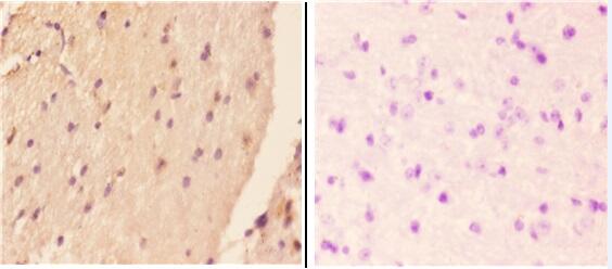
Real IHC data Immunohistochemistry (IHC) analyzes of Amylin (H28) pAb in paraffin-embedded mouse brain tissue at 1:50,showing distinct cytoplasmic membrane and secreted staining.Negative control (the right) Using PBS instead of primary antibody, secondary antibody is Goat Anti-Rabbit IgG-biotin followed by avidin-peroxidase.
Anti-Amylin (H28) IAPP Antibody
Cat # A00414-1
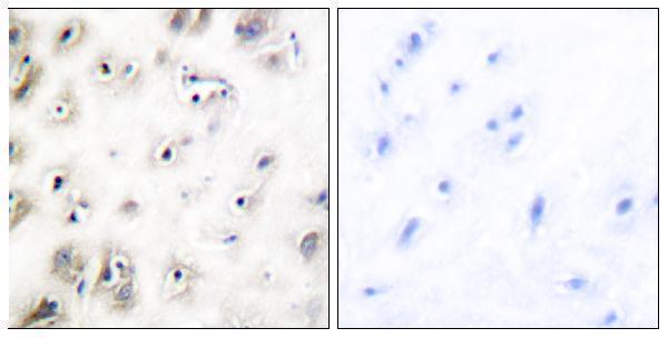
Real IHC data Immunohistochemistry analysis of paraffin-embedded human brain tissue, using Amylin Antibody. The picture on the right is blocked with the synthesized peptide.
Anti-Amylin IAPP Antibody
Cat # A00414

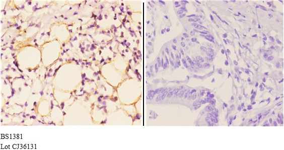
A00414-1 lists IHC and IF and shows paraffin-section IHC in mouse brain and human colon carcinoma (catalog: applications; A00414-1 IHC captions). A00414 lists IHC and IF and shows paraffin-section IHC in human brain with a peptide-blocked comparison (catalog: applications; A00414 IHC caption).

Which to pick: For tissue IHC, choose A00414-1 when its mouse brain or human colon carcinoma examples match your sample, or A00414 for its human brain example; each caption supports paraffin sections, and neither reports the fixative (A00414-1 and A00414 IHC captions). For IF/ICC, both list IF, but neither supplies an IF figure or documents ICC validation, so verify performance in your sample (catalog: applications and IF image alts). Both list human, mouse and rat reactivity, so choose between them using the relevant IHC example and stated dilution range; clonality is unreported for both (catalog: reactivity, IHC captions, IHC dilutions and clone fields).

Each figure is that product's own IHC / IF validation image from its datasheet.

References

  1. UniProt Consortium. UniProt entry P10997 (IAPP_HUMAN, Islet amyloid polypeptide).
  2. Human Protein Atlas. IAPP tissue IHC expression (reliability: Enhanced).
  3. Human Protein Atlas. IAPP subcellular location (ICC-IF): Localized to vesicles..
  4. Human Protein Atlas. IAPP antibody validation summary (2 antibodies).
  5. The overexpression of human amylin in pancreatic β cells facilitate the appearance of amylin aggregates in the kidney contributing to diabetic nephropathy. Scientific reports 2024 — PMC11494195.
  6. Kill two birds with one stone: making multi-transgenic pre-diabetes mouse models through insulin resistance and pancreatic apoptosis pathogenesis. PeerJ 2018 — PMC5909684.
  7. Induction of IAPP amyloid deposition and associated diabetic abnormalities by a prion-like mechanism. The Journal of experimental medicine 2017 — PMC5584114.
  8. Efficacy of IAPP suppression in mouse and human islets by GLP-1 analogue conjugated antisense oligonucleotide. Frontiers in molecular biosciences 2023 — PMC9939749.
  9. PubMed PMID:2651160 — UniProt-cited evidence.
  10. PubMed PMID:2608057 — UniProt-cited evidence.
  11. PubMed PMID:3053705 — UniProt-cited evidence.