IDH3A / Isocitrate dehydrogenase [NAD] subunit alpha, mitochondrial · IHC design guide

Design Immunohistochemistry for IDH3A

Plan IDH3A staining in paraffin sections using the observed granular cytoplasmic tissue pattern (HPA tissue IHC). Use the tissue examples and controls to assess signal, keeping in mind that IDH3A is a mitochondrial protein (UniProt).

Evidence assembled Oct 2026 · For research use; verify linked source records and product datasheet before use
Immunohistochemistry protocol sheet for IDH3A (IHC for IDH3A): expected localisation Granular cytoplasmic staining is observed in most tissues (HPA tissue IHC), antibody A09544-3, validated IHC image, and IHC protocol steps
Printable IDH3A IHC protocol sheet — expected localisation Granular cytoplasmic staining is observed in most tissues (HPA tissue IHC), antibody A09544-3, controls and protocol steps. Open the full IDH3A IHC guide →

IDH3A Immunohistochemistry Experimental Design Guide

Expected localisation, validated protocols, controls and antibodies — the at-a-glance facts below, then the full design guide.

Must know before staining
Expected localisation Granular cytoplasmic staining is observed in most tissues (HPA tissue IHC)
Staining pattern Granular cytoplasmic staining in most tissues (HPA tissue IHC)
Antigen retrieval EDTA pH 8.0 HIER, heat-mediated (datasheet A09544-3)
Positive control ⓘ Adrenal gland+4 more · see all
Negative control ⓘ Liver+2 more · see all
Important caveats
Reasons your staining may differ from the expected pattern.
Fixation Matched tissue-IHC evidence does not establish the fixation claim. Validate the specimen-specific method before use. (selected-SKU IHC image A09544-3)
Caveat Staining and RNA data show medium consistency (HPA tissue IHC)
Regulation RNA expression is enhanced in tongue (HPA tissue RNA)
Isoform / epitope 2 isoforms; their epitope differences are unreported (UniProt)
Section 1

Recommended IDH3A IHC & IF Protocols

The catalog antibody’s IHC-P protocol uses EDTA pH 8.0 heat retrieval (datasheet: A09544-3). Four published IDH3A IHC protocols provide tissue-specific alternatives (PMC6718547; PMC6125878; PMC13564267; PMC11106410).

Recommended immunohistochemistry (IHC-P) protocol parameters
SampleParaffin-embedded human esophageal squamous carcinoma tissue; fixative not specified (datasheet A09544-3)
FixationImage fixative and duration unreported (datasheet A09544-3); verify before use.
Sectioning4–5 µm sections on charged slides (standard)
DeparaffinisationXylene, graded ethanol series to water (standard)
Antigen retrievalHeat retrieval: EDTA pH 8.0 (datasheet A09544-3); 20 min, 95–100 °C (standard)
Peroxidase block3% H2O2, 10 min, room temperature (standard)
Blocking10% goat serum (datasheet A09544-3)
Primary antibodyRabbit anti-IDH3A, 2-5 μg/ml (datasheet A09544-3)
Primary incubationOvernight at 4 °C (datasheet A09544-3)
DetectionHRP-conjugated secondary, DAB chromogen (datasheet A09544-3)
CounterstainHematoxylin, blue, dehydrate and mount (standard)
Expected resultIDH3A-positive staining in glandular cells of adrenal gland (HPA tissue IHC: High). HPA tissue profile: Cytoplasmic expression with a granular pattern in most tissues. No signal in the no-primary control.
💡Decision noteStart with EDTA pH 8.0 for the catalog antibody (datasheet: A09544-3); consider citrate retrieval when adapting a published tissue protocol (PMC6718547; PMC6125878; PMC11106410).
Section 2

What Is the Expected IDH3A Staining Pattern?

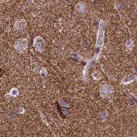
IDH3A localizes to mitochondria and has no transmembrane segment (UniProt P50213 topology; HPA ICC-IF: mitochondria). In paraffin-section IHC, expect granular cytoplasmic staining in many tissues, including strong staining in selected glandular, epithelial, and neuronal cells (HPA tissue IHC). HPA rates its tissue IHC profile Supported, with medium consistency between antibody staining and RNA data and external verification pending (HPA tissue IHC: reliability).

What am I looking at on my slide?
Granular cytoplasmic staining in adrenal glandular cells, bronchial respiratory epithelium, or colon glandular cells.This matches the reported high staining in those specific cell populations and the granular tissue pattern (HPA tissue IHC: High; profile). Assess staining in the named cells, since a tissue-wide score can conceal differences between cell types (standard IHC practice).
Predominantly nuclear or cell-surface staining, without convincing granular cytoplasmic signal.The compartment conflicts with mitochondrial localization and the reported tissue pattern (UniProt P50213: mitochondrion; HPA tissue IHC: granular cytoplasm). Treat it as suspect staining and check antibody-dependent background and detection controls before assigning it to IDH3A (standard IHC practice).
Strong staining in liver cholangiocytes, ovarian stromal cells, or skeletal-muscle myocytes.HPA reports IDH3A as not detected in those particular cell populations (HPA tissue IHC: Not detected). Review morphology and controls for cross-reactivity or endogenous detection activity; the observation alone does not establish either cause (standard IHC practice).
Broad, smooth color over cells and surrounding tissue, obscuring cytoplasmic granules.This is less interpretable than the reported granular cytoplasmic pattern (HPA tissue IHC: profile). Diffuse background may arise during antibody incubation, washing, blocking, or chromogenic detection; inspect a no-primary control and the underlying morphology (standard IHC practice).
No staining in the expected cell population of a high-staining tissue.A blank result in, for example, adrenal glandular cells or colon glandular cells conflicts with their reported high staining (HPA tissue IHC: High). First establish that the cells are present and that the detection run worked before interpreting absence of IDH3A (standard IHC practice).
💡Expected IDH3A appearanceA credible positive is granular cytoplasmic staining in the specified high-staining glandular, epithelial, or neuronal cells (HPA tissue IHC: profile; High), consistent with mitochondrial localization (UniProt P50213: mitochondrion); dominant nuclear or smooth, widespread color is suspect (standard IHC practice).
How each factor affects the staining
Compartment and protein processingIDH3A is mitochondrial, lacks a transmembrane segment, and has an annotated mature chain spanning residues 28–366 (UniProt P50213: localization, topology, chain). These facts guide localization assessment; they do not identify this antibody's epitope or establish a retrieval requirement.
Cell population and staining levelHPA reports high staining in selected glandular, respiratory epithelial, and neuronal cells, while cardiomyocytes, adipocytes, and bone-marrow hematopoietic cells are low (HPA tissue IHC: High; Low). Compare the same named cell population; low staining is a poor basis for declaring an IHC run failed.
Strength of validationThe HPA tissue profile is Supported, with medium agreement between staining and RNA expression and external verification pending (HPA tissue IHC: reliability). HPA041465 has IHC status Supported; its Enhanced status applies to ICC, while HPA062971 has no listed IHC status (HPA antibodies).
IF/ICC Q&A: What pattern should IF show?A mitochondrial signal is expected in ICC-IF (HPA subcellular: mitochondria). HPA lists enhanced ICC validation for HPA041465 and HPA062971 (HPA antibodies). This IF observation supports a compartment check; it does not establish an IHC staining level in a tissue section.
Why is my staining missing, weak or wrong?
SituationLikely causeNext action
High-staining reference cells are blank.The result conflicts with HPA's high staining for the selected cell population; a failed detection run or missed cell population remains possible (HPA tissue IHC: High; standard IHC practice).Confirm morphology, run a known-positive section in the same detection batch, and review retrieval, primary incubation, and detection steps using the applicable antibody instructions (standard IHC practice).
Granules are lost beneath diffuse chromogenic haze.The haze obscures the reported granular cytoplasmic profile; background can arise from antibody or detection steps (HPA tissue IHC: profile; standard IHC practice).Inspect a no-primary control, then review blocking, washes, antibody concentration, and chromogen development as general IHC controls (standard IHC practice).
The dominant signal is nuclear or outlines cell membranes.That pattern disagrees with mitochondrial localization and the granular cytoplasmic tissue profile (UniProt P50213: mitochondrion; HPA tissue IHC: profile).Check compartment against a high-staining reference cell population and compare with no-primary staining before scoring it as IDH3A (HPA tissue IHC: High; standard IHC practice).
A reported negative cell population stains strongly.Cholangiocytes, ovarian stromal cells, and skeletal-muscle myocytes are reported as not detected; cross-reactivity or endogenous detection activity are possible explanations (HPA tissue IHC: Not detected; standard IHC practice).Verify cell identity, examine no-primary controls, and check endogenous enzyme blocking where relevant to the chromogenic system (standard IHC practice).
Cardiomyocytes or adipocytes show only faint staining.Low staining in these cells agrees with HPA's observations and need not indicate assay failure (HPA tissue IHC: Low).Judge run performance using an HPA high-staining cell population processed alongside the sample, then score the low-staining cells separately (HPA tissue IHC: High; Low; standard IHC practice).
Results differ between IHC antibodies or from IF images.HPA reports Supported IHC for HPA041465, no IHC status for HPA062971, and Enhanced ICC status for both; the validation claims cover different applications (HPA antibodies).Record the antibody and application used, compare IHC results within the same cell population, and evaluate IF chiefly for mitochondrial localization (HPA antibodies; HPA subcellular: mitochondria; standard IHC practice).

Sample controls for IDH3A IHC & IF

🧪Run adrenal gland first; its glandular cells should stain for IDH3A (HPA: High in adrenal gland glandular cells). Use liver cholangiocytes as the biological comparison (HPA: Not detected in liver cholangiocytes); no cell population within the adrenal slide is established as IDH3A-negative, so evaluate surrounding cells for background without treating them as a validated negative control.
Positive control tissue: Adrenal gland (Glandular cells, HPA High)
Negative control tissue: Liver (HPA Not detected)
ICC-IF cell lines (HPA subcellular resource): HPA ICC-IF images show IDH3A in A-431, U-251MG, U2OS, A-549, SiHa, KOLF2.1J, with annotated localisation: Mitochondria (enhanced) (HPA subcellular).
Technical controls: Include a no-primary (secondary-only) slide, a host- and isotype-matched control immunoglobulin, and IDH3A-knockout material where available to assess antibody specificity (standard IHC practice). For chromogenic detection in adrenal tissue, quench endogenous peroxidase and, if using avidin–biotin detection, check endogenous biotin background (standard IHC practice).
⚠️Feasibility: No matched IDH3A source supplied reports a target-specific fixation window, fixation effect, or dependence on a particular antigen-retrieval condition; optimize retrieval empirically (supplied IDH3A evidence). The selected-SKU paraffin-section caption concerns ILDR1, and its fixative is unreported, so it cannot establish an IDH3A fixation or retrieval condition (selected-SKU caption: ILDR1; fixative unreported). Frozen sections are not established as easier by these data; IF/ICC offers a mitochondrial localisation check (HPA subcellular: mitochondria), while endogenous peroxidase can add background to chromogenic adrenal IHC (standard IHC practice).

HPA tissue IHC evidence for IDH3A

Comprehensive Human Protein Atlas IHC scoring per tissue (reliability: Supported — Medium consistency between antibody staining and RNA expression data. Pending external verification.). Rows are taken directly from the HPA tissue chart — click any row's HPA link to view the source.

Positive expression · recommended positive controls

TissueCell typeLevelEvidenceSource
Adrenal gland Glandular cells High Protein (IHC) HPA →
Bronchus Respiratory epithelial cells High Protein (IHC) HPA →
Caudate Neuronal cells High Protein (IHC) HPA →
Cerebellum Cells in granular layer High Protein (IHC) HPA →
Cerebral cortex Neuronal cells High Protein (IHC) HPA →

Undetected expression · recommended negative controls

TissueCell typeLevelEvidenceSource
Liver Cholangiocytes Not detected Protein (IHC) HPA →
Ovary Ovarian stroma cells Not detected Protein (IHC) HPA →
Skeletal muscle Myocytes Not detected Protein (IHC) HPA →
Section 3

Advanced IDH3A IHC Tips

Use compartment-aware controls when interpreting IDH3A staining: it is mitochondrial, and tissue IHC commonly shows a granular cytoplasmic pattern (UniProt P50213; HPA tissue IHC).

How should I optimize retrieval when IDH3A staining is weak in paraffin sections?
Start with heat-mediated EDTA pH 8.0 antigen retrieval for paraffin-section IHC (page retrieval setting: datasheet A09544-3). Process matched sections together and compare a short and longer heating interval, such as 10 and 20 minutes, followed by controlled cooling (standard IHC practice). The supplied A09544-3 tissue caption identifies ILDR1, so its image and processing details do not establish IDH3A staining performance (selected tissue-IHC caption). If staining remains weak, test citrate pH 6.0 as a fallback on matched sections while holding detection conditions constant (standard IHC practice). Favor the condition that preserves morphology and yields granular cytoplasmic signal with low background (HPA tissue IHC; standard IHC practice).
Could fixation explain variable IDH3A staining between paraffin blocks?
Target-specific fixation sensitivity for IDH3A is unknown from the supplied evidence, so treat fixation as an experimental variable (evidence payload). Record fixative, fixation duration, tissue thickness, and processing history for each block before comparing staining intensity (standard IHC practice). For a prospective comparison, fix matched samples under the same routine conditions and change only one variable at a time (standard IHC practice). Compare morphology and granular cytoplasmic signal in the same cell type, using a positive-control section in each staining run (HPA tissue IHC; standard IHC practice). The A09544-3 caption reports paraffin embedding but gives no fixative and describes ILDR1, not IDH3A (selected tissue-IHC caption).
Where should convincing IDH3A chromogenic staining appear within a cell?
Expect a granular cytoplasmic pattern in tissue sections, consistent with mitochondrial localisation (HPA tissue IHC; UniProt P50213). Evaluate granules within intact cells at high magnification rather than treating diffuse brown tissue color as equivalent signal (standard IHC practice). IDH3A has no annotated transmembrane segment, so crisp cell-surface staining alone would need separate validation (UniProt P50213 topology). Include a positive-control tissue with reported high staining, such as colon glandular cells, alongside the study sections (HPA tissue IHC). Compare the candidate signal with negative-control staining and cell morphology before calling a compartment-specific result (standard IHC practice).
Can this stain distinguish IDH3A isoforms or a processed mitochondrial form?
Do not assign an isoform from chromogenic staining alone: the record lists 2 IDH3A isoforms, but no antibody epitope is supplied (UniProt P50213; evidence payload). The annotated mature chain spans residues 28–366, so an epitope near the amino terminus could affect recognition after processing if the antibody binds there (UniProt P50213 processing). Annotated modifications include phosphothreonine 101 and acetyllysine 223, but their effect on this antibody is unknown (UniProt P50213; evidence payload). Request the immunogen or mapped epitope before making isoform or modification claims (standard antibody-validation practice). Use an independent antibody or orthogonal expression measurement to investigate discordant staining (standard IHC practice).
How can parallel IF help assess an ambiguous IDH3A IHC pattern?
Use parallel IF on suitable material to check whether signal follows a mitochondrial pattern; the reported subcellular location is mitochondria (HPA subcellular; UniProt P50213). Multiplex with a marker for the expected cell population, such as a glandular epithelial marker when assessing colon glandular cells (HPA tissue IHC; standard IF practice). Choose a fluorophore channel after checking unstained tissue autofluorescence, and include single-label controls when interpreting overlap (standard IF practice). Test mild permeabilisation for antibody access to the mitochondrial antigen; the supplied topology identifies no transmembrane segment but does not map the epitope to a membrane side (UniProt P50213 topology; evidence payload). Keep IF findings as supporting evidence for the chromogenic IHC interpretation (standard assay-validation practice).
What should I adjust when DAB obscures granular IDH3A staining?
First inspect a no-primary control for tissue color and detection-reagent background under the same development conditions (standard IHC practice). Block endogenous peroxidase before applying an HRP detection system, then compare a shorter DAB development time against the current condition (standard chromogenic IHC practice). Titrate the IHC-validated antibody and optimize blocking or washing on matched sections if diffuse staining persists (standard IHC practice). Judge improvement by whether intact cells show discrete cytoplasmic granules, the reported tissue pattern for IDH3A (HPA tissue IHC). Do not import the 2 μg/ml concentration from A09544-3 as an IDH3A starting dilution; that caption identifies ILDR1 (selected tissue-IHC caption).
How should I quantify IDH3A IHC across tissues with different cell mixtures? ⚠ ANSWER MARKED FOR VERIFICATION
Define the cell population before scoring: HPA reports high staining in colon glandular cells and no detection in liver cholangiocytes (HPA tissue IHC). Within each population, score the percentage of positive cells and staining intensity, or calculate an H-score from intensity categories (standard IHC practice). If spatial abundance matters, report positive-cell density per mm² of viable, annotated tissue (standard image-analysis practice). Normalize comparisons to the number or area of the same cell type and keep acquisition, DAB development, and thresholds consistent (standard IHC practice). Record granular cytoplasmic signal separately from diffuse background so the score reflects the reported IDH3A pattern (HPA tissue IHC; standard IHC practice).
How do I distinguish true IDH3A staining from tissue artefact?
A plausible positive shows granular cytoplasmic staining in intact cells, consistent with mitochondrial IDH3A and the reported tissue pattern (UniProt P50213; HPA tissue IHC). Check whether staining is concentrated at section edges, in necrotic areas, or in damaged cells before assigning biological meaning (standard IHC practice). Nuclear-only or sharp membrane-only signal conflicts with the reported mitochondrial location and merits independent validation (HPA subcellular; UniProt P50213 topology). Use no-primary and peroxidase-block controls to assess detection background and endogenous enzyme activity (standard chromogenic IHC practice). Interpret unexpected negatives cautiously because HPA tissue IHC is rated Supported with medium consistency and awaits external verification (HPA tissue IHC).
Boster reagents

Best IDH3A / Isocitrate dehydrogenase [NAD] subunit alpha, mitochondrial IHC Antibodies

The catalog lists A09544-3 as anti-IDH3A for IHC and IF/ICC in human, mouse and rat (catalog applications and reactivity), but its image captions identify ILDR1, leaving IDH3A image validation unresolved (image captions).

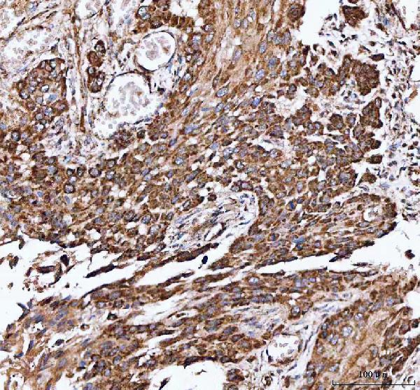
Real IHC data IHC analysis of ILDR1 using anti-ILDR1 antibody (A09544-3). ILDR1 was detected in a paraffin-embedded section of human esophageal squamous carcinoma tissue. Heat mediated antigen retrieval was performed in EDTA buffer (pH 8.0, epitope retrieval solution). The tissue section was blocked with 10% goat serum. The tissue section was then incubated with 2 μg/ml rabbit anti-ILDR1 Antibody (A09544-3) overnight at 4°C. Peroxidase Conjugated Goat Anti-rabbit IgG was used as secondary antibody and incubated for 30 minutes at 37°C. The tissue section was developed using HRP Conjugated Rabbit IgG Super Vision Assay Kit (Catalog # SV0002) with DAB as the chromogen.
Anti-IDH3A Antibody ®
Cat # A09544-3

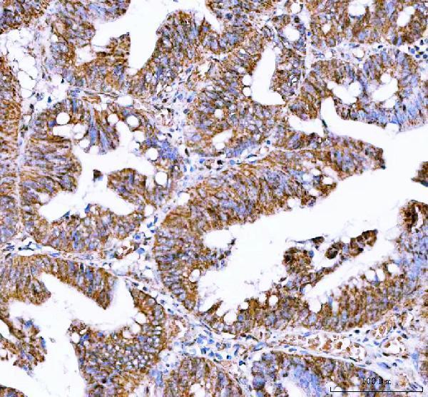
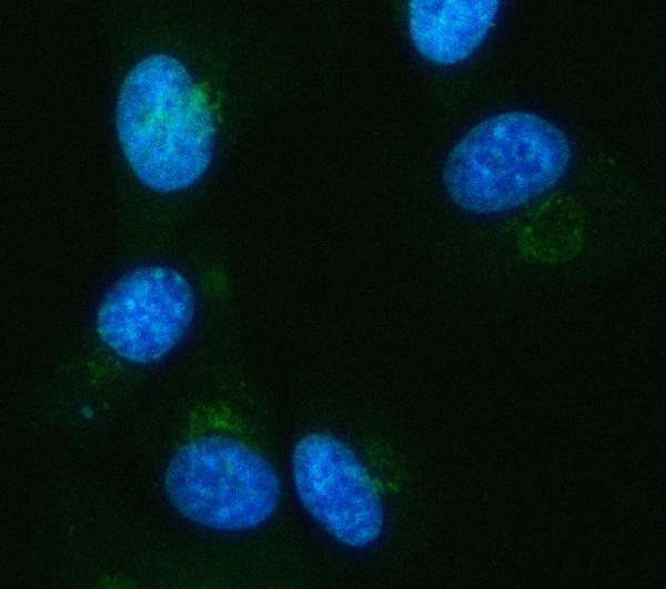
A09544-3 is the sole listed SKU; its IHC captions show paraffin sections of human esophageal squamous carcinoma, human rectum adenocarcinoma and rat heart, but identify the stained target as ILDR1 (IHC image captions). Its IF captions show PC-3 cells and paraffin sections of human liver and intestinal cancers, again identifying ILDR1 (IF image captions).

Which to pick: A09544-3 is the listed option for paraffin-section IHC and IF/ICC (catalog applications); its captions cannot establish IDH3A staining because they identify ILDR1 (image captions). The catalog lists human, mouse and rat reactivity, but the supplied images do not establish IDH3A staining across those species (catalog reactivity; image captions). Clonality and the fixative are unreported (catalog clone field; IHC image captions).

Each figure is that product's own IHC / IF validation image from its datasheet.

References

  1. UniProt Consortium. UniProt entry P50213 (IDH3A_HUMAN, Isocitrate dehydrogenase [NAD] subunit alpha, mitochondrial).
  2. Human Protein Atlas. IDH3A tissue IHC expression (reliability: Supported).
  3. Human Protein Atlas. IDH3A subcellular location (ICC-IF): Localized to the mitochondria..
  4. Human Protein Atlas. IDH3A antibody validation summary (2 antibodies).
  5. Effect of IDH3a on glucose uptake in lung adenocarcinoma: A pilot study based on [(18) F]FDG. Cancer medicine 2019 — PMC6718547.
  6. ERRα suppression enhances the cytotoxicity of the MEK inhibitor trametinib against colon cancer cells. Journal of experimental & clinical cancer research : CR 2018 — PMC6125878.
  7. Nuclear IDH3A Drives Transcriptional Programs in Melanoma via the YBX1-JUN/FOS Axis. Advanced science (Weinheim, Baden-Wurttemberg, Germany) 2026 — PMC13564267.
  8. Screening mitochondria-related biomarkers in skin and plasma of atopic dermatitis patients by bioinformatics analysis and machine learning. Frontiers in immunology 2024 — PMC11106410.
  9. PubMed PMID:7755589 — UniProt-cited evidence.
  10. PubMed PMID:17974005 — UniProt-cited evidence.
  11. PubMed PMID:15489334 — UniProt-cited evidence.