IDI1 / Isopentenyl-diphosphate Delta-isomerase 1 · IHC design guide

Design Immunohistochemistry for IDI1

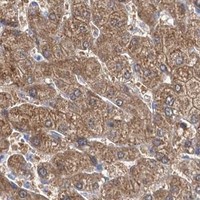
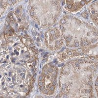
Plan IDI1 paraffin-section IHC with the catalog antibody at 2–5 μg/ml (datasheet A07892-1) and expect cytoplasmic staining (HPA tissue IHC). Adrenal glandular cells stain strongly, while bone-marrow hematopoietic cells have no detected staining (HPA tissue IHC); interpret intensity cautiously because staining and RNA show low consistency (HPA tissue IHC).

Evidence assembled Oct 2026 · For research use; verify linked source records and product datasheet before use
Immunohistochemistry protocol sheet for IDI1 (IHC for IDI1): expected localisation Cytoplasmic tissue staining (HPA tissue IHC), antibody A07892-1, validated IHC image, and IHC protocol steps
Printable IDI1 IHC protocol sheet — expected localisation Cytoplasmic tissue staining (HPA tissue IHC), antibody A07892-1, controls and protocol steps. Open the full IDI1 IHC guide →

IDI1 Immunohistochemistry Experimental Design Guide

Expected localisation, validated protocols, controls and antibodies — the at-a-glance facts below, then the full design guide.

Must know before staining
Expected localisation Cytoplasmic tissue staining (HPA tissue IHC)
Staining pattern Ubiquitous cytoplasmic staining; intensity varies by cell type (HPA tissue IHC)
Antigen retrieval EDTA pH 8.0 HIER, heat-mediated (datasheet A07892-1)
Positive control ⓘ Adrenal gland+4 more · see all
Negative control ⓘ Bone marrow
Important caveats
Reasons your staining may differ from the expected pattern.
Fixation Keep paraffin-section fixation consistent (standard IHC practice; not target-specific); Matched tissue-IHC evidence does not establish the fixation claim. Validate the specimen-specific method before use. (selected-SKU IHC image A07892-1)
Caveat Staining and RNA show low consistency (HPA tissue IHC)
Regulation No specific regulation annotated (UniProt)
Isoform / epitope Two isoforms; epitope coverage undetermined (UniProt)
Section 1

Recommended IDI1 IHC & IF Protocols

The catalog antibody’s IHC-P protocol is accompanied by one published IDI1 tissue-section protocol (PMC10915403).

Recommended immunohistochemistry (IHC-P) protocol parameters
SampleParaffin-embedded human esophageal squamous carcinoma tissue; fixative not specified (datasheet A07892-1)
FixationImage fixative and duration unreported (datasheet A07892-1); verify before use.
Sectioning4–5 µm sections on charged slides (standard)
DeparaffinisationXylene, graded ethanol series to water (standard)
Antigen retrievalHeat retrieval: EDTA pH 8.0 (datasheet A07892-1); 20 min, 95–100 °C (standard)
Peroxidase block3% H2O2, 10 min, room temperature (standard)
Blocking10% goat serum (datasheet A07892-1)
Primary antibodyRabbit anti-IDI1, 2-5 μg/ml (datasheet A07892-1)
Primary incubationOvernight at 4 °C (datasheet A07892-1)
DetectionHRP-conjugated secondary, DAB chromogen (datasheet A07892-1)
CounterstainHematoxylin, blue, dehydrate and mount (standard)
Expected resultIDI1-positive staining in glandular cells of adrenal gland (HPA tissue IHC: High). HPA tissue profile: Ubiquitous cytoplasmic expression. No signal in the no-primary control.
💡Decision noteStart with heat-mediated EDTA pH 8.0 retrieval (datasheet A07892-1); the published protocol used heated EDTA for 30 min (PMC10915403).
Section 2

What Is the Expected IDI1 Staining Pattern?

IDI1 is a peroxisomal protein with no transmembrane segment (UniProt Q13907). In paraffin section IHC, expect cytoplasmic staining across many cell types (HPA: ubiquitous cytoplasmic expression), especially adrenal glandular, bronchial respiratory epithelial, gallbladder glandular and prostate glandular cells (HPA: High). Interpret tissue differences cautiously: HPA rates the IHC evidence Approved but reports low consistency between antibody staining and RNA expression (HPA: reliability summary).

What am I looking at on my slide?
Cytoplasmic stain in adrenal glandular or bronchial respiratory epithelial cells.This matches two HPA High cell populations (HPA: adrenal gland; bronchus). A granular appearance can be compatible with peroxisomal localisation (UniProt Q13907), but routine chromogenic IHC alone cannot establish that individual stained structures are peroxisomes.
Predominantly nuclear, cell-surface or extracellular deposit, with little cytoplasmic stain.That distribution conflicts with the reported cytoplasmic tissue pattern (HPA: tissue IHC) and peroxisomal localisation without a transmembrane segment (UniProt Q13907). Check morphology and controls before interpreting it as IDI1; precipitated chromogen or nonspecific binding may mimic localisation.
Strong staining confined to bone marrow hematopoietic cells.HPA reports IDI1 as Not detected in that population (HPA: bone marrow). Consider cross-reactivity or endogenous detection activity and compare a no-primary control; a single discordant field cannot establish target expression or prove which artefact caused the signal.
Haze spans tissue, stroma and empty spaces without clear cell boundaries.This is difficult to reconcile with cell-associated cytoplasmic staining (HPA: tissue IHC). Treat it as background until the no-primary control, wash conditions and chromogen development are assessed; avoid scoring diffuse deposit as positive cells.
No stain in adrenal glandular cells while section morphology and counterstain are intact.Adrenal glandular cells are a High HPA population (HPA: adrenal gland). Recheck the assay with a known-positive section and its controls before calling the specimen negative; HPA's Approved rating and RNA–staining inconsistency do not guarantee positivity in every section (HPA: reliability summary).
💡Expected IDI1 appearanceCall a section positive when identifiable cells show cytoplasmic chromogen, with strong staining plausible in HPA High glandular or respiratory epithelial populations (HPA: tissue IHC); isolated nuclear, surface or acellular deposit is a suspect positive because IDI1 is peroxisomal (UniProt Q13907).
How each factor affects the staining
Compartment and topology (UniProt Q13907)IDI1 is peroxisomal and has no transmembrane segment (UniProt Q13907). Assess intracellular cytoplasmic staining; the HPA tissue summary describes the pattern as ubiquitous cytoplasmic (HPA: tissue IHC). Chromogenic resolution limits any claim of organelle-level colocalisation.
Tissue and cell population (HPA: tissue IHC)HPA records High staining in adrenal glandular, bronchial respiratory epithelial, gallbladder glandular and prostate glandular cells; several other listed populations are Medium or Low, and bone marrow hematopoietic cells are Not detected (HPA: tissue IHC). Compare like cell types when judging intensity.
Strength of tissue evidence (HPA: reliability summary)The IHC profile is Approved, with low consistency between antibody staining and RNA expression data (HPA: reliability summary). Use the observed staining pattern as a guide, while treating unexpected intensity or absence as a result to check with controls rather than a definitive expression change.
Antibody and isoform coverage (HPA: HPA039169; UniProt Q13907)HPA039169 is a rabbit polyclonal antibody with Approved IHC status (HPA: antibody record); UniProt lists two IDI1 isoforms (UniProt Q13907). No epitope or isoform coverage is supplied, so the observed IHC pattern cannot be assigned to one isoform.
IF evidence boundary (HPA: subcellular and antibody records)HPA supplies no ICC-IF images or main ICC-IF location for IDI1, and lists no ICC status for HPA039169 (HPA: subcellular; antibody record). IF localisation or an IF protocol cannot be inferred from those entries; evaluate IF separately.
Why is my staining missing, weak or wrong?
SituationLikely causeNext action
Expected cytoplasmic signal is absent in an adrenal glandular positive control.The run may have inadequate detection, or the selected section may not reproduce the HPA High observation (HPA: adrenal gland). The supplied sources do not identify a target-specific fixation or retrieval sensitivity.Check section morphology, reagent sequence and detection controls; confirm that the positive control contains glandular cells. Review retrieval and antibody conditions as general IHC variables, without assuming a validated IDI1-specific adjustment.
Strong stain appears mainly in nuclei or along cell borders.The pattern is discordant with cytoplasmic HPA staining (HPA: tissue IHC) and peroxisomal UniProt localisation (UniProt Q13907); chromogen deposit or nonspecific binding is possible.Inspect a no-primary control and an adjacent section, then review blocking, washes and chromogen development. Score the result only after cell-associated cytoplasmic staining can be distinguished from deposit.
Bone marrow hematopoietic cells stain strongly.Those cells are Not detected in HPA tissue IHC (HPA: bone marrow). Cross-reactivity or endogenous detection activity is possible; the staining pattern alone does not distinguish them.Compare no-primary and detection-only controls, check endogenous enzyme blocking if using enzyme detection, and assess whether the signal follows identifiable cells rather than diffuse tissue deposit.
Every area of the slide has a diffuse brown haze.Broad acellular deposit does not match the reported cytoplasmic cell pattern (HPA: tissue IHC). Excess detection background or chromogen development is a general IHC possibility.Inspect blank spaces and a no-primary section; review washes, blocking and development time. Reassess the tissue only after cellular boundaries are readable.
A low-staining population is judged negative beside a High population.HPA reports differing levels by cell type, including Low thyroid glandular cells and High adrenal glandular cells (HPA: tissue IHC). A single intensity threshold may hide weak cellular staining.Score the named cell population and its cytoplasmic localisation separately, using the same run's controls. Record weak staining distinctly from no detectable staining rather than extrapolating between tissues.
IF shows puncta, but their identity or specificity is uncertain.Peroxisomal localisation is reported by UniProt (UniProt Q13907), while HPA provides no ICC-IF images or main ICC-IF location for IDI1 (HPA: subcellular record). Puncta alone cannot verify peroxisomes.Treat IF as a separate application requiring its own validation and localisation controls; do not transfer the Approved IHC status of HPA039169 to ICC-IF (HPA: antibody record).

Sample controls for IDI1 IHC & IF

🧪Run adrenal gland first and score its glandular cells, where IDI1 staining is High (HPA: High in adrenal gland glandular cells). Run bone marrow as the negative tissue, where hematopoietic cells are Not detected (HPA: Not detected in bone marrow hematopoietic cells); use unstained cells outside the adrenal glandular compartment as an internal background reference, without assuming they are IDI1-negative (HPA: adrenal gland result specifies glandular cells).
Positive control tissue: Adrenal gland (Glandular cells, HPA High)
Negative control tissue: Bone marrow (HPA Not detected)
ICC-IF cell lines (HPA subcellular resource): HPA carries no ICC-IF cell line for IDI1; derive a cell-line control from the positive tissue's cell type (Glandular cells) and confirm it by RNA or western blot first.
Technical controls: Include no-primary (secondary-only) and host-matched rabbit IgG isotype controls, matching the primary antibody’s monoclonal or polyclonal format when known; use IDI1 knockout material as a biological negative if available (selected-SKU caption: rabbit anti-IDI1 antibody). Block endogenous peroxidase and check adrenal tissue pigment before interpreting DAB staining (selected-SKU caption: peroxidase secondary and DAB detection).
⚠️Feasibility: A target-specific fixation window or fixation effect is unreported, and the selected-SKU tissue-IHC caption does not report its fixative (selected-SKU caption: fixative not stated). Heat retrieval with EDTA at pH 8.0 was used for the reported paraffin-section IHC image, but its necessity for adrenal gland is unreported (selected-SKU caption: EDTA retrieval in a paraffin-embedded carcinoma section). Paraffin-section IHC has a reported example, while the supplied HPA subcellular record lists no ICC-IF image-bearing cell lines; the evidence therefore does not establish that frozen sections or IF are easier for IDI1, and adrenal pigment may complicate chromogenic scoring (selected-SKU caption: paraffin-section IHC; HPA subcellular: no ICC-IF image-bearing cell lines).

HPA tissue IHC evidence for IDI1

Comprehensive Human Protein Atlas IHC scoring per tissue (reliability: Approved — Low consistency between antibody staining and RNA expression data.). Rows are taken directly from the HPA tissue chart — click any row's HPA link to view the source.

Positive expression · recommended positive controls

TissueCell typeLevelEvidenceSource
Adrenal gland Glandular cells High Protein (IHC) HPA →
Bronchus Respiratory epithelial cells High Protein (IHC) HPA →
Gallbladder Glandular cells High Protein (IHC) HPA →
Prostate Glandular cells High Protein (IHC) HPA →
Appendix Glandular cells Medium Protein (IHC) HPA →

Undetected expression · recommended negative controls

TissueCell typeLevelEvidenceSource
Bone marrow Hematopoietic cells Not detected Protein (IHC) HPA →
Section 3

Advanced IDI1 IHC Tips

Troubleshooting guidance for IDI1 chromogenic IHC in paraffin sections, with one Q&A on adapting the assay for IF.

What retrieval should I try first if IDI1 staining is weak in paraffin sections?
Start with heat-mediated antigen retrieval in EDTA at pH 8.0 (datasheet A07892-1). The catalog antibody stained a paraffin section of human esophageal squamous carcinoma after that retrieval, using 2 μg/ml primary antibody overnight at 4°C (datasheet A07892-1). If signal remains weak, check deparaffinisation, retrieval temperature and heating consistency before changing the buffer (standard IHC practice). Compare any revised retrieval condition on adjacent sections with the same detection and exposure conditions (standard IHC practice). Record both signal and tissue morphology, because excessive heating can impair interpretation even when chromogen intensity increases (standard IHC practice).
Can I adjust fixation to rescue inconsistent IDI1 staining?
Target-specific fixation sensitivity for IDI1 is unknown from the supplied evidence; the catalog image identifies a paraffin section but does not state its fixative (datasheet A07892-1). Record fixative, fixation duration, tissue thickness and processing history for each specimen before attributing variable staining to the antibody (standard IHC practice). Compare sections processed under matched conditions, then assess whether weak regions track a processing gradient or tissue damage (standard IHC practice). Keep the validated EDTA retrieval at pH 8.0 constant during that comparison (datasheet A07892-1). Do not infer a fixation effect from IDI1’s peroxisomal annotation, acetylation site or HPA tissue staining (UniProt Q13907; HPA tissue IHC).
How should I interpret diffuse cytoplasmic IDI1 staining versus discrete puncta?
IDI1 is annotated as peroxisomal and has no transmembrane segment, so assess cytoplasmic staining in relation to that expected compartment (UniProt Q13907). HPA describes ubiquitous cytoplasmic tissue staining, but its IHC assessment reports low consistency between antibody staining and RNA expression (HPA tissue IHC). At brightfield resolution, fine peroxisomal signal may appear diffuse; morphology alone cannot establish organelle identity (standard IHC practice; UniProt Q13907). Compare staining within intact cells across adjacent sections and include a no-primary control to assess nonspecific chromogen deposition (standard IHC practice). Strong staining confined to nuclei or extracellular material needs independent validation before assignment to IDI1 (UniProt Q13907 topology and localisation).
Can this antibody distinguish IDI1 isoforms or reveal an acetylation-dependent epitope?
The record lists 2 IDI1 isoforms, but the supplied catalog image does not identify an antibody epitope or establish isoform selectivity (UniProt Q13907; datasheet A07892-1). It also lists acetyllysine at residue 176 and a Nudix hydrolase domain spanning residues 49–199 (UniProt Q13907). Those annotations do not establish whether this antibody detects one isoform preferentially or responds to acetylation (UniProt Q13907; datasheet A07892-1). For an isoform-specific conclusion, obtain the immunogen or epitope map and compare it with both isoform sequences before interpreting section staining (standard antibody-validation practice). Report the result as IDI1 immunoreactivity until that specificity is demonstrated (standard antibody-validation practice).
How can I adapt the tissue IHC result to multiplex IDI1 immunofluorescence?
Treat IF as a separate assay: the supplied catalog evidence shows chromogenic staining in a paraffin section, without an IF validation result (datasheet A07892-1). For a cell-type check, pair IDI1 with an epithelial marker such as cytokeratin when examining bronchial epithelium, where HPA reports high IDI1 staining in respiratory epithelial cells (HPA tissue IHC; standard IF practice). Choose a fluorophore channel away from strong tissue autofluorescence and inspect unstained controls in every channel (standard IF practice). Optimise permeabilisation for whether the antibody’s epitope is cytosol-facing or inside an organelle; the supplied peroxisomal and no-transmembrane annotations do not resolve epitope accessibility (UniProt Q13907).
What should I check when IDI1 DAB staining appears widespread or muddy?
First compare the stained section with a no-primary control and inspect where DAB accumulated relative to intact cells (standard IHC practice). The catalog example uses a peroxidase-conjugated secondary and DAB; those are detection components, not evidence that every brown deposit is IDI1 (datasheet A07892-1). Apply a peroxidase block and assess nonspecific secondary binding with appropriate controls, especially when staining tracks blood-rich or damaged areas (standard IHC practice). The example blocked with 10% goat serum and used 2 μg/ml primary antibody overnight at 4°C (datasheet A07892-1). If background persists, titrate primary antibody and detection development while keeping retrieval and section thickness matched (standard IHC practice).
How should I score IDI1 staining across specimens with different cell composition? ⚠ ANSWER MARKED FOR VERIFICATION
Define the cell population and region of interest before scoring, because IDI1 staining varies among annotated cell types (HPA tissue IHC; standard IHC practice). For chromogenic sections, report an H-score or percentage of positive cells with an explicit intensity threshold; use positive-cell density per mm² when cell counts vary across regions (standard IHC practice). Normalise within the same annotated cell compartment and comparable viable tissue area, rather than pooling epithelium, stroma and damaged regions (standard IHC practice). Keep retrieval, antibody concentration, DAB development and imaging settings consistent across comparison batches (standard IHC practice). Include control sections and report the scoring rule so weak cytoplasmic staining is handled consistently (standard IHC practice).
How can I distinguish genuine IDI1 signal from staining artefacts?
Favour staining in intact cytoplasm that follows plausible cellular boundaries, consistent with HPA’s cytoplasmic profile and UniProt’s peroxisomal annotation (HPA tissue IHC; UniProt Q13907). HPA reports high staining in bronchial respiratory epithelial cells and no detection in bone-marrow hematopoietic cells, useful reference patterns rather than absolute controls for every specimen (HPA tissue IHC). Treat nuclear-only deposits, tissue-edge gradients and necrotic areas cautiously, and compare them with a no-primary section (UniProt Q13907 localisation; standard IHC practice). Check whether brown deposits persist after peroxidase blocking, since endogenous enzyme activity can mimic DAB signal (standard IHC practice). HPA’s approved assessment still notes low staining-to-RNA consistency, so confirm consequential interpretations independently (HPA tissue IHC).
Boster reagents

Best IDI1 / Isopentenyl-diphosphate Delta-isomerase 1 IHC Antibodies

The catalog antibody has real IHC data from human paraffin sections (catalog IHC captions); its listed reactivity includes human, mouse and rat (catalog: reactivity). No IF image is supplied (catalog: IF images).

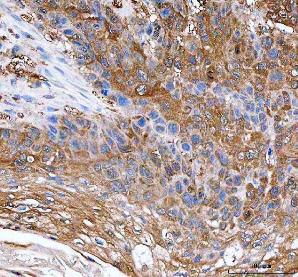
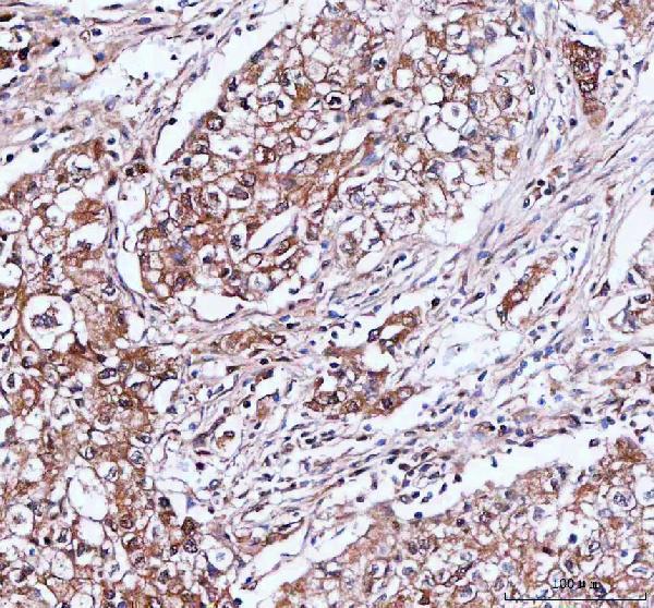
Real IHC data IHC analysis of IDI1 using anti-IDI1 antibody (A07892-1). IDI1 was detected in a paraffin-embedded section of human esophageal squamous carcinoma tissue. Heat mediated antigen retrieval was performed in EDTA buffer (pH 8.0, epitope retrieval solution). The tissue section was blocked with 10% goat serum. The tissue section was then incubated with 2 μg/ml rabbit anti-IDI1 Antibody (A07892-1) overnight at 4°C. Peroxidase Conjugated Goat Anti-rabbit IgG was used as secondary antibody and incubated for 30 minutes at 37°C. The tissue section was developed using HRP Conjugated Rabbit IgG Super Vision Assay Kit (Catalog # SV0002) with DAB as the chromogen.
Anti-IDI1 Antibody ®
Cat # A07892-1

A07892-1 is listed for IHC and has images from human esophageal squamous carcinoma, lung cancer and rectum adenocarcinoma paraffin sections (catalog: applications; IHC captions). It is rabbit-derived and lists human, mouse and rat reactivity; no IF image is supplied (catalog: host, reactivity and IF images).

Which to pick: Choose A07892-1 for paraffin-section tissue IHC: its own captions show human tissue stained after EDTA pH 8.0 retrieval at 2 μg/ml (catalog: A07892-1 IHC captions). The fixative is unreported (catalog: A07892-1 IHC captions). For IF/ICC, this payload provides no validation; for mouse or rat IHC, A07892-1 lists reactivity with those species, but its supplied IHC images show human tissue only (catalog: applications, reactivity, IF images and IHC captions).

Each figure is that product's own IHC / IF validation image from its datasheet.

References

  1. UniProt Consortium. UniProt entry Q13907 (IDI1_HUMAN, Isopentenyl-diphosphate Delta-isomerase 1).
  2. Human Protein Atlas. IDI1 tissue IHC expression (reliability: Approved).
  3. Human Protein Atlas. IDI1 subcellular location (ICC-IF): Highest expression in RPTEC/TERT1: 189.2 nTPM.
  4. Human Protein Atlas. IDI1 antibody validation summary (1 antibodies).
  5. Prevention of high-fat/high-sugar diet-induced type 2 diabetes mellitus-associated non-alcoholic fatty liver disease in rats with fermented and raw Rosa roxburghii Tratt (Cili) juice. Frontiers in nutrition 2025 — PMC12127171.
  6. IDI1 inhibits the cGAS-Sting signaling pathway in hepatocellular carcinoma. Heliyon 2024 — PMC10915403.
  7. The IDI1/SREBP2 axis drives intrahepatic cholestasis and is a treatment target of San-Huang-Cai-Zhu formula identified by sequencing and experiments. Frontiers in pharmacology 2023 — PMC9944032.
  8. TEAD1 is crucial for developmental myelination, Remak bundles, and functional regeneration of peripheral nerves. eLife 2024 — PMC10959528.
  9. PubMed PMID:8020941 — UniProt-cited evidence.
  10. PubMed PMID:14702039 — UniProt-cited evidence.
  11. PubMed PMID:15164054 — UniProt-cited evidence.