IFI44 / Interferon-induced protein 44 · IHC design guide

Design Immunohistochemistry for IFI44

Plan IFI44 staining in paraffin sections using the catalog antibody’s IHC dilution of 1:100–1:200 (datasheet). Compare staining with the reported nuclear and cytoplasmic tissue pattern, and interpret results cautiously because antibody staining has low concordance with RNA expression (HPA tissue IHC).

Evidence assembled Oct 2026 · For research use; verify linked source records and product datasheet before use
Immunohistochemistry protocol sheet for IFI44 (IHC for IFI44): expected localisation Nuclear and cytoplasmic staining in several tissues (HPA tissue IHC), antibody A10907, validated IHC image, and IHC protocol steps
Printable IFI44 IHC protocol sheet — expected localisation Nuclear and cytoplasmic staining in several tissues (HPA tissue IHC), antibody A10907, controls and protocol steps. Open the full IFI44 IHC guide →

IFI44 Immunohistochemistry Experimental Design Guide

Expected localisation, validated protocols, controls and antibodies — the at-a-glance facts below, then the full design guide.

Must know before staining
Expected localisation Nuclear and cytoplasmic staining in several tissues (HPA tissue IHC)
Staining pattern Several tissues show nuclear and cytoplasmic staining (HPA tissue IHC)
Antigen retrieval Citrate pH 6.0 HIER, 95–98 °C, 20 min (rule: cytoplasmic / membrane antigen)
Positive control ⓘ Gallbladder+4 more · see all
Negative control ⓘ Adipose tissue+4 more · see all
Important caveats
Reasons your staining may differ from the expected pattern.
Fixation Matched tissue-IHC evidence does not establish the fixation claim. Validate the specimen-specific method before use. (selected-SKU IHC image A10907)
Caveat Staining has low concordance with RNA; verify specificity (HPA tissue IHC)
Regulation Interferon induced (UniProt)
Isoform / epitope 2 isoforms; confirm epitope coverage for both (UniProt)
Section 1

Recommended IFI44 IHC & IF Protocols

The catalog antibody protocol is followed by published IFI44 IHC protocols for renal biopsies, breast cancer clinical samples, and rat cochlea and kidney (PMC11921169; PMC13071160; PMC5647021).

Recommended immunohistochemistry (IHC-P) protocol parameters
SampleParaffin-embedded human colon tissue; fixative not specified (datasheet A10907)
FixationImage fixative and duration unreported (datasheet A10907); verify before use.
Sectioning4–5 µm sections on charged slides (standard)
DeparaffinisationXylene, graded ethanol series to water (standard)
Antigen retrievalHeat-induced epitope retrieval in citrate buffer, pH 6.0, 20 min at 95–98 °C (standard rule: cytoplasmic / membrane antigen)
Peroxidase block3% H2O2, 10 min, room temperature (standard)
Blocking10% normal serum of the secondary host, 30 min, room temperature (standard)
Primary antibodyRabbit anti-IFI44, 1:100-1:200 (datasheet A10907)
Primary incubationOvernight at 4 °C (standard)
DetectionHRP-polymer secondary, DAB chromogen 5–10 min (standard)
CounterstainHematoxylin, blue, dehydrate and mount (standard)
Expected resultIFI44-positive staining in glandular cells of gallbladder (HPA tissue IHC: High). HPA tissue profile: Nuclear and ctyoplasmic expression in several tissues. No signal in the no-primary control.
💡Decision noteStart with citrate pH 6.0 HIER at 95–98 °C for 20 min (page antigen retrieval); use the article’s stated conditions when reproducing its protocol (PMC11921169).
Section 2

What Is the Expected IFI44 Staining Pattern?

In paraffin sections, expect IFI44 staining in the cytoplasm and sometimes nuclei of the cell populations reported by HPA, with strongest listed staining in gallbladder glandular cells (HPA tissue IHC). IFI44 is cytoplasmic and has no transmembrane segment (UniProt Q8TCB0). HPA rates tissue IHC “Approved,” while reporting low agreement with RNA data and pending external verification (HPA tissue IHC).

What am I looking at on my slide?
Gallbladder glandular cells stain strongly; appendix or rectum glandular cells stain less strongly.This follows HPA’s High gallbladder and Medium appendix and rectum IHC observations (HPA tissue IHC). Assess staining within the named cells rather than treating every cell in the section as an expected positive.
Signal appears in cytoplasm, nuclei, or both within an expected positive cell population.HPA describes nuclear and cytoplasmic tissue staining (HPA tissue IHC); UniProt assigns IFI44 to cytoplasm (UniProt Q8TCB0). Nucleoplasmic localisation is separately reported by ICC-IF (HPA subcellular), so nuclear tissue signal merits cautious interpretation.
Staining is confined to cell surfaces, vessel outlines, or extracellular material, without convincing intracellular signal.A surface-only pattern conflicts with the reported intracellular locations (UniProt Q8TCB0; HPA tissue IHC). Check morphology and controls for nonspecific deposition or detection artefact before calling those structures IFI44-positive (general IHC practice).
The strongest staining occurs in cells other than the named positive population, or in listed negative cells.Adipocytes in adipose tissue and cervix glandular cells were Not detected by HPA IHC (HPA tissue IHC). Unexpected staining can reflect cross-reactivity or endogenous detection activity; establish which with reagent and detection controls (general IHC practice).
Diffuse staining obscures cell borders, or gallbladder glandular cells have no detectable signal.Diffuse colour cannot establish the cell-specific pattern (general IHC practice). Absent gallbladder signal disagrees with HPA’s High observation (HPA tissue IHC), but HPA’s low RNA–staining agreement limits its use as an absolute benchmark (HPA tissue IHC).
💡Expected IFI44 appearanceCall a result positive when gallbladder glandular cells show clear intracellular, predominantly cytoplasmic or mixed nuclear–cytoplasmic staining near HPA’s High reference level; isolated surface deposits or diffuse colour without identifiable positive cells are suspect (HPA tissue IHC; UniProt Q8TCB0; general IHC practice).
How each factor affects the staining
Tissue and cell selectionHPA reports High staining in gallbladder glandular cells and Medium staining in bone marrow hematopoietic cells (HPA tissue IHC). Choose a control that tests the cell population being scored; neighboring cells need separate interpretation (general IHC practice).
Compartment evidenceUniProt reports cytoplasm, HPA tissue IHC reports nuclear and cytoplasmic expression, and HPA ICC-IF reports approved nucleoplasm localisation (UniProt Q8TCB0; HPA tissue IHC; HPA subcellular). These observations come from different assays; score tissue sections against tissue IHC morphology.
Antibody and assay validationHPA027148 is Approved for IHC, while HPA043858 is Approved for ICC-IF in the supplied antibody record (HPA antibodies). The ICC-IF status does not establish IHC performance for that antibody; HPA also flags low tissue RNA–staining consistency (HPA tissue IHC).
Retrieval and detection controlsAntigen retrieval and detection can alter the visibility of staining in routine paraffin IHC (general IHC practice). No supplied source establishes IFI44-specific retrieval conditions or fixation sensitivity; compare control sections under the same conditions before attributing a change to IFI44.
Why is my staining missing, weak or wrong?
SituationLikely causeNext action
No stain appears in gallbladder glandular cells.The run may have failed, or the chosen conditions may give too little detectable signal (general IHC practice); HPA reports these cells as High (HPA tissue IHC).Check tissue morphology and a working run control, then review retrieval, primary-antibody and detection steps (general IHC practice). Treat the HPA reference cautiously because its tissue IHC has low RNA–staining agreement (HPA tissue IHC).
Most of the section has diffuse chromogen.Background from primary antibody, secondary detection or endogenous activity can obscure cell-specific staining (general IHC practice).Compare a section without primary antibody and the run’s detection control; review blocking, washing and detection conditions (general IHC practice). Score IFI44 only where cellular signal can be distinguished from background.
Only membrane-like outlines or extracellular deposits stain.That distribution does not match cytoplasmic UniProt localisation or HPA’s intracellular tissue pattern (UniProt Q8TCB0; HPA tissue IHC).Inspect a matched negative control and confirm the signal lies within intact cells before reporting positivity (general IHC practice). Do not use the surface pattern alone as evidence of IFI44 localisation.
Nuclear staining is present, but cytoplasmic staining is weak.Nuclear staining occurs in HPA tissue IHC, although UniProt lists cytoplasm; HPA ICC-IF separately reports nucleoplasm (HPA tissue IHC; UniProt Q8TCB0; HPA subcellular).Describe nuclear and cytoplasmic components separately, compare the expected cell population and run controls, and avoid transferring the ICC-IF localisation claim directly to the paraffin section.
Listed negative cells stain as strongly as the chosen positive cells.The result conflicts with HPA’s Not detected calls for those cells and could reflect cross-reactivity or detection background (HPA tissue IHC; general IHC practice).Compare sections processed together with and without primary antibody, inspect cell identity, and repeat the comparison before assigning IFI44 positivity (general IHC practice).
A medium-level tissue shows little staining while gallbladder remains positive.HPA reports different levels across tissues: gallbladder is High, whereas appendix, rectum and bone marrow are Medium in their named cells (HPA tissue IHC).Score the correct cell population and compare sections processed in the same run (general IHC practice). Record the discrepancy without inferring an IFI44-specific fixation effect; none is supplied by UniProt or HPA.

Sample controls for IFI44 IHC & IF

🧪Run gallbladder first and score its glandular cells for IFI44 staining (HPA: High in gallbladder glandular cells). Use duodenum glandular cells as a negative comparator (HPA: Not detected in duodenum glandular cells); on the gallbladder slide, non-glandular cells should show only background staining if independently confirmed negative, since the HPA row establishes positivity only for glandular cells (HPA: gallbladder glandular cells).
Positive control tissue: Gallbladder (Glandular cells, HPA High)
Negative control tissue: Adipose tissue (HPA Not detected)
ICC-IF cell lines (HPA subcellular resource): HPA ICC-IF images show IFI44 in A-431, HeLa, U2OS, with annotated localisation: Nucleoplasm (approved) (HPA subcellular).
Technical controls: Include a no-primary, secondary-only control, an isotype control matched to the primary antibody’s host species and immunoglobulin class, and a biological negative such as IFI44 knockout tissue or cells (standard IHC practice). Check gallbladder sections for endogenous peroxidase and tissue pigment before interpreting chromogenic signal; use an endogenous-biotin control if detection relies on biotin (standard IHC practice).
⚠️Feasibility: A target-specific fixation window or fixation effect is unreported, and the selected A10907 colon IHC caption does not state its fixative (catalog caption: fixative not stated). That caption uses microwave retrieval in 10 mM PBS, pH 7.2, at 1:100, but it does not establish whether retrieval is required under other conditions (catalog caption: colon tissue IHC). The supplied evidence does not establish whether frozen sections or IF are easier; assess pigment and endogenous-peroxidase background when scoring gallbladder chromogenic staining (standard IHC practice).

HPA tissue IHC evidence for IFI44

Comprehensive Human Protein Atlas IHC scoring per tissue (reliability: Approved — Low consistency between antibody staining and RNA expression data. Pending external verification.). Rows are taken directly from the HPA tissue chart — click any row's HPA link to view the source.

Positive expression · recommended positive controls

TissueCell typeLevelEvidenceSource
Gallbladder Glandular cells High Protein (IHC) HPA →
Appendix Glandular cells Medium Protein (IHC) HPA →
Bone marrow Hematopoietic cells Medium Protein (IHC) HPA →
Cerebellum Cells in granular layer Medium Protein (IHC) HPA →
Cerebral cortex Neuronal cells Medium Protein (IHC) HPA →

Undetected expression · recommended negative controls

TissueCell typeLevelEvidenceSource
Adipose tissue Adipocytes Not detected Protein (IHC) HPA →
Breast Adipocytes Not detected Protein (IHC) HPA →
Cervix Glandular cells Not detected Protein (IHC) HPA →
Duodenum Glandular cells Not detected Protein (IHC) HPA →
Esophagus Squamous epithelial cells Not detected Protein (IHC) HPA →
Section 3

Advanced IFI44 IHC Tips

Troubleshoot IFI44 staining in paraffin section chromogenic IHC, with one question on IF/ICC.

Which retrieval conditions should I try when IFI44 staining is weak?
Start with citrate buffer at pH 6.0, heated to 95–98 °C for 20 minutes (page retrieval setting). If staining remains weak, compare a separate section using microwave retrieval in 10 mM PBS, pH 7.2, the condition shown for the catalog antibody at 1:100 in paraffin embedded human colon (A10907 IHC caption). Keep section thickness, detection chemistry, and exposure to chromogen consistent across conditions so the comparison isolates retrieval (standard IHC practice). Include a no primary control, and judge signal in intact cells while checking whether stronger retrieval also increases diffuse background or damages tissue (standard IHC practice).
How can I troubleshoot fixation related loss of IFI44 signal?
IFI44 specific fixation sensitivity is unknown from the supplied evidence, so do not infer an optimal fixative or fixation duration from the tissue staining pattern (supplied record; HPA tissue IHC). The catalog image documents paraffin embedded human colon, but its caption does not state the fixative (A10907 IHC caption). Record the fixative, fixation time, processing history, and section age for each specimen, then compare matched sections processed under documented conditions (standard IHC practice). Apply the same retrieval and antibody conditions across those sections, and use a no primary control to distinguish altered antigen detection from background caused by processing (standard IHC practice).
Should IFI44 staining appear in the cytoplasm or nucleus?
Inspect cytoplasmic staining first because IFI44 is annotated as cytoplasmic and has no transmembrane segment (UniProt Q8TCB0 subcellular annotation; UniProt Q8TCB0 topology). Nuclear staining also deserves assessment: tissue IHC reports nuclear and cytoplasmic expression, while an approved IF/ICC subcellular annotation places IFI44 in the nucleoplasm (HPA tissue IHC; HPA subcellular). Score compartments separately in intact, identifiable cells rather than treating all brown signal as equivalent (standard IHC practice). If a sample shows only nuclear or only cytoplasmic signal, verify the pattern with controls and an independent antibody where available, given the reported low consistency between tissue staining and RNA expression (HPA tissue IHC reliability).
Could antibody epitope choice explain different IFI44 staining patterns?
IFI44 has 2 annotated isoforms and a TLDc domain spanning residues 1–152, but the supplied evidence does not map this antibody’s epitope to either isoform (UniProt Q8TCB0 isoforms; UniProt Q8TCB0 domains; supplied A10907 caption). Check the antibody’s documented immunogen or epitope before interpreting divergent staining as isoform selective, and compare results with an independently mapped antibody if one is available (standard IHC validation practice). IFI44 has no annotated signal peptide, propeptide, glycosylation sites, or modified residues in this record (UniProt Q8TCB0 processing; UniProt Q8TCB0 PTMs). Therefore, document retrieval and compartment specific staining before proposing an epitope masking mechanism for any specimen (standard IHC practice).
How should I assess IFI44 by multiplex IF/ICC?
For the separate IF/ICC workflow, pair IFI44 with a marker identifying the cell population being examined; HPA tissue IHC reports glandular cells in gallbladder and hematopoietic cells in bone marrow among stained populations (HPA tissue IHC). Choose fluorophores in channels with low measured tissue autofluorescence, and collect single stain and no primary controls before assessing overlap (standard IF practice). Plan permeabilisation for an intracellular epitope: UniProt places IFI44 in the cytoplasm without a transmembrane segment, while HPA IF/ICC reports nucleoplasmic localisation (UniProt Q8TCB0 subcellular annotation; UniProt Q8TCB0 topology; HPA subcellular). Optimise permeabilisation and fixation on matched specimens rather than importing the paraffin section retrieval conditions into IF/ICC (standard IF practice; A10907 IHC caption).
What should I check when IFI44 IHC shows widespread brown background?
Run a no primary control through the same chromogenic detection sequence to identify secondary reagent, endogenous peroxidase, or substrate related signal (standard IHC practice). Block endogenous peroxidase before DAB development and optimise protein blocking and washes if background persists; these are general chromogenic IHC steps, not IFI44 specific evidence (standard IHC practice). Titrate the catalog antibody around the documented 1:100 use in paraffin embedded human colon, recording whether cell associated signal improves relative to diffuse staining (A10907 IHC caption; standard IHC practice). Compare background across intact tissue, damaged edges, and low signal reference areas before calling broadly distributed brown pigment IFI44 positive (standard IHC practice).
How should I quantify IFI44 staining across paraffin sections? ⚠ ANSWER MARKED FOR VERIFICATION
Define the scored cell population and cytoplasmic and nuclear compartments before analysis, because both compartments appear in the supplied localisation evidence (HPA tissue IHC; HPA subcellular). For chromogenic IHC, report the percentage of positive cells and an H-score based on intensity categories, or report positive cell density per mm² when cell counts are the endpoint (standard IHC practice). Normalise counts to the number of eligible intact cells or measured viable tissue area, and keep thresholds, imaging settings, and section selection rules fixed across samples (standard IHC practice). Report compartment scores separately and exclude folds, necrosis, and section edges from the analysis area (standard IHC practice).
How can I distinguish convincing IFI44 staining from artefact?
A convincing result has reproducible cell associated staining in an interpretable compartment, with a clean no primary control and preservation of tissue structure (standard IHC practice). Evaluate cytoplasmic signal against UniProt’s annotation and nuclear signal against the HPA tissue and IF/ICC observations, while recognising that these sources differ in emphasis (UniProt Q8TCB0 subcellular annotation; HPA tissue IHC; HPA subcellular). Check whether stained cells match the population being scored; HPA reports high staining in gallbladder glandular cells but no detection in duodenal glandular cells (HPA tissue IHC). Treat staining confined to edges, necrotic areas, or the no primary control as suspect, and interpret tissue comparisons cautiously because HPA reports low agreement between staining and RNA expression (standard IHC practice; HPA tissue IHC reliability).
Boster reagents

Best IFI44 / Interferon-induced protein 44 IHC Antibodies

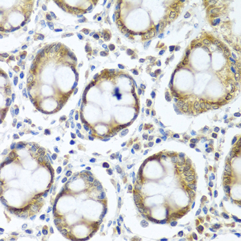
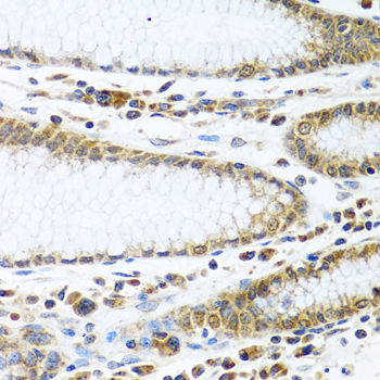
A10907 has IHC images from paraffin-embedded human colon and stomach (catalog image captions); IF/ICC is listed for human, mouse and rat (catalog applications and reactivity).

Real IHC data Immunohistochemistry of paraffin-embedded human colon using IFI44 antibody at dilution of 1:100 .Perform microwave antigen retrieval with 10 mM PBS buffer pH 7.2 before commencing with IHC staining protocol.
Anti-Interferon-induced protein 44 IFI44 Antibody
Cat # A10907

A10907 will render with its own human colon IHC figure at 1:100 (catalog figure caption). A second caption documents human stomach IHC at 1:100; IF/ICC is listed, but no IF image is supplied (catalog image captions and applications).

Which to pick: Choose A10907 for paraffin-section tissue IHC: its colon and stomach captions report microwave antigen retrieval in 10 mM PBS, pH 7.2 (catalog IHC image captions). A10907 is also the listed choice for IF/ICC and for human, mouse or rat samples (catalog applications and reactivity); the supplied images document human IHC only (catalog image captions). Its host is rabbit, while clone status and the tissue fixative are unreported (catalog host, clone field and IHC image captions).

Each figure is that product's own IHC / IF validation image from its datasheet.

References

  1. UniProt Consortium. UniProt entry Q8TCB0 (IFI44_HUMAN, Interferon-induced protein 44).
  2. Human Protein Atlas. IFI44 tissue IHC expression (reliability: Approved).
  3. Human Protein Atlas. IFI44 subcellular location (ICC-IF): Localized to the nucleoplasm..
  4. Human Protein Atlas. IFI44 antibody validation summary (2 antibodies).
  5. Determining IFI44 as a key lupus nephritis's biomarker through bioinformatics and immunohistochemistry. Renal failure 2025 — PMC11921169.
  6. IFI44 Orchestrates an IL-10-Driven M2 Macrophage Program in Breast Cancer: An Immune Prognostic Signature. Breast cancer (Dove Medical Press) 2026 — PMC13071160.
  7. Altered gene expression profile in a rat model of gentamicin-induced ototoxicity and nephrotoxicity, and the potential role of upregulated Ifi44 expression. Molecular medicine reports 2017 — PMC5647021.
  8. Cell-type-specific expression analysis of liver transcriptomics with clinical parameters to decipher the cause of intrahepatic inflammation in chronic hepatitis B. iMeta 2024 — PMC11316924.
  9. PubMed PMID:7925411 — UniProt-cited evidence.
  10. PubMed PMID:14702039 — UniProt-cited evidence.
  11. PubMed PMID:16710414 — UniProt-cited evidence.