This website uses cookies to ensure you get the best experience on our website.


- Table of Contents
Plan paraffin-section IFI44L IHC with the IHC-validated antibody at 2–5 μg/ml (datasheet A10975-1). Assess cytoplasmic staining alongside HPA-described high and undetected cell populations, bearing in mind that tissue-IHC reliability is uncertain (HPA tissue IHC).

Expected localisation, validated protocols, controls and antibodies — the at-a-glance facts below, then the full design guide.
| Expected localisation | Cytoplasmic staining in most tissues (HPA tissue IHC) | |
| Staining pattern | Cytoplasmic staining in glandular and myoepithelial cells (HPA tissue IHC) | |
| Antigen retrieval | EDTA pH 8.0 HIER, heat-mediated (datasheet A10975-1) | |
| Positive control | Breast+4 more · see all | |
| Negative control | Endometrium+4 more · see all |
| Fixation | Matched tissue-IHC evidence does not establish the fixation claim. Validate the specimen-specific method before use. (selected-SKU IHC image A10975-1) | |
| Caveat | Low consistency between IHC staining and RNA expression (HPA tissue IHC) | |
| Regulation | Type I interferon inducible (UniProt) | |
| Isoform / epitope | 2 isoforms; antibody epitope coverage is unknown (UniProt) |
The catalog antibody’s IHC-P protocol (datasheet: A10975-1) and a published rat-heart IHC protocol (PMC8657967) provide starting conditions for IFI44L staining.
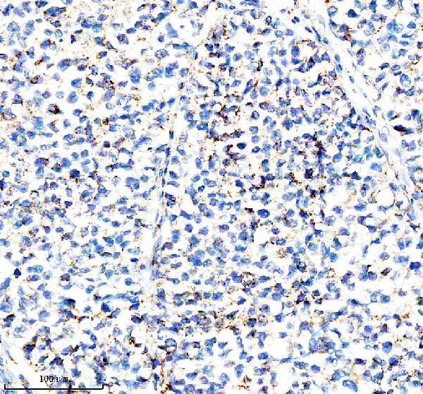
| Sample | Paraffin-embedded human non-small cell lung cancer tissue; fixative not specified (datasheet A10975-1) |
| Fixation | Image fixative and duration unreported (datasheet A10975-1); verify before use. |
| Sectioning | 4–5 µm sections on charged slides (standard) |
| Deparaffinisation | Xylene, graded ethanol series to water (standard) |
| Antigen retrieval | Heat retrieval: EDTA pH 8.0 (datasheet A10975-1); 20 min, 95–100 °C (standard) |
| Peroxidase block | 3% H2O2, 10 min, room temperature (standard) |
| Blocking | 10% goat serum (datasheet A10975-1) |
| Primary antibody | Rabbit anti-IFI44L, 2-5 μg/ml (datasheet A10975-1) |
| Primary incubation | Overnight at 4 °C (datasheet A10975-1) |
| Detection | HRP-conjugated secondary, DAB chromogen (datasheet A10975-1) |
| Counterstain | Hematoxylin, blue, dehydrate and mount (standard) |
| Expected result | IFI44L-positive staining in myoepithelial cells of breast (HPA tissue IHC: High). HPA tissue profile: Cytoplasmic expression in most tissues. No signal in the no-primary control. |
For paraffin-section IHC, expect mainly cytoplasmic IFI44L staining in the cell types where HPA reports protein expression, including breast myoepithelial cells and duodenal glandular cells (HPA: tissue IHC). UniProt lists IFI44L as cytoplasmic with no transmembrane segment (UniProt Q53G44). Interpret this pattern cautiously: HPA rates its tissue IHC profile Uncertain because antibody staining and RNA expression show low consistency (HPA: tissue IHC reliability).
| Cell-associated cytoplasmic chromogen is strong in breast myoepithelial cells or duodenal glandular cells. | This matches two HPA High observations and its broader cytoplasmic tissue profile (HPA: breast and duodenum IHC). Compare the labelled cells with tissue morphology; the HPA profile remains Uncertain and cannot establish that every positive cell represents specific IFI44L detection (HPA: tissue IHC reliability). |
| Staining is predominantly nuclear, membranous, or extracellular in a paraffin section. | Membranous or extracellular staining conflicts with UniProt's cytoplasmic location and lack of a transmembrane segment (UniProt Q53G44). Nuclear IHC warrants antibody and control review, but is not automatically artefact: HPA reports approved nucleoplasmic ICC-IF staining, while its tissue IHC profile is cytoplasmic and Uncertain (HPA: subcellular; HPA: tissue IHC). |
| Strong chromogen appears in cells that HPA lists as Not detected, such as lung alveolar cells. | Check whether the signal follows cell boundaries and survives detection controls; cross-reactivity or endogenous detection activity can mimic cellular staining (general IHC practice). HPA's Not detected finding applies to the specified cells in its sampled tissue, not to every cell in the organ or every biological state (HPA: lung IHC). |
| Colour spreads across stroma, lumens, section edges, or many unrelated cell types. | A diffuse field is less persuasive than cell-associated cytoplasmic staining in HPA High populations (HPA: tissue IHC). Excess background can arise from nonspecific antibody binding, endogenous enzyme activity, or detection chemistry; interpret it using appropriate reagent controls and the tissue's morphology (general IHC practice). |
| No signal appears in a reference section containing an HPA High cell population. | First confirm that the expected cells are present: HPA reports High staining in prostate glandular cells and smooth muscle cells, among others (HPA: tissue IHC). An absent result may reflect assay performance or specimen biology; HPA's Uncertain reliability prevents treating one reference section as a definitive IFI44L-positive control (HPA: tissue IHC reliability). |
| Tissue-profile confidence | HPA calls its cytoplasmic tissue IHC profile Uncertain because staining and RNA expression have low consistency; treat cell-specific levels as reference observations that need independent confirmation (HPA: tissue IHC reliability). |
| Cell identity within a tissue | HPA reports High duodenal glandular-cell staining but Not detected lung alveolar-cell staining; assess the named cell population rather than assigning one expected intensity to an entire organ (HPA: duodenum and lung IHC). |
| Interferon-response biology | IFI44L is a type I interferon-stimulated gene with roles in infection responses (UniProt Q53G44). Biological context may inform sample selection, but these annotations do not predict staining intensity in a particular paraffin section. |
| Isoforms and antibody recognition | UniProt lists 2 isoforms (UniProt Q53G44). Their existence alone does not identify which form an antibody detects; use documented epitope information if available, without assuming isoform-specific staining. |
| IHC versus IF/ICC compartment | Does IF/ICC have the same expected compartment? HPA reports approved nucleoplasmic ICC-IF images in HeLa, PC-3, and U2OS, whereas its tissue IHC profile is cytoplasmic and Uncertain (HPA: subcellular; HPA: tissue IHC). Assess each application separately. |
| Situation | Likely cause | Next action |
|---|---|---|
| A proposed positive section is blank. | The section may lack the specific HPA High cell population, or the IHC workflow may have failed (HPA: tissue IHC; general IHC practice). | Verify cell identity and section quality; run an independently characterized positive control alongside the specimen, then review retrieval, primary-antibody conditions, and detection using the assay's validated settings (general IHC practice). |
| Chromogen is strong in an HPA Not detected population. | Nonspecific binding or endogenous detection activity is possible; HPA lists endometrial stromal cells as Not detected in its tissue IHC samples (HPA: endometrium IHC; general IHC practice). | Compare with an appropriate primary-antibody omission or detection control, check morphology, and reassess specificity with independent evidence before calling these cells IFI44L-positive (general IHC practice). |
| Most of the section shows diffuse background. | Nonspecific reagent binding, inadequate blocking, or endogenous enzyme activity can obscure cell-associated staining (general IHC practice). | Review blocking, washes, primary-antibody concentration, and detection controls within the validated IHC workflow; score only interpretable cell-associated signal (general IHC practice). |
| Nuclear staining dominates tissue IHC. | The result differs from HPA's cytoplasmic tissue profile, although approved nucleoplasmic ICC-IF staining makes a blanket artefact call unjustified (HPA: tissue IHC; HPA: subcellular). | Document the compartment and antibody used, inspect negative controls, and seek independent antibody or orthogonal confirmation before assigning nuclear IHC signal to IFI44L (general IHC practice). |
| Different reference tissues give inconsistent intensities. | HPA reports distinct cell-specific levels and rates its tissue IHC profile Uncertain (HPA: tissue IHC reliability). | Compare matching cell populations and processing conditions, record staining intensity by cell type, and avoid using HPA levels as universal acceptance thresholds (HPA: tissue IHC; general IHC practice). |
| IF/ICC and IHC suggest different compartments. | HPA reports approved nucleoplasmic ICC-IF localization but an Uncertain cytoplasmic tissue IHC profile (HPA: subcellular; HPA: tissue IHC). | Report each application and its validation status separately; use the dedicated IF/ICC guide for that application and seek independent confirmation before reconciling the locations (HPA: subcellular; HPA: tissue IHC). |


Comprehensive Human Protein Atlas IHC scoring per tissue (reliability: Uncertain — Low consistency between antibody staining and RNA expression data. Pending external verification.). Rows are taken directly from the HPA tissue chart — click any row's HPA link to view the source.
| Tissue | Cell type | Level | Evidence | Source |
|---|---|---|---|---|
| Breast | Myoepithelial cells | High | Protein (IHC) | HPA → |
| Duodenum | Glandular cells | High | Protein (IHC) | HPA → |
| Prostate | Glandular cells | High | Protein (IHC) | HPA → |
| Rectum | Glandular cells | High | Protein (IHC) | HPA → |
| Smooth muscle | Smooth muscle cells | High | Protein (IHC) | HPA → |
| Tissue | Cell type | Level | Evidence | Source |
|---|---|---|---|---|
| Endometrium | Cells in endometrial stroma | Not detected | Protein (IHC) | HPA → |
| Esophagus | Squamous epithelial cells | Not detected | Protein (IHC) | HPA → |
| Hippocampus | Glial cells | Not detected | Protein (IHC) | HPA → |
| Liver | Cholangiocytes | Not detected | Protein (IHC) | HPA → |
| Lung | Alveolar cells | Not detected | Protein (IHC) | HPA → |
Troubleshoot IFI44L staining in paraffin sections by checking retrieval, compartment, cell identity, controls, and the limits of the available tissue evidence.
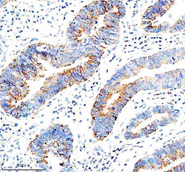
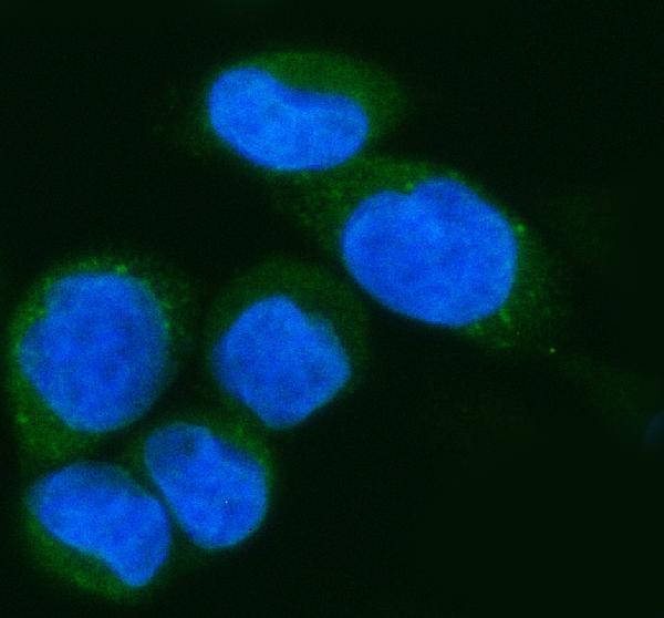
A10975-1 has IHC images from human paraffin-embedded lung and stomach cancer sections and an IF image from SiHa cells (catalog image captions). Reactivity is listed for human, monkey, mouse and rat (catalog).
A10975-1 will render with its own IHC figure from a human paraffin-embedded non-small cell lung cancer section (IHC image caption). Its additional IHC caption shows human stomach cancer tissue, and its IF caption shows SiHa cells (catalog image captions).
Which to pick: Choose A10975-1 for paraffin-section IHC: its figure used heat retrieval in EDTA at pH 8.0 and 2 μg/ml primary antibody; the fixative is unreported (IHC image caption). Choose the same SKU for IF/ICC because its IF caption shows SiHa cells at 5 μg/ml (IF image caption). For other species, its catalog lists monkey, mouse and rat reactivity, while the supplied IHC and IF images show human material only (catalog reactivity; image captions).