This website uses cookies to ensure you get the best experience on our website.


- Table of Contents
Plan paraffin-section IFIT3 IHC around cytoplasmic staining in most tissues (HPA tissue IHC). Use kidney tubule cells as a high-staining reference and adipocytes as an undetected reference (HPA tissue IHC); the IHC-validated antibody A03920 is used at 5 μg/mL (datasheet).

Expected localisation, validated protocols, controls and antibodies — the at-a-glance facts below, then the full design guide.
| Expected localisation | Cytoplasmic staining in most tissues (HPA tissue IHC) | |
| Staining pattern | Most tissues show cytoplasmic staining (HPA tissue IHC) | |
| Antigen retrieval | Citrate pH 6.0 HIER, 95–98 °C, 20 min (rule: cytoplasmic / membrane antigen) | |
| Positive control | Epididymis+4 more · see all | |
| Negative control | Adipose tissue+4 more · see all |
| Fixation | Keep fixation consistent between sections. (standard IHC practice; not target-specific) | |
| Caveat | Antibody staining has low consistency with RNA data (HPA tissue IHC) | |
| Regulation | Interferon-induced expression (UniProt) | |
| Isoform / epitope | No annotated isoforms; one 1–490 chain (UniProt) |
Compare the catalog antibody’s IHC-P protocol with the published IFIT3 chromogenic IHC protocol for ESCC tissue microarrays (PMC12847741).
| Sample | Tissue sections; selected-image fixative not specified (standard IHC workflow) |
| Fixation | Image fixative and duration unreported (datasheet A03920); verify before use. |
| Sectioning | 4–5 µm sections on charged slides (standard) |
| Deparaffinisation | Xylene, graded ethanol series to water (standard) |
| Antigen retrieval | Heat-induced epitope retrieval in citrate buffer, pH 6.0, 20 min at 95–98 °C (standard rule: cytoplasmic / membrane antigen) |
| Peroxidase block | 3% H2O2, 10 min, room temperature (standard) |
| Blocking | 10% normal serum of the secondary host, 30 min, room temperature (standard) |
| Primary antibody | Rabbit anti-IFIT3, 5 μg/mL (datasheet A03920) |
| Primary incubation | Overnight at 4 °C (standard) |
| Detection | HRP-polymer secondary, DAB chromogen 5–10 min (standard) |
| Counterstain | Hematoxylin, blue, dehydrate and mount (standard) |
| Expected result | IFIT3-positive staining in glandular cells of epididymis (HPA tissue IHC: High). HPA tissue profile: Cytoplasmic expression in most tissues. No signal in the no-primary control. |
IFIT3 should appear predominantly cytoplasmic in paraffin-section IHC, including glandular cells in epididymis and seminal vesicle, kidney tubular cells, and skin melanocytes (HPA tissue IHC). Cytosolic and mitochondrial localisation is supported by ICC-IF (HPA subcellular; UniProt O14879). IFIT3 has no transmembrane segment (UniProt O14879 topology). HPA rates the tissue IHC pattern Approved, while reporting low consistency between antibody staining and RNA expression (HPA tissue IHC).
| Clear cytoplasmic staining in kidney tubular cells or epididymal glandular cells. | This matches cell types scored High by HPA and the reported cytoplasmic tissue pattern (HPA tissue IHC). Judge staining within the named cells, alongside morphology and counterstain. A section-wide positive impression can conceal unstained neighbouring cell types; it does not establish that every cell in the tissue expresses IFIT3. |
| Predominantly nuclear staining, with little or no cytoplasmic signal. | A nuclear-only result conflicts with IFIT3's reported cytoplasmic and mitochondrial locations (UniProt O14879; HPA subcellular). Treat it as suspect staining and review controls, counterstain alignment, and detection before assigning it to IFIT3. The supplied sources do not support a nuclear staining pattern as the expected IHC result. |
| Strong staining in adipocytes, skeletal-muscle myocytes, or pancreatic exocrine cells. | Those specific cell types are scored Not detected, so strong signal there is unexpected under the HPA tissue pattern (HPA tissue IHC). Consider antibody cross-reactivity or endogenous detection activity; check the omission and detection controls. Do not call the entire tissue negative when HPA identifies only a particular cell type. |
| Diffuse colour over cells, extracellular areas, and the whole section. | Broad, poorly localised deposition cannot establish the reported cytoplasmic pattern (HPA tissue IHC). Uneven blocking, washing, or detection can produce background (standard IHC practice). Compare the negative control and look for cell boundaries before scoring intensity; widespread colour alone is weak evidence for IFIT3. |
| No staining in kidney tubular cells or seminal-vesicle glandular cells. | An absent result conflicts with HPA's High staining in these named cells (HPA tissue IHC), but one negative section cannot establish biological absence. Check whether the expected cells are present and whether a positive control worked; then review the antibody, retrieval, and detection steps as general IHC workflow checks. |
| Cell type and tissue context | HPA scores epididymal and seminal-vesicle glandular cells, kidney tubular cells, and melanocytes High; several other named cell types are Not detected (HPA tissue IHC). Select and score the specified cells rather than treating a tissue label as a uniform positive or negative control. |
| Compartment and optical resolution | IFIT3 is reported in cytoplasm and mitochondria (UniProt O14879; HPA subcellular). Routine chromogenic IHC supports a cytoplasmic readout; it should not be used to claim resolved mitochondrial colocalisation without a suitable method (standard IHC practice). IFIT3 has no annotated transmembrane segment (UniProt O14879 topology). |
| Strength of tissue-pattern evidence | HPA labels its tissue IHC reliability Approved but notes low consistency between antibody staining and RNA expression (HPA tissue IHC). Its listed antibody HPA059914 is IHC Approved, without an Enhanced designation in the supplied record (HPA antibodies). Interpret an unexpected pattern with independent controls rather than treating approval as proof of specificity. |
| Biological expression context | IFIT3 is interferon induced, and protein expression is reported higher in PBMCs and monocytes from SLE patients than healthy individuals (UniProt O14879). This supports context-dependent expression, but it supplies no expected staining intensity for a particular paraffin section. Record specimen context when comparing cases. |
| Situation | Likely cause | Next action |
|---|---|---|
| Expected positive cells are blank. | The named cells may be absent from the section, or an IHC workflow step may have failed; HPA reports High staining in kidney tubules and epididymal glandular cells (HPA tissue IHC). | Confirm morphology and a working positive control. Review the validated IHC procedure's retrieval, antibody incubation, and detection steps without assuming a target-specific fixation effect (standard IHC practice). |
| Signal is mainly nuclear. | The compartment conflicts with the reported cytoplasmic and mitochondrial localisation (UniProt O14879; HPA subcellular); counterstain or nonspecific signal may complicate the readout (standard IHC practice). | Compare the primary-antibody omission control, check nuclear versus cytoplasmic boundaries, and reassess the staining before scoring those nuclei as IFIT3 (standard IHC practice). |
| HPA-negative cell types stain strongly. | Adipocytes and skeletal-muscle myocytes are Not detected in the supplied HPA tissue pattern (HPA tissue IHC). Cross-reactivity or endogenous detection activity is possible, rather than established. | Check a primary-antibody omission control and the detection system's relevant endogenous-activity control; compare cell morphology with an HPA-positive cell type (standard IHC practice; HPA tissue IHC). |
| The section has uniform haze or patchy precipitate. | Background can arise from blocking, washing, or detection conditions (standard IHC practice). It obscures the cell-restricted cytoplasmic pattern described by HPA (HPA tissue IHC). | Inspect the negative control and reagent quality, then optimise blocking and washes within the validated IHC workflow; score only interpretable cellular staining (standard IHC practice). |
| Bronchial staining looks concentrated near cilia. | HPA lists Medium staining in bronchial ciliated cells at ciliary rootlets (HPA tissue IHC). This restricted observation needs cell-level interpretation. | Check that staining follows ciliated-cell morphology and compare with the control; avoid treating every apical deposit as specific IFIT3 staining (HPA tissue IHC; standard IHC practice). |
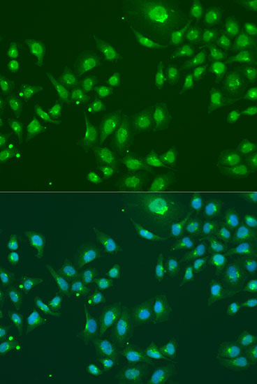
| Can IF/ICC help check the compartment? | HPA reports supported cytosolic and mitochondrial ICC-IF localisation and lists A-431, HeLa, and ASC52telo images (HPA subcellular). | Use the separate IF/ICC guide for that application; compare its compartment readout with the IHC cytoplasmic pattern. The supplied ICC-IF evidence does not define an IHC protocol option (HPA subcellular; HPA tissue IHC). |


Comprehensive Human Protein Atlas IHC scoring per tissue (reliability: Approved — Low consistency between antibody staining and RNA expression data.). Rows are taken directly from the HPA tissue chart — click any row's HPA link to view the source.
| Tissue | Cell type | Level | Evidence | Source |
|---|---|---|---|---|
| Epididymis | Glandular cells | High | Protein (IHC) | HPA → |
| Kidney | Cells in tubules | High | Protein (IHC) | HPA → |
| Seminal vesicle | Glandular cells | High | Protein (IHC) | HPA → |
| Skin | Melanocytes | High | Protein (IHC) | HPA → |
| Adrenal gland | Glandular cells | Medium | Protein (IHC) | HPA → |
| Tissue | Cell type | Level | Evidence | Source |
|---|---|---|---|---|
| Adipose tissue | Adipocytes | Not detected | Protein (IHC) | HPA → |
| Cerebellum | Cells in granular layer | Not detected | Protein (IHC) | HPA → |
| Esophagus | Squamous epithelial cells | Not detected | Protein (IHC) | HPA → |
| Hippocampus | Glial cells | Not detected | Protein (IHC) | HPA → |
| Oral mucosa | Squamous epithelial cells | Not detected | Protein (IHC) | HPA → |
Troubleshoot IFIT3 staining in paraffin sections by checking retrieval, compartment, cell identity and controls before interpreting chromogenic signal.
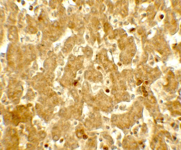
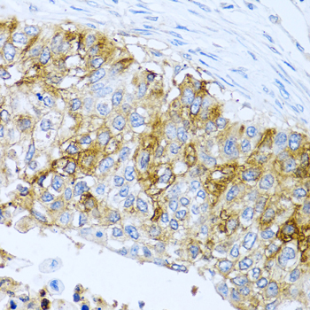
Two anti-IFIT3 antibodies have real human tissue IHC images: liver for A03920 (catalog image caption) and paraffin-embedded lung cancer for A03920-1 (catalog image caption). IF is listed for A03920-1 (catalog applications).
A03920 is listed for human IHC-P and pictured in human liver tissue at 5 μg/mL (catalog applications, reactivity, and image caption). A03920-1 is listed for human, mouse, and rat IHC and IF, with an IHC image from paraffin-embedded human lung cancer; no IF image is supplied (catalog applications, reactivity, and image captions).
Which to pick: For human paraffin-section IHC, A03920 lists IHC-P and 5 μg/mL; its image shows human liver tissue, but the caption does not report processing or fixative (catalog applications, dilution, and image caption). A03920-1 has an IHC image from paraffin-embedded human lung cancer and lists 1:100–1:200 for IHC; the caption does not report the fixative (catalog image caption and dilution). Choose A03920-1 for an IF starting point or mouse and rat work because those applications and species are listed; ICC validation and antibody clonality are unreported (catalog applications, reactivity, and clone fields).