This website uses cookies to ensure you get the best experience on our website.


- Table of Contents
Use high staining in bone marrow hematopoietic cells and undetected staining in adipocytes as IHC reference points (HPA tissue IHC). Expect cytoplasmic staining, but interpret it cautiously because antibody staining and RNA expression show low consistency (HPA tissue IHC).

Expected localisation, validated protocols, controls and antibodies — the at-a-glance facts below, then the full design guide.
| Expected localisation | Cytoplasmic tissue staining (HPA tissue IHC) | |
| Staining pattern | Cytoplasmic staining in several tissue cell types (HPA tissue IHC) | |
| Antigen retrieval | EDTA pH 8.0 HIER, heat-mediated (datasheet A04964-2) | |
| Positive control | Bone marrow+4 more · see all | |
| Negative control | Adipose tissue+4 more · see all |
| Fixation | Keep fixation consistent across paraffin sections (standard IHC practice; not target-specific) | |
| Caveat | Antibody may detect more than one gene product (HPA tissue IHC) | |
| Regulation | Interferon inducible (UniProt) | |
| Isoform / epitope | No annotated isoforms; cytoplasmic and extracellular regions (UniProt) |
The catalog antibody protocol is paired with the published IFITM2 IHC setup for breast tissue and carcinoma specimens (PMC11673214).
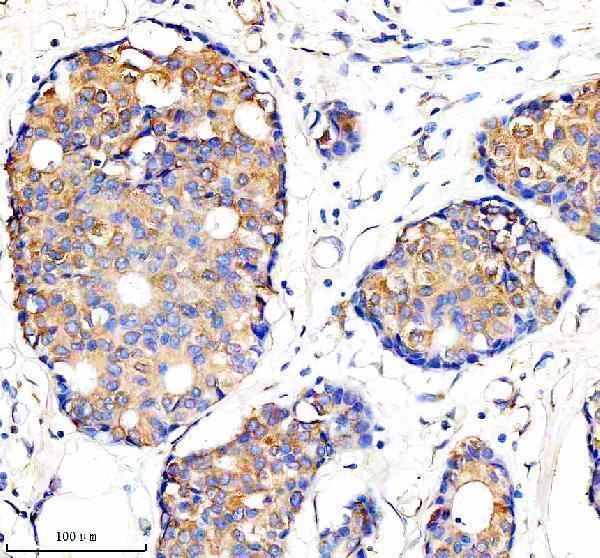
| Sample | Paraffin-embedded human breast cancer tissue; fixative not specified (datasheet A04964-2) |
| Fixation | Image fixative and duration unreported (datasheet A04964-2); verify before use. |
| Sectioning | 4–5 µm sections on charged slides (standard) |
| Deparaffinisation | Xylene, graded ethanol series to water (standard) |
| Antigen retrieval | Heat retrieval: EDTA pH 8.0 (datasheet A04964-2); 20 min, 95–100 °C (standard) |
| Peroxidase block | 3% H2O2, 10 min, room temperature (standard) |
| Blocking | 10% goat serum (datasheet A04964-2) |
| Primary antibody | Rabbit anti-IFITM2, 1:50 recommended; image 1:100 (datasheet A04964-2) |
| Primary incubation | Overnight at 4 °C (datasheet A04964-2) |
| Detection | HRP-conjugated secondary, DAB chromogen (datasheet A04964-2) |
| Counterstain | Hematoxylin, blue, dehydrate and mount (standard) |
| Expected result | IFITM2-positive staining in hematopoietic cells of bone marrow (HPA tissue IHC: High). HPA tissue profile: Cytoplasmic expression in several tissues. No signal in the no-primary control. |
In paraffin-section IHC, expect chiefly cytoplasmic staining in selected cells, including bone-marrow hematopoietic cells, endometrial stromal cells, and kidney glomerular cells (HPA: high in these cells; cytoplasmic expression in several tissues). Membrane-associated staining is biologically plausible because IFITM2 occupies cell, late-endosome, and lysosome membranes and has one transmembrane segment (UniProt Q01629 topology/localization). Treat the pattern as provisional: HPA rates tissue IHC Uncertain, citing poor agreement with RNA and possible recognition of more than one gene (HPA: tissue IHC reliability).
| Granular or rim-like cytoplasmic signal in hematopoietic cells of bone marrow, with limited nuclear signal. | Consistent with HPA's high staining in those cells and its broad cytoplasmic tissue profile (HPA: bone marrow; tissue profile). Membrane-associated compartments are plausible (UniProt Q01629 localization), but chromogenic IHC alone cannot identify individual endosomes or lysosomes (general IHC practice). Judge the cell-specific pattern alongside controls because tissue IHC reliability is uncertain (HPA: reliability). |
| Predominant, crisp nuclear staining in a paraffin section while the expected cytoplasmic pattern is absent. | This does not match HPA's reported tissue IHC profile (HPA: cytoplasmic expression). Review controls and antibody specificity before assigning it to IFITM2. HPA reports an additional nucleoplasmic ICC-IF location with uncertain status, so a nuclear observation alone cannot establish a genuine tissue IHC pattern (HPA: subcellular ICC-IF). |
| Strong staining of adipocytes, despite little signal in nearby expected-positive cells. | HPA reports IFITM2 as not detected in adipocytes, making this pattern unexpected for that cell type (HPA: adipose tissue). Consider cross-reactivity or endogenous chromogen activity and inspect negative controls (general IHC practice). The HPA tissue result is an uncertain reference pattern, not proof that every adipocyte must be negative (HPA: reliability). |
| Brown signal spread across tissue, lumen, and slide background without a clear cellular pattern. | Treat the distribution as background until control slides show otherwise (general IHC practice). It does not resemble the reported cell-specific cytoplasmic staining (HPA: tissue IHC profile). Review blocking, washing, detection background, and tissue pigment with suitable controls (general IHC practice); do not infer IFITM2 abundance from a uniform deposit. |
| No staining in bone-marrow hematopoietic cells on a run intended to demonstrate a positive control. | HPA reports high staining in these cells, so a blank control warrants a run-level check (HPA: bone marrow). Verify tissue identity, retrieval and detection performance, then assess the catalog antibody against a second validated reagent or orthogonal evidence when available (general IHC practice). HPA's uncertain reliability limits how decisively one negative slide can be interpreted (HPA: reliability). |
| Membrane topology | IFITM2 has a transmembrane segment at residues 107–127 and cytoplasmic regions at 1–56 and 78–106 (UniProt Q01629 topology). These positions inform interpretation of a known epitope, but the supplied record gives no catalog-antibody epitope; do not predict retrieval needs from topology alone. |
| Tissue and cell selection | HPA reports high staining in endometrial stromal cells and kidney glomerular cells, medium staining in skin fibroblasts, and no detection in adipocytes (HPA: tissue IHC). Choose a documented positive cell population for run assessment and compare the named cell type, not merely the tissue label (general IHC practice). |
| Strength of the IHC reference | The tissue profile has Uncertain reliability because antibody staining agrees poorly with RNA and may include protein from more than one gene (HPA: reliability description). A matching pattern supports an interpretation, but specificity still needs appropriate controls or independent confirmation (general IHC practice). |
| ICC-IF versus tissue IHC | HPA's ICC-IF summary places IFITM2 mainly at cell junctions with an additional uncertain nucleoplasmic location; the tissue IHC summary describes cytoplasmic expression (HPA: subcellular ICC-IF; tissue IHC). Keep these assay-specific observations distinct when assessing a chromogenic paraffin section. |
| Endogenous detection activity | Endogenous enzyme activity or tissue pigment can resemble chromogenic signal, so use detection and negative controls to locate non-antibody color (general IHC practice). No supplied HPA or UniProt evidence assigns a particular endogenous activity to IFITM2-positive cells. |
| Situation | Likely cause | Next action |
|---|---|---|
| Expected-positive cells are blank across the whole section. | A failed retrieval or detection step, an inactive primary, or an unsuitable section can erase the run's usable signal (general IHC practice); HPA reports high staining in bone-marrow hematopoietic cells (HPA: bone marrow). | Check a positive-control section and reagent controls, confirm the selected cells are present, and review the catalog antibody's IHC-P instructions before changing conditions (general IHC practice). |
| Signal is weak in an HPA-listed high cell population. | The run may have low analytical sensitivity, or the reference pattern may not reproduce with this antibody; HPA rates tissue IHC Uncertain (HPA: reliability). | Compare a same-run positive control, inspect detection and retrieval records, and score localization separately from intensity (general IHC practice). Avoid calling a weak signal definitively negative. |
| Unexpected cells stain strongly while the documented cell population does not. | Cross-reactivity or nonspecific detection is possible (general IHC practice); HPA warns that tissue staining may include protein from more than one gene (HPA: reliability description). | Identify the stained cell type, compare the relevant HPA cell-level entry, and seek an independent antibody or orthogonal result before assigning IFITM2 (HPA: tissue IHC; general IHC practice). |
| A diffuse brown haze obscures cellular boundaries. | Nonspecific reagent binding, incomplete washing, or detection background can produce haze (general IHC practice). HPA describes a cytoplasmic tissue pattern rather than a featureless field (HPA: tissue profile). | Review negative and detection-only controls, then optimize blocking, washing, and chromogen exposure within the validated IHC workflow (general IHC practice). |
| Signal looks exclusively nuclear. | The result conflicts with the HPA cytoplasmic tissue IHC profile; HPA's nucleoplasmic ICC-IF annotation is uncertain and comes from a different assay (HPA: tissue IHC; subcellular ICC-IF). | Check the counterstain and negative controls, then confirm with an independent reagent before scoring nuclear staining as IFITM2 (general IHC practice). |
| IF/ICC Q&A: should cell-junction fluorescence dictate the paraffin IHC score? | HPA reports approved cell-junction localization in ICC-IF, whereas its tissue IHC profile is cytoplasmic (HPA: subcellular ICC-IF; tissue IHC). The observations come from different assay contexts. | Score the paraffin section against its tissue-specific IHC cell and compartment pattern, and consult the separate IF/ICC guide for fluorescence interpretation (HPA: tissue IHC; general IHC practice). |


Comprehensive Human Protein Atlas IHC scoring per tissue (reliability: Uncertain — Low consistency between antibody staining and RNA expression data. Caution, targets protein from more than one gene.). Rows are taken directly from the HPA tissue chart — click any row's HPA link to view the source.
| Tissue | Cell type | Level | Evidence | Source |
|---|---|---|---|---|
| Bone marrow | Hematopoietic cells | High | Protein (IHC) | HPA → |
| Endometrium | Cells in endometrial stroma | High | Protein (IHC) | HPA → |
| Epididymis | Glandular cells | High | Protein (IHC) | HPA → |
| Fallopian tube | Glandular cells | High | Protein (IHC) | HPA → |
| Kidney | Cells in glomeruli | High | Protein (IHC) | HPA → |
| Tissue | Cell type | Level | Evidence | Source |
|---|---|---|---|---|
| Adipose tissue | Adipocytes | Not detected | Protein (IHC) | HPA → |
| Appendix | Glandular cells | Not detected | Protein (IHC) | HPA → |
| Cerebellum | Cells in granular layer | Not detected | Protein (IHC) | HPA → |
| Duodenum | Glandular cells | Not detected | Protein (IHC) | HPA → |
| Gallbladder | Glandular cells | Not detected | Protein (IHC) | HPA → |
Troubleshoot IFITM2 staining in paraffin sections by checking retrieval, compartment, cell identity, and controls before comparing signal across samples.
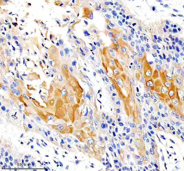
A04964-2 has IHC images from human breast and lung cancer paraffin sections (catalog image captions). IF and ICC are listed applications, but no IF image is supplied (catalog applications; image payload).
A04964-2 will render with its IHC figure from a human breast cancer paraffin section (card image caption). Its catalog also shows IHC on a human lung cancer paraffin section and lists IF and ICC applications for human samples (catalog image caption; applications; reactivity).
Which to pick: Choose A04964-2 for paraffin-section IHC: its own captions document heat retrieval in EDTA at pH 8.0, while the fixative is unreported (catalog image captions). The same rabbit polyclonal antibody is listed for IF/ICC, although no IF image is supplied (catalog host; dilution record; applications; image payload). Cross-species use is unsupported because the catalog lists only human reactivity (catalog reactivity).