This website uses cookies to ensure you get the best experience on our website.


- Table of Contents
Plan chromogenic IHC for IFT20 around its cytoplasmic tissue staining (HPA tissue IHC). Compare high-staining appendix glandular cells with low-staining liver cholangiocytes (HPA tissue IHC), and start within the catalog antibody’s 2–5 μg/ml IHC range (datasheet A07253).

Expected localisation, validated protocols, controls and antibodies — the at-a-glance facts below, then the full design guide.
| Expected localisation | Cytoplasmic tissue staining (HPA tissue IHC) | |
| Staining pattern | Cytoplasmic staining across tissues (HPA tissue IHC) | |
| Antigen retrieval | EDTA pH 8.0 HIER, heat-mediated (datasheet A07253) | |
| Positive control | Appendix+4 more · see all | |
| Negative control | None in HPA (detected in all 45 tissues); use no-primary + isotype controls |
| Fixation | Keep fixation consistent across sections (standard IHC practice; not target-specific) | |
| Caveat | Staining and RNA show medium consistency (HPA tissue IHC) | |
| Regulation | Low tissue specificity (HPA tissue RNA) | |
| Isoform / epitope | Four isoforms; epitope coverage is unreported (UniProt) |
The catalog antibody protocol uses heat-mediated EDTA pH 8.0 retrieval (datasheet A07253). One published IHC protocol provides a comparison in human lung tissue (PMC9308367).
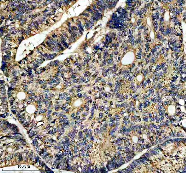
| Sample | Paraffin-embedded human colon cancer tissue; fixative not specified (datasheet A07253) |
| Fixation | Image fixative and duration unreported (datasheet A07253); verify before use. |
| Sectioning | 4–5 µm sections on charged slides (standard) |
| Deparaffinisation | Xylene, graded ethanol series to water (standard) |
| Antigen retrieval | Heat retrieval: EDTA pH 8.0 (datasheet A07253); 20 min, 95–100 °C (standard) |
| Peroxidase block | 3% H2O2, 10 min, room temperature (standard) |
| Blocking | 10% goat serum (datasheet A07253) |
| Primary antibody | Rabbit anti-IFT20, 2-5 μg/ml (datasheet A07253) |
| Primary incubation | Overnight at 4 °C (datasheet A07253) |
| Detection | HRP-conjugated secondary, DAB chromogen (datasheet A07253) |
| Counterstain | Hematoxylin, blue, dehydrate and mount (standard) |
| Expected result | IFT20-positive staining in glandular cells of appendix (HPA tissue IHC: High). HPA tissue profile: Cytoplasmic expression in all tissues. No signal in the no-primary control. |
IFT20 is a cytoplasmic protein associated with the Golgi, centrosomes, basal bodies and cilia (UniProt Q8IY31 localization; no transmembrane segment). In paraffin-section IHC, expect cytoplasmic staining across many tissues, including strong staining in several glandular and epithelial cell groups (HPA: cytoplasmic expression in all tissues; selected groups High). HPA rates the tissue IHC evidence Approved, with medium consistency between staining and RNA expression (HPA: reliability).
| Clear cytoplasmic stain in appendix or breast glandular cells, with cellular outlines still readable. | This fits the reported IHC pattern: both cell groups are High, within a broader cytoplasmic tissue profile (HPA: appendix and breast glandular cells High; cytoplasmic expression in all tissues). Judge it against controls on the same run; a single positive slide does not establish antibody specificity (standard IHC practice). |
| Fine cytoplasmic or perinuclear emphasis, without a distinct ciliary dot at routine IHC magnification. | Golgi and centrosomal association can contribute to local emphasis (UniProt Q8IY31 localization). A visible cilium is not required to call paraffin-section IHC positive: HPA describes tissue staining as cytoplasmic, while its resolved ciliary and microtubule locations come from ICC-IF (HPA: tissue IHC profile; subcellular ICC-IF). |
| Predominantly nuclear, crisp surface-membrane or extracellular staining with little cytoplasmic signal. | That distribution conflicts with the reported cytoplasmic IHC profile and the lack of a transmembrane segment (HPA: tissue IHC profile; UniProt Q8IY31 topology). Check morphology and controls before calling it target staining; chromogen deposits or nonspecific binding can mimic compartment-restricted signal (standard IHC practice). |
| Strong stain confined to an unexpected cell group while expected positive cells in the same section are blank. | Consider cross-reactivity or endogenous detection activity, especially if the discordance persists in controls (standard IHC practice). Broad expression alone cannot identify a cell group as truly negative: HPA reports cytoplasmic expression across tissues and provides no negative tissue group here (HPA: tissue IHC profile). |
| Uniform haze, or no signal in a known High group such as bronchial respiratory epithelium. | Haze obscures cell localization; absence in a reported High group makes a negative result hard to interpret (HPA: bronchus respiratory epithelial cells High). Assess the no-primary control, tissue integrity and run performance before assigning either result to IFT20 biology (standard IHC practice). |
| Tissue and cell-group selection | Appendix, breast, endometrium, epididymis and gallbladder glandular cells are High; bronchial respiratory epithelium is also High (HPA: tissue IHC levels). Cholangiocytes and the listed muscle cell groups are Low, so weak staining there is less decisive than a failure in a High group (HPA: tissue IHC levels). |
| Compartment and optical resolution | The tissue IHC summary is cytoplasmic; ICC-IF additionally resolves microtubules and primary cilia (HPA: tissue IHC profile; subcellular ICC-IF). Golgi, centrosome and basal-body association gives biological context, but paraffin IHC need not display each small structure separately (UniProt Q8IY31 localization; standard IHC practice). |
| Strength of validation | The listed antibody HPA021376 is Approved for IHC and ICC, while tissue staining has medium consistency with RNA expression (HPA: antibody status; reliability description). Treat a matching pattern as supportive evidence, and use controls to evaluate staining in the particular run (standard IHC practice). |
| Isoforms and epitope coverage | Four isoforms are listed, but this record gives no antibody epitope or isoform-specific IHC pattern (UniProt Q8IY31 isoforms; supplied HPA antibody record). Do not interpret regional intensity differences as isoform switching without separate evidence (standard IHC interpretation). |
| Topology and processing | IFT20 has no annotated transmembrane segment, signal peptide, propeptide or glycosylation site; its annotated chain spans residues 1–132 (UniProt Q8IY31 topology and processing). These facts support an intracellular interpretation but do not establish antigen-retrieval needs or fixation sensitivity (UniProt Q8IY31; supplied source scope). |
| Situation | Likely cause | Next action |
|---|---|---|
| A reported High tissue is completely blank. | A failed staining run, unsuitable section or detection problem is possible; HPA's High label does not guarantee every specimen or section will stain (HPA: selected High groups; standard IHC practice). | Inspect tissue preservation and the run's positive control; verify retrieval, primary-antibody application and chromogenic detection against the validated IHC procedure before interpreting absence (standard IHC practice). |
| Brown signal blankets tissue and obscures cell borders. | Background from detection chemistry, endogenous activity or insufficient washing can hide the expected cytoplasmic pattern (HPA: tissue IHC profile; standard IHC practice). | Compare no-primary and reagent controls; check the relevant endogenous-activity block, washing and detection development, then reassess identifiable cells (standard IHC practice). |
| Stain is mainly nuclear, extracellular or sharply membrane-limited. | The compartment disagrees with the cytoplasmic tissue pattern; IFT20 has no annotated transmembrane segment (HPA: tissue IHC profile; UniProt Q8IY31 topology). | Review section morphology and negative controls, and repeat with the IHC-validated antibody under its validated conditions if the mismatch persists (standard IHC practice). |
| A Low cell group appears stronger than nearby reported High cells. | Sample variation, nonspecific binding or endogenous detection activity may contribute; HPA labels cholangiocytes and listed muscle groups Low, not negative (HPA: tissue IHC levels; standard IHC practice). | Score cell groups separately, compare matched controls and avoid treating one intensity reversal as proof of altered IFT20 expression (HPA: tissue IHC levels; standard IHC practice). |
| Only scattered dots are visible, with no convincing cellular cytoplasm. | IFT20 associates with centrosomes and basal bodies, but isolated dots may also be deposits or background (UniProt Q8IY31 localization; standard IHC practice). | Check whether dots follow cell morphology, compare no-primary controls and seek a reproducible cytoplasmic pattern in a reported High group (HPA: tissue IHC profile; standard IHC practice). |
| Q: Should IF/ICC show the same broad chromogenic pattern? | A: IF/ICC offers finer compartment resolution; HPA reports approved microtubule and primary-cilium localization, with additional approved sites including basal body and mitotic spindle (HPA: subcellular ICC-IF). | Use the separate IF/ICC guide to interpret fluorescence localization; do not require individually resolved cilia in paraffin-section IHC (HPA: subcellular ICC-IF; tissue IHC profile; standard microscopy practice). |
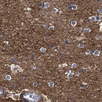


Comprehensive Human Protein Atlas IHC scoring per tissue (reliability: Approved — Medium consistency between antibody staining and RNA expression data.). Rows are taken directly from the HPA tissue chart — click any row's HPA link to view the source.
| Tissue | Cell type | Level | Evidence | Source |
|---|---|---|---|---|
| Appendix | Glandular cells | High | Protein (IHC) | HPA → |
| Breast | Glandular cells | High | Protein (IHC) | HPA → |
| Bronchus | Respiratory epithelial cells | High | Protein (IHC) | HPA → |
| Cerebral cortex | Neuropil | High | Protein (IHC) | HPA → |
| Endometrium | Glandular cells | High | Protein (IHC) | HPA → |
| Tissue | Cell type | Level | Evidence | Source |
|---|---|---|---|---|
| None in HPA: IFT20 is detected in all 45 scored tissues. Use a no-primary (secondary-only) and an isotype control instead. | ||||
Troubleshoot IFT20 staining in paraffin section IHC using the catalog antibody’s tested retrieval conditions and compartment aware controls.
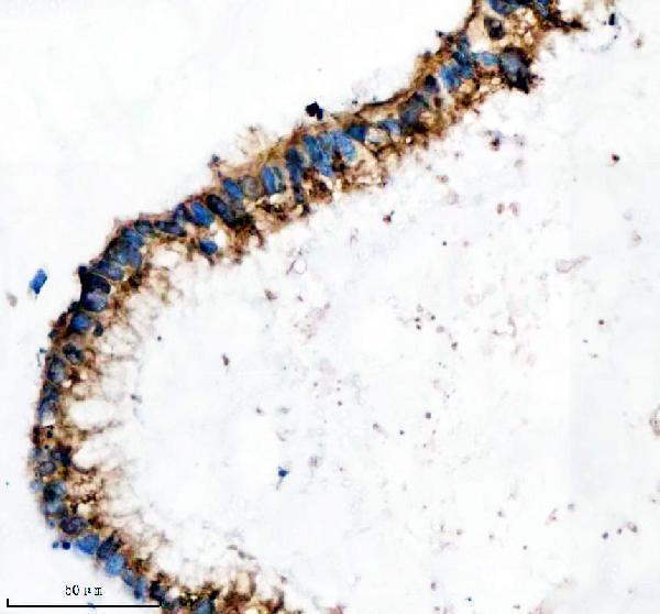
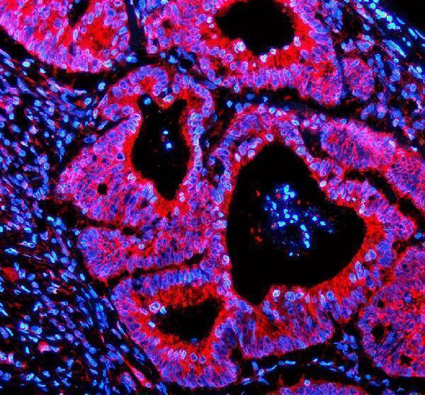
A07253 has IHC and IF images from paraffin-embedded human colon cancer tissue, plus an IHC image from human testis cancer tissue (catalog image captions).
A07253 is listed for IHC and IF and for human, mouse, and rat reactivity (catalog: applications and reactivity). Its IHC captions show paraffin-embedded human colon and testis cancer sections; its IF caption shows a paraffin-embedded human colon cancer section (catalog image captions).
Which to pick: Choose A07253 for paraffin-section IHC: its colon cancer caption documents EDTA retrieval at pH 8.0 and 2 μg/ml primary antibody (A07253 IHC caption). For IF, A07253 has a paraffin-section example at 5 μg/ml; ICC validation and clonality are unreported (A07253 IF caption; catalog: applications and clone). For mouse or rat work, A07253 lists both species as reactive, while its IHC images show human tissue only; the fixative is unreported (catalog: reactivity; A07253 IHC captions).