This website uses cookies to ensure you get the best experience on our website.


- Table of Contents
Real validated IGF1 Western blot protocols, expected-band and isoform facts, troubleshooting for weak or shifted signal, and recommended anti-IGF1 WB antibodies. Everything you need to plan the experiment before you commit precious samples.
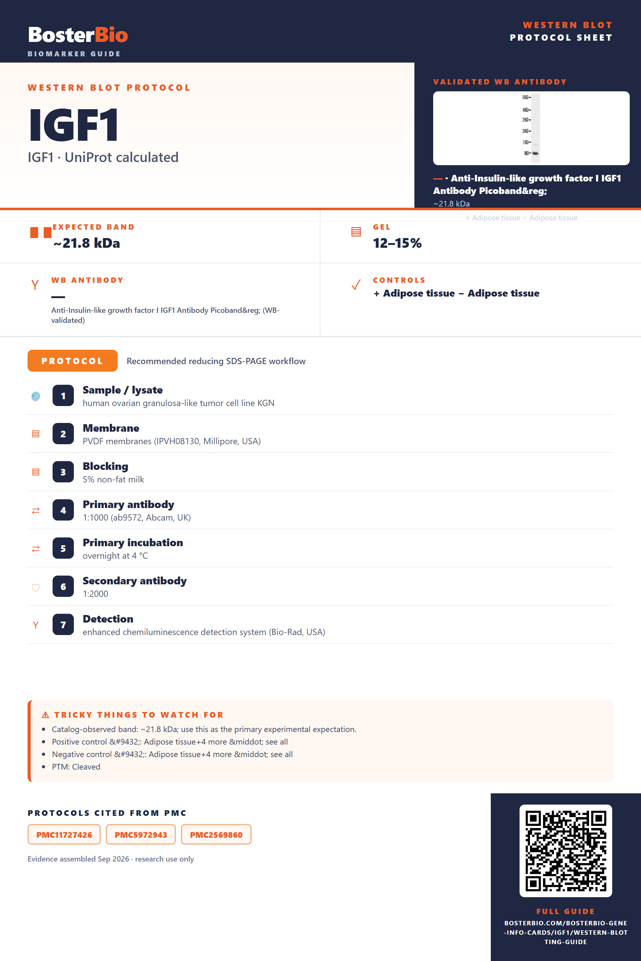
Expected bands, validated protocols, controls and antibodies — the at-a-glance facts below, then the full design guide.
| Expected band | ~21.8 kDa | |
| Gel | 12–15% | |
| Positive control | Adipose tissue+4 more · see all | |
| Negative control | Adipose tissue+4 more · see all |
| PTM | Cleaved | |
| Caveat | Propeptide cleavage variability | |
| Regulation | Myogenesis | |
| Isoform | 4 isoform(s) |
Literature-validated Western blot parameters for IGF1 — gel percentage, transfer, blocking, antibody incubation and detection, extracted from published methods.
| Sample / lysate | human ovarian granulosa-like tumor cell line KGN |
| Membrane | PVDF membranes (IPVH08130, Millipore, USA) |
| Blocking | 5% non-fat milk |
| Primary antibody | 1:1000 (ab9572, Abcam, UK) |
| Primary incubation | overnight at 4 °C |
| Secondary antibody | 1:2000 |
| Detection | enhanced chemiluminescence detection system (Bio-Rad, USA) |
IGF-1's 21.8 kDa precursor is cleaved of its signal peptide and propeptide during secretion, so the mature circulating protein runs as a much smaller band than the full-length prediction.
| Single band near 21.8 kDa in whole-cell/intracellular lysate | represents the unprocessed 195-aa precursor before signal peptide and propeptide removal |
| Much smaller band well below 21.8 kDa in mature/secreted preparations | reflects loss of the 21-residue signal peptide and the 22-48 propeptide during maturation, leaving a markedly smaller mature chain |
| Little or no band in whole-cell lysate | IGF-1 is annotated as secreted, so little full-length or mature protein is retained intracellularly |
| Sharp single band with no smearing | no glycosylation sites are annotated, so no glycoform-driven mass heterogeneity is expected |
| Slight mobility shift between non-reduced and reduced samples | the three annotated intrachain disulfide bonds compact the mature domain, so non-reduced protein can migrate slightly faster than fully reduced protein |
| Multiple bands at different apparent masses | four annotated splice isoforms can yield precursors of differing length, appearing as extra bands depending on which isoform is expressed |
| Predicted precursor mass (21.8 kDa, 195 aa) | sets the baseline size for the unprocessed full-length chain seen before maturation |
| Signal peptide (residues 1-21) | removed during secretion, reducing the apparent size of the mature band relative to the precursor |
| Propeptide (residues 22-48) | cleaved off during processing, further shrinking the mature chain well below the 21.8 kDa precursor mass |
| Three intrachain disulfide bonds (54-96, 66-109, 95-100) | compact the folded domain, so non-reduced samples can migrate slightly faster than fully reduced samples |
| Four splice isoforms | isoforms of differing precursor length may appear as additional bands at distinct apparent masses, described only qualitatively as relatively larger or smaller |
| No annotated glycosylation sites | band should run as a sharp single species rather than a smeared or shifted-higher glycoform pattern |
| Secreted subcellular location | mature band is best detected in conditioned media, serum, or plasma rather than whole-cell lysate |
| Situation | Likely cause | Next action |
|---|---|---|
| No band in lysate | IGF-1 is secreted, so little full-length or mature protein remains inside cells | probe conditioned media, serum, or plasma rather than whole-cell lysate |
| Band higher than expected | antibody is detecting the uncleaved precursor that still carries its signal peptide and/or propeptide | determine whether the sample reflects intracellular precursor versus secreted mature protein and interpret size accordingly |
| Band lower than expected | mature, processed IGF-1 lacking the signal peptide and propeptide runs much smaller than the 21.8 kDa full-length prediction | compare the band against the expected mature-form size rather than the precursor predicted mass |
| Multiple bands | four annotated splice isoforms can generate precursors of different lengths | confirm isoform expression by sequencing or RT-PCR, or use an antibody targeting a region common to all isoforms |
| Fragments below expected size | the small, disulfide-stabilized mature IGF-1 chain is susceptible to proteolytic degradation during handling | keep samples cold, add protease inhibitors, and minimize freeze-thaw cycles |
| Weak or no signal | secreted mature IGF-1 is a small, low-abundance peptide that can be under-loaded or run off low-percentage gels | use a high-percentage or gradient gel, a low-MWCO transfer membrane, and concentrated serum/media samples |

Comprehensive Human Protein Atlas IHC scoring per tissue. Rows are taken directly from the HPA tissue chart — click any row's HPA link to view the source.
| Tissue | Cell type | Level | Evidence | Source |
|---|---|---|---|---|
| Adipose tissue | adipocytes | Not detected | Protein (IHC) | HPA → |
| Adrenal gland | glandular cells | Not detected | Protein (IHC) | HPA → |
| Appendix | glandular cells | Not detected | Protein (IHC) | HPA → |
| Bone marrow | hematopoietic cells | Not detected | Protein (IHC) | HPA → |
| Breast | adipocytes | Not detected | Protein (IHC) | HPA → |
| Tissue | Cell type | Level | Evidence | Source |
|---|---|---|---|---|
| Adipose tissue | adipocytes | Not detected | Protein (IHC) | HPA → |
| Adrenal gland | glandular cells | Not detected | Protein (IHC) | HPA → |
| Appendix | glandular cells | Not detected | Protein (IHC) | HPA → |
| Bone marrow | hematopoietic cells | Not detected | Protein (IHC) | HPA → |
| Breast | adipocytes | Not detected | Protein (IHC) | HPA → |
Deeper troubleshooting and optimisation questions for IGF1, answered from its protein features.
BosterBio's IGF1 antibodies are among the best-performing WB antibodies on the market — well cited, thoroughly validated, and orthogonally cross-validated against negative tissues and complementary methods.
For IGF-1 Western blots, Boster offers PA1374, an anti-IGF-1 antibody with a documented WB image showing detection of recombinant human IGF-1 protein (2.5ng) — a useful starting point for confirming assay sensitivity before running experimental samples.
Which to pick: Only one Boster IGF-1 antibody is catalogued here, PA1374. It has a published WB validation image (recombinant human IGF-1, 2.5ng), so it's the default choice; no alternative SKUs are available to compare reactivity or format against.