This website uses cookies to ensure you get the best experience on our website.
- Table of Contents
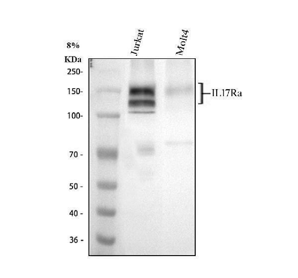
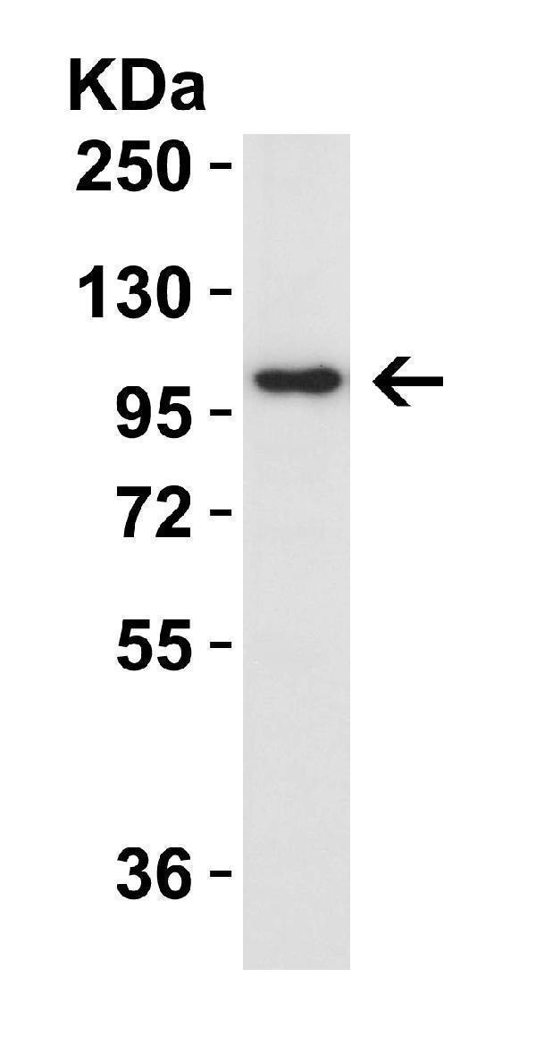
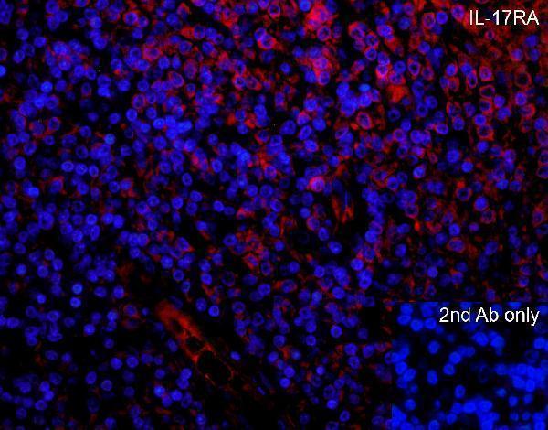
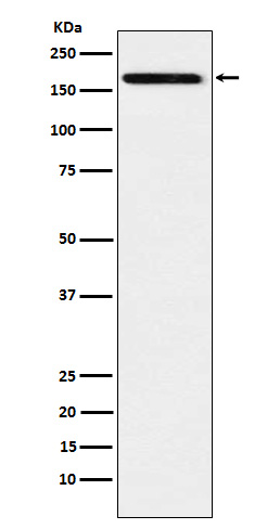
Facts about Interleukin-17 receptor A.
Binds IL17A and IL17F homodimers as part of a heterodimeric complex with IL17RC (By similarity). Also binds heterodimers formed by IL17A and IL17F as part of a heterodimeric complex with IL17RC (By similarity).
Mouse | |
---|---|
Gene Name: | Il17ra |
Uniprot: | Q60943 |
Entrez: | 16172 |
Belongs to: |
---|
No superfamily |
CD217 antigen; CD217; Cdw217; CDw217interleukin 17 receptor; hIL-17R; IL-17 R; IL-17 RA; IL-17 receptor A; IL17RA; IL-17RA; IL-17RAMGC10262; IL17Rinterleukin-17 receptor A; interleukin 17 receptor A
Mass (kDA):
97.808 kDA
Mouse | |
---|---|
Location: | 6 F1|6 56.95 cM |
Sequence: | 6; |
This article provides all the details about IL-17, markers IL-17RA and I17F and their roles in research into biology. This information is helpful to researchers from all over the world. Scientists must be able distinguish between these markers in order to make the right decisions regarding their research. Continue reading to learn more about IL-17F and IL17RA.
IL17RA is also known as interleukin-17 receptor alpha, is a protein which interacts with the SARS coronavirus. The protein is known for its ability to activate the NFKappa-B and MAPkinase pathways. IL17RA is also crucial in the recruitment of neutrophils at infection sites as well as for phagocyte killing of fungi and bacteria that are not in the extracellular. IL17RA is also involved in germinal center development and in the selection of plasma cells from B cells.
IL17RA is a novel molecular target that could be utilized to treat immune system disorders as well as to develop new medications. It is a proinflammatory cytokine produced by T cells that have been activated. It induces maturation of CD34-positive precursors into neutrophils. The IL17RA gene codes for the IL17RA molecular, which is a type I membrane glycoprotein with the lowest affinity for interleukin-17A.
The IL-17RA gene belongs to the cytokine family IL-17 and connects to both IL-17RB, as well as IL-17E. It may also associate with the IL-17RD, but the corresponding ligand to the IL-17RA/RD compound hasn't been identified. The IL-17RA/RD fusion complex is highly expressed in hematopoietic cells.
Although IL-17A has been linked to allergic disorders More evidence is needed. IL-17A expression causes monocyte infiltration and infiltration of lymphocytes CD4+/B220+ and PAS staining. However, IL-17A overexpression did not correlate significantly with the increase in Th2-related gene expression. It also has been reported to increase the production of mucus from the inflamed lung tissue.
Boster Bio's IL17RA antibody contains a blocking peptide. The antibody can be stored at -20C for a year and frozen for a further six month. The data sheet assures that the peptide will satisfy all requirements. Furthermore, the antibody is designed to react with IL17RA and is suitable for both IF and WB tests. The IL17RA antibody is completely free of BSA.
ADSC-Exo was given subcutaneously on the third day of the experiment. After the treatments digital images were taken of the wounds on days 3, 5, 7 and 10. The animals were killed after two weeks, and wound tissues were collected for histological analysis. The tissue samples were stained with antibodies for the IL17RA. In three instances, the methods were successful with positive cells.
Humans suffering from severe asthma are connected to the expression of IL-17A. In mice, IL-17A is also important in the development of allergic-related AHR. C57Bl/6 and BALB/c mice produce more IL-17A compared to controls. This suggests that IL-17A could be involved in allergic asthma. There are no FDA approved products for the marker IL17A but it is a crucial protein to understand the causes of allergic asthma.
In addition to its role in promoting M2-like phenotype in THP-1-derived macrophages In addition, IL-17RA is an important marker for research into the immune system. The marker IL-17RA is an important instrument in immunological studies and diagnosis of certain types of cancer. For this purpose, IL17RA has been used for more than two decades.
The Interleukin-17A (IL-17A) gene encodes a protein produced by activated T cells. It regulates the activity of NFkappaB and mitogen-activated proteins kinases. It also enhances the expression of IL-6 and nitric oxide. Psoriasis, as well as multiple sclerosis, have been linked to IL-17A. Both affinity and specificity have been confirmed for the Boster antibody.
For these studies, the HSFs were cultivated in glass dishes 14 mm at an amount of 4 percent paraformaldehyde. After the cells had reached 50% confluence, the samples were fixed in paraformaldehyde 4% for 30 minutes. After fixation, the sections were rinsed with 2% BSA-PBS. After blocking the sections, they were then incubated with primary antibodies against IL-17RA and a SMA. The sections were then incubated overnight at 4 degC.
IL-17RA is an immunoreactive molecule that connects to IL-17RB, or IL-17E. The IL-17RA protein is also found in tissues that respond to IL-25, although its exact receptor is not known. It is linked to the IL-17RD protein to create the complex. This complex is found in nearly every type of cell. Its amino acid sequence is 69% similar to that of mouse's IL-17R.
It is widely expressed and is widely expressed. IL-17RA activation is triggered by an essential adaptor molecule, Act1, and the transforming growth factor ss. Once activated, IL-17RA signalizes the recruitment of NF-kB TAK1 and the TAK2 and IL-17F via p65. It has been proven that it regulates the production of IL-17.
TRAF3IP2 adapter, TRAF6 proteins and IL17RA interact with IL17RA by activating the NFKappa B/MAP-kinase pathway. It also encourages the recruitment of neutrophils in the presence of infection sites, encourages the killing of fungi and bacteria in extracellular environments and is involved in germinal center formation. Additionally, IL-17RA encourages the transfer of B cells to plasma cells.
THP-1-derived macrophages are stimulated to display a M2-like phenotype through IL-17. It also causes NF-kB-independent Phosphorylation. In NSCLC patients, high concentrations of IL-17 is associated with low survival. It is also an indicator of risk for HIV development and a crucial aspect in the progression of cancer.
In the study, two pathologists analyzed sections of stained tissue. They utilized light microscopy at 400x and 200x magnifications. Five representative fields were recorded in each case. Image-Pro Plus v6.0 software was used to count positive cells. Positive cells were counted and the number of cells was calculated using the chi squared method. The chi-squared statistic for the percentage of positive cells was calculated by multiplying the number cells by the total number nuclei.
Another indicator for osteoclastogenic activity is the IL-17A. It is linked to osteoclastogenesis, bone immunopathology, and bone immunopathology. Neutrophil activation could lead to loss of bone. Neutralization of IL-17 might be an alternative treatment for periodontitis. The results of the clinical trial are encouraging, but more research is required to determine if this type of biological treatment is viable for periodontitis.
The marker IL17RA is an inflammation T cell-produced cytokine in the immune system. It plays a key role in many autoimmune diseases. It is highly expressed by T cells that are activated and is 57% identical to the 17-26 kD secretory glycoprotein encoded by the herpesvirus saimiri gene. It is a proinflammatory, anti-apoptotic and mitogenic agent and triggers nuclear factor-kappa-B.
HSCs were capable of expressing the IL17RA gene on days 1 2, 3 and 5. The mRNA was reverse-transcribed with a cDNA synthesis kit (Promega). The mRNA was subjected to real-time qPCR using SYBR Green detection. GAPDH was used to normalize the results. Six-well plates containing various levels of IL-17A were used to determine IL-17RA expression and IL-6 expression. The levels of exposure were monitored using enzyme-linked immunosorbent assay kits (R&D Systems).
The IL-17A marker is associated with various inflammatory diseases of the liver. The receptor for IL-17A may be found in many tissues and cells including the liver. Liver diseases, like hepatitis B, is linked to high levels of IL-17A. Hepatitis C and alcoholic liver disease are linked to elevated levels of IL-17A in the liver. Research hotspots include reducing liver injury and preventing fibrosis.
HSCs secrete IL-17A, which stimulates the production of the inflammatory cytokine IL-6. When IL-17RA levels are reduced in HSCs, IL-6 expression is inhibited, reducing the inflammation response. Therefore decreasing IL-17A expression could be beneficial to control the inflammatory response in the liver. There are a variety of uses for IL-17A and IL-17RA in Boster Bio.
To study the human immune system, IL-17RA can be used to monitor inflammation and autoimmune disease. This antibody is produced in accordance with the Boster Bio data sheet. It is guaranteed to be 100 100% accurate. In addition, boster antibodies are guaranteed to conform to their specifications. Additionally, they're also free of BSA. You can buy blocking peptides from Boster Bio for further research.
PMID: 8777726 by Yao Z., et al. Herpesvirus Saimiri encodes a new cytokine, IL-17, which binds to a novel cytokine receptor.
PMID: 17911633 by Kuestner R.E., et al. Identification of the IL-17 receptor related molecule IL-17RC as the receptor for IL-17F.