This website uses cookies to ensure you get the best experience on our website.


- Table of Contents
Plan chromogenic IL21R IHC in paraffin sections using spleen as a documented positive tissue (HPA tissue IHC; datasheet A00808-1). Interpret cytoplasmic lymphoid staining with HPA's uncertain staining reliability and IL21R's membrane topology in mind (HPA tissue IHC; UniProt).

Expected localisation, validated protocols, controls and antibodies — the at-a-glance facts below, then the full design guide.
| Expected localisation | Cytoplasmic in tissue IHC (HPA tissue IHC); membrane receptor (UniProt) | |
| Staining pattern | Cytoplasmic staining in lymphoid tissue cells (HPA tissue IHC) | |
| Antigen retrieval | EDTA pH 8.0 HIER, heat-mediated (datasheet A00808-1) | |
| Positive control | Bone marrow+3 more · see all | |
| Negative control | Adipose tissue+4 more · see all |
| Fixation | Keep fixation consistent across sections (standard IHC practice; not target-specific) | |
| Caveat | Presumed off-target staining; verify cell specificity (HPA tissue IHC) | |
| Regulation | No regulation data in record (UniProt) | |
| Isoform / epitope | No isoforms listed; extracellular vs cytoplasmic epitope matters (UniProt) |
The catalog antibody’s IHC-P protocol (datasheet A00808-1) is followed by four published IL21R chromogenic IHC protocols (PMC11992169; PMC7390201; PMC5429142; PMC13376877).
| Sample | Paraffin-embedded human spleen tissue; fixative not specified (datasheet A00808-1) |
| Fixation | Image fixative and duration unreported (datasheet A00808-1); verify before use. |
| Sectioning | 4–5 µm sections on charged slides (standard) |
| Deparaffinisation | Xylene, graded ethanol series to water (standard) |
| Antigen retrieval | Heat retrieval: EDTA pH 8.0 (datasheet A00808-1); 20 min, 95–100 °C (standard) |
| Peroxidase block | 3% H2O2, 10 min, room temperature (standard) |
| Blocking | 10% goat serum (datasheet A00808-1) |
| Primary antibody | Rabbit anti-IL21R, 2-5 μg/ml (datasheet A00808-1) |
| Primary incubation | Overnight at 4 °C (datasheet A00808-1) |
| Detection | HRP-conjugated secondary, DAB chromogen (datasheet A00808-1) |
| Counterstain | Hematoxylin, blue, dehydrate and mount (standard) |
| Expected result | IL21R-positive staining in hematopoietic cells of bone marrow (HPA tissue IHC: High). HPA tissue profile: Cytoplasmic expression in lymphoid tissues. No signal in the no-primary control. |
IL21R is a membrane receptor with an extracellular region, one transmembrane segment and a cytoplasmic region (UniProt Q9HBE5 topology). In tissue IHC, expect staining in hematopoietic and lymphoid cell populations, often cytoplasmic (HPA: tissue IHC profile). Interpret apparent positives cautiously: HPA rates the tissue pattern Uncertain because antibody staining and RNA expression have low consistency, and presumed off-target staining was disregarded (HPA: tissue reliability).
| Clear staining in bone marrow hematopoietic cells or spleen red-pulp cells. | These are the strongest reported tissue IHC positives, both rated High (HPA: bone marrow and spleen). Assess the cell population and compartment together; a strong chromogen deposit alone does not establish IL21R specificity, given the Uncertain tissue reliability (HPA: tissue reliability). |
| Staining in lymph-node or tonsil germinal-center cells. | A Medium signal fits the reported tissue pattern (HPA: lymph node and tonsil). HPA describes tissue staining as cytoplasmic, while UniProt places IL21R in the membrane (HPA: tissue IHC profile; UniProt Q9HBE5 topology). Record the observed compartment instead of treating either location alone as definitive. |
| Predominantly nuclear staining, with no convincing signal in expected cell populations. | A nuclear pattern is outside the reported membrane and cytosol locations (UniProt Q9HBE5 subcellular location; HPA: ICC-IF subcellular location). Treat it as suspect and review the matched negative control, tissue morphology and detection background before scoring it as IL21R (general IHC practice). |
| Broad staining of adipocytes or respiratory epithelial cells, or colour deposited across many unrelated cells. | HPA reports IL21R as Not detected in adipocytes of adipose tissue and respiratory epithelial cells of bronchus (HPA: tissue IHC). Such staining may reflect cross-reactivity or endogenous detection activity; investigate with appropriate staining controls (general IHC practice). |
| No staining in bone marrow hematopoietic cells or spleen red-pulp cells. | Both are reported High positives (HPA: bone marrow and spleen), so first check tissue preservation, control performance and the staining workflow (general IHC practice). A negative result is not proof of absent IL21R: the tissue IHC profile itself has Uncertain reliability (HPA: tissue reliability). |
| Topology and antibody epitope | IL21R spans the membrane at residues 233–253, with extracellular residues 20–232 and cytoplasmic residues 254–538 (UniProt Q9HBE5 topology). No epitope position is supplied, so topology cannot predict this antibody's exact IHC compartment. |
| Tissue evidence strength | HPA labels tissue IHC reliability Uncertain and reports presumed off-target binding that it disregarded (HPA: tissue reliability). Give the reported cell-level distribution more weight than isolated staining in an unexpected population. |
| IF/ICC Q: What location is reported? | A: Plasma membrane and cytosol are both approved ICC-IF locations (HPA: ICC-IF subcellular location). That finding can guide compartment review, but it does not establish an IHC paraffin-section protocol or validate a tissue-positive cell type. |
| Antigen retrieval | Retrieval is an optimization variable in paraffin IHC (general IHC practice). No supplied evidence establishes an IL21R-specific retrieval condition or effect; interpret changes alongside tissue and detection controls. |
| Glycosylation | UniProt lists 6 glycosylation sites in the extracellular region (UniProt Q9HBE5 glycosylation and topology). Their presence does not establish whether this antibody's epitope is affected in a paraffin section. |
| Situation | Likely cause | Next action |
|---|---|---|
| Expected positive tissue is blank. | The staining run may have failed, or signal may be below detection; HPA reports High staining in bone marrow hematopoietic cells and spleen red-pulp cells (HPA: tissue IHC). | Check a known-positive section processed in the same run, then review retrieval, primary-antibody use and detection steps against the validated IHC workflow (general IHC practice). |
| Signal appears mainly in nuclei. | Nuclei fall outside the supplied membrane and cytosol locations (UniProt Q9HBE5 subcellular location; HPA: ICC-IF subcellular location). | Compare the slide with a negative control and reassess whether nuclear colour is specific staining or detection artefact before reporting IL21R (general IHC practice). |
| Adipocytes or bronchial respiratory epithelium stain strongly. | Those cell types are reported Not detected, and HPA flags presumed off-target binding in the tissue dataset (HPA: tissue IHC and reliability). | Treat the finding as unconfirmed. Check cell identity and negative controls, and seek independent support before assigning the signal to IL21R (general IHC practice). |
| Diffuse colour obscures cell boundaries. | Background from detection chemistry, insufficient blocking or overly concentrated primary antibody can obscure a cellular pattern (general IHC practice). | Review negative-control staining and adjust blocking, washing or antibody concentration within the validated workflow; rescore only when cells and compartments are distinguishable (general IHC practice). |
| Germinal-center signal is weaker than splenic red-pulp signal. | That relative pattern is consistent with Medium staining in lymph-node and tonsil germinal centers versus High staining in spleen red pulp (HPA: tissue IHC). | Score each cell population against its reported level and the run's controls; avoid calling a Medium germinal-center result negative solely because spleen stains more strongly (HPA: tissue IHC; general IHC practice). |
| Membrane and cytoplasmic staining seem inconsistent. | UniProt describes a membrane receptor, HPA tissue IHC describes cytoplasmic expression, and HPA ICC-IF approves both plasma membrane and cytosol (UniProt Q9HBE5; HPA: tissue IHC and ICC-IF). | Document each compartment and cell type separately. Use tissue controls and the HPA Uncertain reliability rating when judging whether the pattern supports IL21R (HPA: tissue reliability; general IHC practice). |
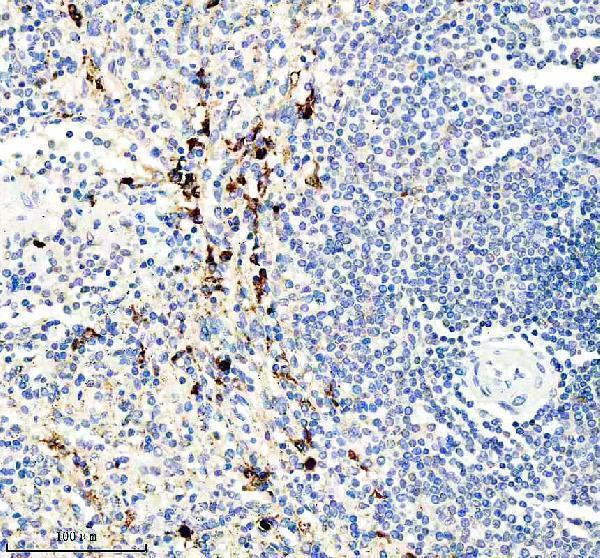
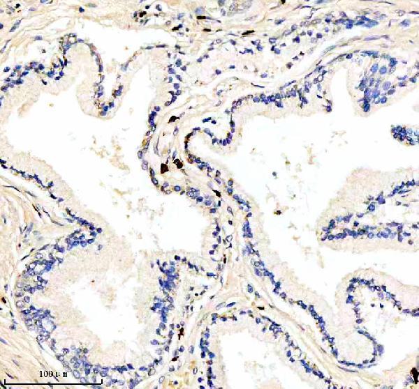


Comprehensive Human Protein Atlas IHC scoring per tissue (reliability: Uncertain — Low consistency between antibody staining and RNA expression data. Presumed off target binding observed and disregarded.). Rows are taken directly from the HPA tissue chart — click any row's HPA link to view the source.
| Tissue | Cell type | Level | Evidence | Source |
|---|---|---|---|---|
| Bone marrow | Hematopoietic cells | High | Protein (IHC) | HPA → |
| Spleen | Cells in red pulp | High | Protein (IHC) | HPA → |
| Lymph node | Germinal center cells | Medium | Protein (IHC) | HPA → |
| Tonsil | Germinal center cells | Medium | Protein (IHC) | HPA → |
| Tissue | Cell type | Level | Evidence | Source |
|---|---|---|---|---|
| Adipose tissue | Adipocytes | Not detected | Protein (IHC) | HPA → |
| Adrenal gland | Glandular cells | Not detected | Protein (IHC) | HPA → |
| Breast | Adipocytes | Not detected | Protein (IHC) | HPA → |
| Bronchus | Respiratory epithelial cells | Not detected | Protein (IHC) | HPA → |
| Caudate | Glial cells | Not detected | Protein (IHC) | HPA → |
Troubleshoot IL21R staining in paraffin sections by checking retrieval, cell identity, localisation and controls before scoring chromogenic signal.
A00808-1 has real IHC data from human spleen and prostate cancer paraffin sections (catalog image captions); no IF/ICC data are supplied (catalog applications and images).
A00808-1 is human-reactive and listed for IHC (catalog: reactivity and applications). Its IHC captions show staining in human spleen and prostate cancer paraffin sections (catalog image captions).
Which to pick: Choose A00808-1 for human paraffin-section IHC: its spleen caption reports EDTA retrieval at pH 8.0 and 2 μg/ml primary antibody (A00808-1 IHC caption); the fixative is unreported (catalog image captions). No SKU is validated for IF/ICC here (catalog: applications and IF images). No SKU is supported for cross-species use because A00808-1 lists human reactivity only (catalog: reactivity).