This website uses cookies to ensure you get the best experience on our website.


- Table of Contents
Real validated IL32 Western blot protocols, expected-band and isoform facts, troubleshooting for weak or shifted signal, and recommended anti-IL32 WB antibodies. Everything you need to plan the experiment before you commit precious samples.
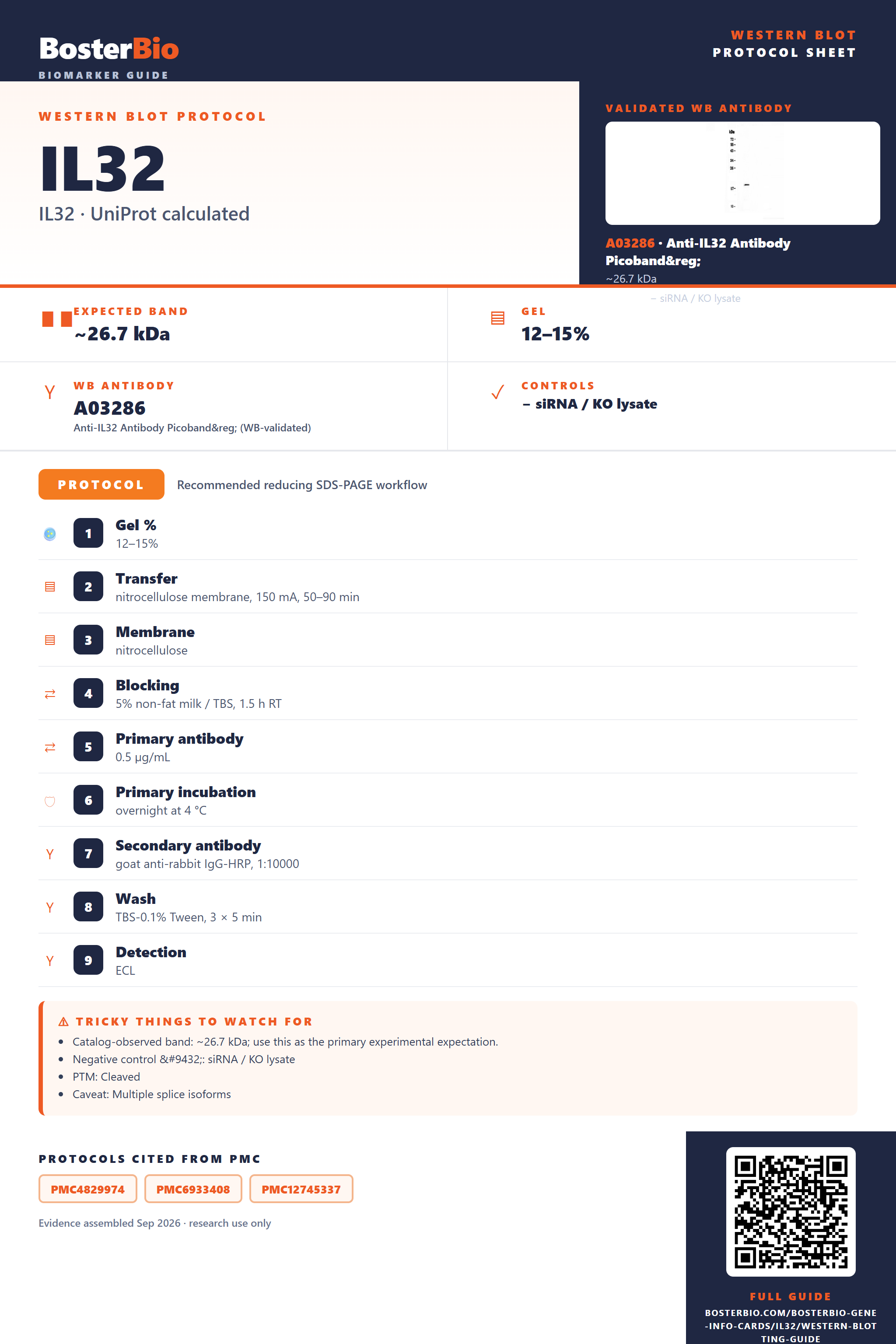
Expected bands, validated protocols, controls and antibodies — the at-a-glance facts below, then the full design guide.
| Expected band | ~26.7 kDa | |
| Gel | 12–15% | |
| Negative control | siRNA / KO lysate |
| PTM | Cleaved | |
| Caveat | Multiple splice isoforms | |
| Regulation | Epithelial mesenchymal transition | |
| Isoform | 7 isoform(s) |
Literature-validated Western blot parameters for IL32 — gel percentage, transfer, blocking, antibody incubation and detection, extracted from published methods.
| Gel % | 12–15% |
| Transfer | nitrocellulose membrane, 150 mA, 50–90 min |
| Membrane | nitrocellulose |
| Blocking | 5% non-fat milk / TBS, 1.5 h RT |
| Primary antibody | 0.5 µg/mL |
| Primary incubation | overnight at 4 °C |
| Secondary antibody | goat anti-rabbit IgG-HRP, 1:10000 |
| Wash | TBS-0.1% Tween, 3 × 5 min |
| Detection | ECL |
| Exposure / imaging | Tanon 5200 |
| Observed band | 19 kDa |
IL32 has a 26.7 kDa calculated precursor mass but the secreted mature protein runs smaller after cleavage of its 30-residue signal peptide.
| single band in the low-to-mid 20s kDa range in secreted or conditioned-media fractions | represents the mature IL32 monomer after signal peptide cleavage, running below the 26.7 kDa full-length calculated mass |
| little or no band in whole-cell lysate | IL32 is annotated as secreted, so most protein leaves the cell and intracellular lysate may under-represent it |
| band running lower than the 26.7 kDa calculated precursor mass | cleavage of the 30-residue N-terminal signal peptide removes several kDa to generate the mature secreted form |
| multiple bands at different apparent masses | seven annotated splice isoforms of IL32 can produce protein products of differing length |
| no shift in band position between reducing and non-reducing gels | IL32 has no annotated disulfide-linked oligomer, so its monomer size should be unaffected by reducing conditions |
| predicted full-length mass (26.7 kDa, 234 aa) | sets the calculated baseline size before any processing is accounted for |
| N-terminal signal peptide (residues 1-30) | cleaved off in the mature secreted protein, lowering the apparent band relative to the calculated precursor mass |
| secreted subcellular localization | shifts detectable protein into conditioned media or secreted fractions rather than whole-cell lysate |
| seven annotated splice isoforms | different isoforms may run at different apparent sizes, producing multiple bands depending on which isoform(s) are expressed and recognized |
| absence of glycosylation or disulfide-linked oligomer | no glycan-driven upward smear or dimer band expected; the monomer band should stay sharp and reducing-condition independent |
| Situation | Likely cause | Next action |
|---|---|---|
| No band in lysate | IL32 is secreted and exported from the cell, leaving little protein in the intracellular pool | probe conditioned media or a secreted-protein fraction instead of relying solely on whole-cell lysate |
| Band lower than expected | removal of the 30-residue signal peptide during secretion lowers the mature protein below the 26.7 kDa calculated precursor mass | compare the observed band against the mature-form size rather than the unprocessed precursor calculation |
| Multiple bands | seven annotated splice isoforms of IL32 can generate protein products of differing length | check which isoform(s) the antibody epitope targets and confirm which isoforms are expressed in the sample |
| Weak or no signal | as a secreted cytokine, IL32 may be diluted across a large media volume or degraded once released | concentrate conditioned media, use fresh protease-inhibited samples, and load a larger volume |
| Fragments below expected size | the secreted mature protein can be susceptible to extracellular proteolytic degradation after release | add protease inhibitors, process samples promptly, and avoid repeated freeze-thaw of secreted fractions |
| Band higher than expected | incomplete cleavage of the signal peptide can leave residual unprocessed precursor in the sample | confirm cleavage state using a mature-specific antibody or compare lysate versus secreted-fraction samples |
Comprehensive Human Protein Atlas IHC scoring per tissue. Rows are taken directly from the HPA tissue chart — click any row's HPA link to view the source.
| Tissue | Cell type | Level | Evidence | Source |
|---|
| Tissue | Cell type | Level | Evidence | Source |
|---|
Deeper troubleshooting and optimisation questions for IL32, answered from its protein features.
BosterBio's IL32 antibodies are among the best-performing WB antibodies on the market — well cited, thoroughly validated, and orthogonally cross-validated against negative tissues and complementary methods.
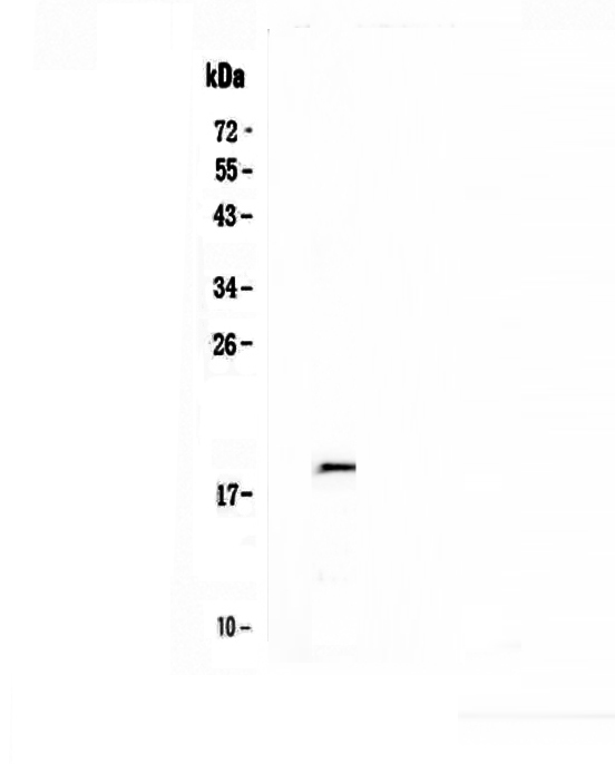
Our top-recommended anti-IL32 antibody for Western blot is a best-performing, widely cited reagent, rigorously validated across multiple studies and orthogonally cross-checked against negative-tissue controls and complementary detection methods, ensuring confident, specific, and reproducible IL32 detection in your Western blot experiments.
Which to pick: Only one anti-IL32 antibody is catalogued here: A03286. It includes an authentic Western blot validation image using recombinant human IL32, making it the clear, and only, choice for your Western blot experiments.