This website uses cookies to ensure you get the best experience on our website.


- Table of Contents
Plan chromogenic IL9R IHC in paraffin sections using the membrane topology and tissue staining evidence (UniProt; HPA tissue IHC). The guide highlights high staining in round or early spermatids, lower staining in several other cell populations, and a reported off-target binding concern (HPA tissue IHC).

Expected localisation, validated protocols, controls and antibodies — the at-a-glance facts below, then the full design guide.
| Expected localisation | Cell membrane; secreted form also annotated (UniProt) | |
| Staining pattern | Subset of immune cells; compartment unreported (HPA tissue IHC) | |
| Antigen retrieval | Citrate pH 6.0 HIER, 95–98 °C, 20 min (rule: cytoplasmic / membrane antigen) | |
| Positive control | Testis | |
| Negative control | Adipose tissue+4 more · see all |
| Fixation | Keep fixation consistent across sections (standard IHC practice; not target-specific). Selected-image fixative and duration unreported (datasheet A07490-1); verify before use. | |
| Caveat | Presumed off-target binding may mimic IL9R staining (HPA tissue IHC) | |
| Regulation | Expression regulation unreported (UniProt) | |
| Isoform / epitope | 3 isoforms; check extracellular versus cytoplasmic epitope (UniProt) |
The catalog antibody protocol is paired with a published IL9R IHC protocol for paraffin-embedded skin sections (PMC4967550).
| Sample | Paraffin-embedded human liver carcinoma tissue; fixative not specified (datasheet A07490-1) |
| Fixation | Image fixative and duration unreported (datasheet A07490-1); verify before use. |
| Sectioning | 4–5 µm sections on charged slides (standard) |
| Deparaffinisation | Xylene, graded ethanol series to water (standard) |
| Antigen retrieval | Heat-induced epitope retrieval in citrate buffer, pH 6.0, 20 min at 95–98 °C (standard rule: cytoplasmic / membrane antigen) |
| Peroxidase block | 3% H2O2, 10 min, room temperature (standard) |
| Blocking | 10% normal serum of the secondary host, 30 min, room temperature (standard) |
| Primary antibody | Rabbit anti-IL9R, 1:50-1:200 (datasheet A07490-1) |
| Primary incubation | Overnight at 4 °C (standard) |
| Detection | HRP-polymer secondary, DAB chromogen 5–10 min (standard) |
| Counterstain | Hematoxylin, blue, dehydrate and mount (standard) |
| Expected result | IL9R-positive staining in round or early spermatids of testis (HPA tissue IHC: High). HPA tissue profile: Expression in a subset of immune cells. No signal in the no-primary control. |
IL9R is a membrane receptor with an extracellular region and cytoplasmic tail (UniProt Q01113 topology); UniProt also annotates a secreted location (UniProt Q01113 subcellular). In paraffin tissue IHC, look first for staining in round or early spermatids, reported as high, and a subset of immune cells (HPA tissue IHC). HPA rates the tissue profile Approved, pending external verification, and notes presumed off-target binding that was disregarded (HPA tissue IHC).
| Distinct staining in round or early spermatids, especially near cell outlines. | This fits the high testis signal reported by HPA (HPA tissue IHC). A membrane-associated appearance is consistent with receptor topology (UniProt Q01113 topology), but HPA's tissue record does not establish the exact stained compartment in these cells. |
| Predominantly nuclear staining, with little staining near cell outlines. | A nuclear-only pattern does not fit the membrane topology or HPA membrane summary (UniProt Q01113 topology; HPA subcellular). Check the counterstain and detection controls before treating nuclear color as IL9R. |
| Strong staining in nearly every cell of a tissue reported as negative. | That conflicts with HPA's cell-specific observations; adipocytes in adipose tissue and respiratory epithelial cells in bronchus are examples reported as not detected (HPA tissue IHC). Cross-reactivity or endogenous chromogenic activity is plausible; neither can be diagnosed from appearance alone. |
| Diffuse color across cells, stroma, and slide background. | This does not resolve into the cell-specific profile described by HPA (HPA tissue IHC). As a general IHC check, inspect reagent-only and detection controls, washing, blocking, and antibody concentration before scoring positive cells. |
| No visible staining in round or early spermatids on an otherwise interpretable section. | That misses the strongest supplied positive reference (HPA tissue IHC: high in round or early spermatids). Review section quality, retrieval and detection controls as general IHC checks; the supplied sources give no IL9R-specific fixation sensitivity or working dilution. |
| Receptor topology and epitope location | IL9R has extracellular residues 41–270, a transmembrane segment at 271–291, and a cytoplasmic region at 292–521 (UniProt Q01113 topology). These positions guide interpretation of a documented antibody epitope; no epitope for the catalog antibody is supplied here. |
| Processing and secreted annotation | The signal peptide spans residues 1–40, and the annotated chain begins at residue 41 (UniProt Q01113 processing). UniProt also lists IL9R as secreted (UniProt Q01113 subcellular); these facts alone cannot establish that diffuse tissue staining is a shed receptor. |
| Glycosylation and isoforms | UniProt lists glycosylation sites at residues 117 and 156 and 3 isoforms (UniProt Q01113). Their effect on this antibody's paraffin-section staining is not supplied, so do not assign a staining difference to either feature without antibody-specific evidence. |
| Strength and scope of tissue evidence | HPA reports high staining in round or early spermatids, low staining in macrophages and selected glandular or urothelial cells, and no detection in listed negative cells (HPA tissue IHC). Its Approved rating is pending external verification, with presumed off-target binding disregarded (HPA tissue IHC). |
| Antibody validation and IF evidence | Both listed antibodies, HPA063240 and HPA064557, have IHC Approved status; neither has an ICC status in the supplied record (HPA antibodies). HPA summarizes the subcellular location as membrane but supplies no ICC-IF image cell lines (HPA subcellular). |
| Situation | Likely cause | Next action |
|---|---|---|
| The testis positive reference is blank. | The result conflicts with HPA's high signal in round or early spermatids (HPA tissue IHC); a failed stain or unsuitable section remains possible. | Confirm the expected cells are present, then review retrieval, primary-antibody application and chromogenic detection with routine positive and reagent controls. No source here identifies an IL9R-specific retrieval condition. |
| Staining looks mainly nuclear. | A nuclear-only result is discordant with membrane topology (UniProt Q01113 topology; HPA subcellular); nonspecific color or counterstain overlap may be involved. | Compare with the no-primary control and inspect cell boundaries at higher magnification. Score only resolved cell staining, and do not infer a new nuclear location from this section alone. |
| Reported negative cell types stain broadly. | Uniform color in adipocytes or bronchial respiratory epithelium conflicts with HPA's not-detected entries (HPA tissue IHC). Cross-reactivity or endogenous enzyme activity may contribute. | Check a no-primary detection control and the routine endogenous-activity block for the chosen chromogen. Compare the distribution with a reported positive cell population (HPA tissue IHC). |
| Background obscures individual cells. | Diffuse background cannot be matched reliably to HPA's subset-of-immune-cells profile (HPA tissue IHC). In routine IHC, excessive antibody, insufficient blocking or incomplete washing can contribute. | Use routine negative controls, verify blocking and washing, and titrate the antibody against a positive reference. The supplied record provides no catalog dilution or IL9R-specific adjustment. |
| Low-level lung or gut staining seems weaker than testis. | HPA reports low macrophage staining in lung and low glandular staining in duodenum, small intestine, colon and rectum, versus high staining in round or early spermatids (HPA tissue IHC). | Score the named cell populations separately and compare runs with the same positive reference. Avoid calling an entire organ positive or negative from a small, low-signal cell population. |
| Can this IHC pattern be used as an IF/ICC protocol? | HPA's membrane summary provides no ICC-IF image cell lines, and the listed antibodies have no ICC status in this record (HPA subcellular; HPA antibodies). | Treat membrane localization as an interpretation cue (UniProt Q01113 topology; HPA subcellular). Use the separate IF/ICC guide for method design; do not transfer an IHC-P condition as a validated IF setting. |
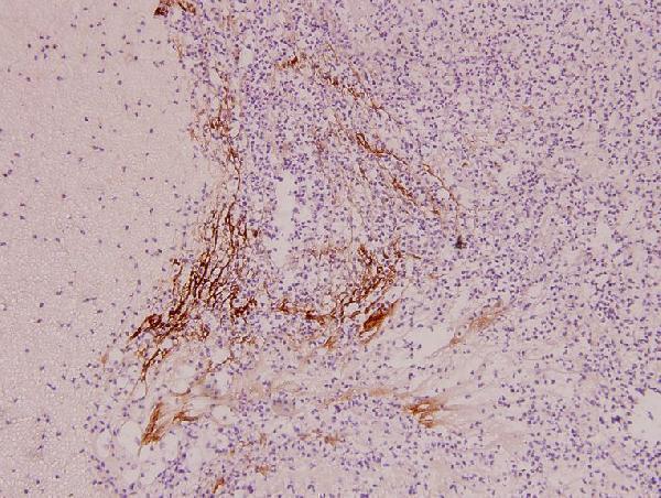


Comprehensive Human Protein Atlas IHC scoring per tissue (reliability: Approved — Pending external verification. Presumed off target binding observed and disregarded.). Rows are taken directly from the HPA tissue chart — click any row's HPA link to view the source.
| Tissue | Cell type | Level | Evidence | Source |
|---|---|---|---|---|
| Testis | Round or early spermatids | High | Protein (IHC) | HPA → |
| Tissue | Cell type | Level | Evidence | Source |
|---|---|---|---|---|
| Adipose tissue | Adipocytes | Not detected | Protein (IHC) | HPA → |
| Adrenal gland | Glandular cells | Not detected | Protein (IHC) | HPA → |
| Bone marrow | Hematopoietic cells | Not detected | Protein (IHC) | HPA → |
| Breast | Adipocytes | Not detected | Protein (IHC) | HPA → |
| Bronchus | Respiratory epithelial cells | Not detected | Protein (IHC) | HPA → |
Troubleshoot IL9R staining in paraffin sections by checking retrieval, cell identity and compartment before interpreting signal intensity.
A07490-1 has a real IHC image from paraffin-embedded human liver carcinoma tissue (catalog image caption). Human, mouse, and rat reactivity is listed (catalog reactivity); no IF image is supplied (catalog images).
A07490-1 is listed for IHC and WB in human, mouse, and rat (catalog applications and reactivity). Its IHC image shows paraffin-embedded human liver carcinoma tissue at 1:100 (catalog image caption).
Which to pick: Choose A07490-1 for paraffin-section tissue IHC: its image shows human liver carcinoma tissue at 1:100, and its listed IHC range is 1:50–1:200 (catalog image caption; catalog dilution). For cross-species IHC, A07490-1 lists mouse and rat reactivity, though its supplied IHC image is human only (catalog reactivity; catalog image caption). No IF/ICC application or image is supplied for A07490-1, so there is no supported IF/ICC pick here; the IHC caption does not report the fixative (catalog applications and images; catalog image caption).