IMMT / MICOS complex subunit MIC60 · IHC design guide

Design Immunohistochemistry for IMMT

Plan IMMT chromogenic IHC-P around cytoplasmic tissue staining (HPA tissue IHC) from a mitochondrial inner membrane target (UniProt). Use cardiomyocytes, gastric glandular cells or kidney collecting ducts as positive references (HPA tissue IHC), and start the catalog antibody at 2–5 μg/ml (datasheet A04102-2).

Evidence assembled Oct 2026 · For research use; verify linked source records and product datasheet before use
Immunohistochemistry protocol sheet for IMMT (IHC for IMMT): expected localisation Cytoplasmic staining (HPA tissue IHC); mitochondrial inner membrane (UniProt), antibody A04102-2, validated IHC image, and IHC protocol steps
Printable IMMT IHC protocol sheet — expected localisation Cytoplasmic staining (HPA tissue IHC); mitochondrial inner membrane (UniProt), antibody A04102-2, controls and protocol steps. Open the full IMMT IHC guide →

IMMT Immunohistochemistry Experimental Design Guide

Expected localisation, validated protocols, controls and antibodies — the at-a-glance facts below, then the full design guide.

Must know before staining
Expected localisation Cytoplasmic staining (HPA tissue IHC); mitochondrial inner membrane (UniProt)
Staining pattern Cytoplasmic in cardiomyocytes, gastric glands and collecting ducts (HPA tissue IHC)
Antigen retrieval EDTA pH 8.0 HIER, heat-mediated (datasheet A04102-2)
Positive control ⓘ Heart muscle+4 more · see all
Negative control ⓘ Adipose tissue+4 more · see all
Important caveats
Reasons your staining may differ from the expected pattern.
Fixation Keep fixation consistent across sections (standard IHC practice; not target-specific)
Caveat Staining varies by cell type; adipocytes are not detected (HPA tissue IHC)
Regulation Expression regulation is not established (UniProt)
Isoform / epitope 4 isoforms; mature chain 34–758; epitope coverage unknown (UniProt)
Section 1

Recommended IMMT IHC & IF Protocols

The catalog antibody has an IHC-P protocol (datasheet A04102-2); two published IMMT studies provide additional paraffin-section conditions (PMC6825906; PMC11150385).

Recommended immunohistochemistry (IHC-P) protocol parameters
SampleParaffin-embedded human esophageal squamous carcinoma tissue; fixative not specified (datasheet A04102-2)
FixationImage fixative and duration unreported (datasheet A04102-2); verify before use.
Sectioning4–5 µm sections on charged slides (standard)
DeparaffinisationXylene, graded ethanol series to water (standard)
Antigen retrievalHeat retrieval: EDTA pH 8.0 (datasheet A04102-2); 20 min, 95–100 °C (standard)
Peroxidase block3% H2O2, 10 min, room temperature (standard)
Blocking10% goat serum (datasheet A04102-2)
Primary antibodyRabbit anti-IMMT, 2-5 μg/ml (datasheet A04102-2)
Primary incubationOvernight at 4 °C (datasheet A04102-2)
DetectionHRP-conjugated secondary, DAB chromogen (datasheet A04102-2)
CounterstainHematoxylin, blue, dehydrate and mount (standard)
Expected resultIMMT-positive staining in cardiomyocytes of heart muscle (HPA tissue IHC: High). HPA tissue profile: General cytoplasmic expression, most abundant in heart, stomach and the renal tubules. No signal in the no-primary control.
💡Decision noteStart with heat-mediated EDTA pH 8.0 retrieval for the catalog antibody (datasheet A04102-2); citrate pH 6.0 is a published alternative (PMC6825906).
Section 2

What Is the Expected IMMT Staining Pattern?

IMMT is an inner mitochondrial membrane protein with residues 65–758 facing the intermembrane space (UniProt Q16891 topology). In paraffin IHC, expect cytoplasmic staining in cardiomyocytes, renal collecting ducts, and stomach glandular cells (HPA: High). HPA describes general cytoplasmic expression, with Supported tissue IHC reliability and medium consistency between staining and RNA data (HPA: tissue IHC).

What am I looking at on my slide?
Cytoplasmic staining is strong in cardiomyocytes, renal collecting ducts, or stomach glandular cells.This matches the high-staining cell populations in the tissue survey (HPA: High). A granular cytoplasmic appearance is compatible with mitochondrial localisation, though chromogenic IHC may not resolve individual mitochondria (UniProt Q16891 location; general IHC practice).
A crisp nuclear or cell-surface signal dominates while cytoplasmic staining is absent.That compartment conflicts with the mitochondrial inner membrane assignment and HPA cytoplasmic profile (UniProt Q16891 location; HPA: tissue IHC). Treat it as suspect and compare it with the slide’s positive tissue and detection controls (general IHC practice).
Strong staining appears in adipocytes or oral squamous epithelial cells.HPA reports IMMT as Not detected in those cells (HPA: adipocytes; HPA: oral mucosa). Check for antibody cross-reactivity or endogenous detection activity; a mismatch is a warning, rather than proof of either cause (general IHC practice).
Colour spreads across stroma, nuclei, and empty regions without clear cell boundaries.This is diffuse background, not the cell-associated cytoplasmic pattern reported for IMMT (HPA: tissue IHC). Review the no-primary control and detection steps before scoring cells as positive (general IHC practice).
Cardiomyocytes or renal collecting ducts show no discernible cytoplasmic signal.Both are high-staining populations in HPA’s tissue IHC survey (HPA: High). A blank result there warrants a check of staining controls and assay performance before interpreting a study sample as IMMT-negative (general IHC practice).
💡Expected IMMT appearanceCall a result positive when high-staining cardiomyocytes, renal collecting ducts, or stomach glandular cells show cell-associated cytoplasmic colour (HPA: High; HPA: cytoplasmic); dominant nuclear colour or diffuse background is suspect (UniProt Q16891 location; general IHC practice).
How each factor affects the staining
Tissue and cell selectionHPA reports High staining in cardiomyocytes and collecting ducts, but Not detected in adipocytes and bone marrow hematopoietic cells; score the specified cell population, not the whole section (HPA: tissue IHC).
Membrane topology and epitopeIMMT has one transmembrane segment at residues 46–64, with residues 65–758 facing the intermembrane space (UniProt Q16891 topology). Epitope location for the chosen antibody is unspecified; no retrieval response can be predicted from topology alone.
Isoforms and processingUniProt lists four isoforms and a mature chain spanning residues 34–758 (UniProt Q16891). Without an antibody epitope assignment, the record cannot establish which isoforms contribute to the observed stain.
Strength of tissue evidenceHPA rates tissue IHC Supported, with medium consistency between antibody staining and RNA expression; its antibody entries are IHC Supported (HPA: tissue IHC; HPA: antibodies). Use cell-level controls when judging unexpected staining (general IHC practice).
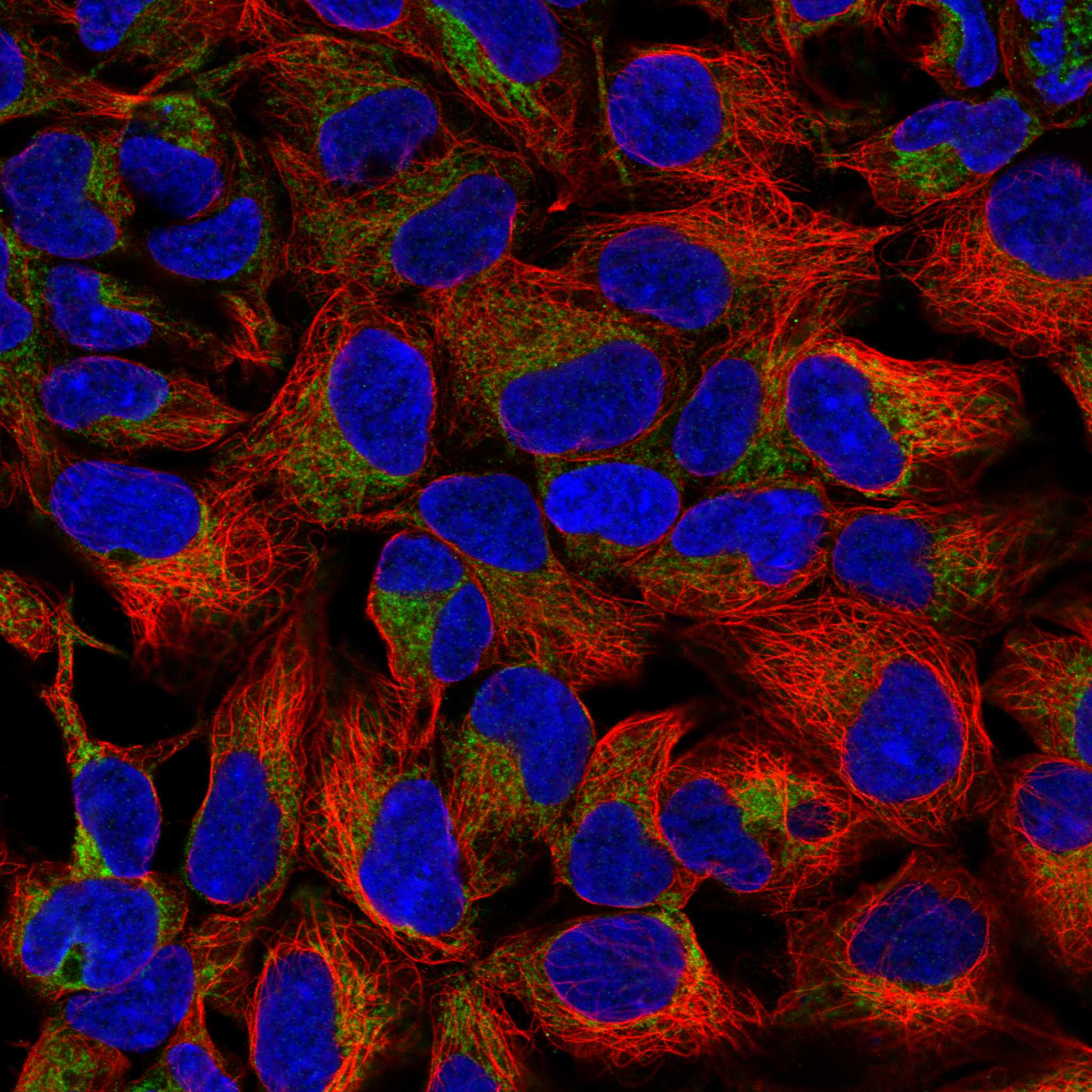
IF/ICC cross-check?Yes: HPA reports enhanced mitochondrial localisation by ICC-IF and ICC Enhanced validation for HPA036164 and HPA036165 (HPA: subcellular; HPA: antibodies). This supports localisation, without specifying an IHC retrieval or staining protocol.
Why is my staining missing, weak or wrong?
SituationLikely causeNext action
Known-positive tissue is blank.The run may have failed; HPA reports High staining in cardiomyocytes, renal collecting ducts, and stomach glandular cells (HPA: tissue IHC).Check the positive control, primary-antibody step, retrieval record, and detection reagents before scoring the sample negative (general IHC practice).
Signal is faint only in a low-staining cell population.Some populations have Low staining, including cerebral cortex neuronal cells and salivary gland glandular cells (HPA: tissue IHC).Compare with a High-staining cell population in the same run; record intensity by cell type (HPA: tissue IHC; general IHC practice).
Nuclear staining dominates.Nuclear localisation conflicts with the mitochondrial assignment (UniProt Q16891 location; HPA: subcellular).Compare with the no-primary control and a known-positive tissue; review detection background before accepting the signal (general IHC practice).
A Not detected cell population stains strongly.Cross-reactivity or endogenous detection activity is possible when staining conflicts with HPA’s adipocyte or oral mucosa entries (HPA: tissue IHC; general IHC practice).Inspect the no-primary control and confirm the scored cell type; investigate the detection system if the control also stains (general IHC practice).
Diffuse colour obscures cell boundaries.Background can prevent assessment of the cytoplasmic pattern reported by HPA (HPA: tissue IHC; general IHC practice).Review blocking, washing, primary-antibody concentration, and the no-primary control; rescore only interpretable cells (general IHC practice).
IF/ICC appears mitochondrial, but IHC is blank.HPA’s enhanced mitochondrial ICC-IF localisation and Supported tissue IHC are distinct observations; agreement is not guaranteed in a given run (HPA: subcellular; HPA: tissue IHC).Use the IHC positive tissue and run controls to assess the paraffin assay; do not infer its retrieval conditions from IF/ICC (general IHC practice).

Sample controls for IMMT IHC & IF

🧪Run heart muscle first: cardiomyocytes should show IMMT staining (HPA: High in cardiomyocytes). Use adipose tissue as the negative comparator (HPA: Not detected in adipocytes); on the heart slide, assess any unstained interstitial cells as an internal background reference without assuming they are IMMT-negative.
Positive control tissue: Heart muscle (Cardiomyocytes, HPA High)
Negative control tissue: Adipose tissue (HPA Not detected)
ICC-IF cell lines (HPA subcellular resource): HPA ICC-IF images show IMMT in HEK293, MCF-7, U2OS, Sperm, NIH 3T3, with annotated localisation: Mitochondria (enhanced) (HPA subcellular).
Technical controls: Include no-primary (secondary-only) and matched rabbit IgG controls (caption: rabbit anti-IMMT antibody), plus IMMT-knockout material or peptide competition if the immunizing peptide is available. Quench endogenous peroxidase in heart sections before HRP/DAB detection (caption: HRP/DAB detection; standard IHC practice).
⚠️Feasibility: A target-specific fixation window or fixation effect is unreported, and the selected A04102-2 paraffin-section caption does not state the fixative. The caption uses heat retrieval in EDTA at pH 8.0, but does not establish that retrieval is required; the supplied evidence does not establish whether frozen sections or IF are easier (caption: paraffin-section retrieval; HPA: mitochondrial ICC-IF localisation). In heart muscle, endogenous peroxidase can complicate chromogenic interpretation, so compare staining with the control slides (standard IHC practice).

HPA tissue IHC evidence for IMMT

Comprehensive Human Protein Atlas IHC scoring per tissue (reliability: Supported — Medium consistency between antibody staining and RNA expression data.). Rows are taken directly from the HPA tissue chart — click any row's HPA link to view the source.

Positive expression · recommended positive controls

TissueCell typeLevelEvidenceSource
Heart muscle Cardiomyocytes High Protein (IHC) HPA →
Kidney Collecting ducts High Protein (IHC) HPA →
Parathyroid gland Glandular cells High Protein (IHC) HPA →
Stomach Glandular cells High Protein (IHC) HPA →
Adrenal gland Glandular cells Medium Protein (IHC) HPA →

Undetected expression · recommended negative controls

TissueCell typeLevelEvidenceSource
Adipose tissue Adipocytes Not detected Protein (IHC) HPA →
Bone marrow Hematopoietic cells Not detected Protein (IHC) HPA →
Epididymis Glandular cells Not detected Protein (IHC) HPA →
Oral mucosa Squamous epithelial cells Not detected Protein (IHC) HPA →
Ovary Follicle cells Not detected Protein (IHC) HPA →
Section 3

Advanced IMMT IHC Tips

Troubleshoot IMMT chromogenic IHC by checking retrieval, antibody conditions, mitochondrial staining patterns, and cell type matched controls.

Which retrieval conditions should I start with for weak IMMT staining?
Start with heat mediated retrieval in EDTA at pH 8.0 for the catalog antibody (datasheet A04102-2). The documented paraffin section workflow then used 2 μg/ml primary antibody overnight at 4°C, so keep those conditions fixed while checking retrieval first (IHC caption A04102-2). Compare an adequately heated section with one processed without retrieval, using matched sections and the same development time (standard IHC practice). If staining remains weak, test another retrieval condition as a fallback on adjacent sections while monitoring tissue damage and background (standard IHC practice). Do not interpret a stronger DAB signal alone as improved specificity; check whether its cellular distribution remains credible (standard IHC practice).
How should I troubleshoot inconsistent IMMT staining after fixation?
The selected paraffin section caption does not state a fixative, so target specific sensitivity to fixation is unknown (IHC caption A04102-2). Record each specimen's fixative, fixation interval, processing history, and section thickness before comparing stain intensity (standard IHC practice). On matched sections, hold EDTA retrieval at pH 8.0 and primary incubation overnight at 4°C constant while evaluating processing differences (datasheet A04102-2; IHC caption A04102-2). Include a reference section in every run and compare staining within the same cell type, since uneven fixation can create spatial intensity gradients (standard IHC practice). Do not attribute a fixation effect to IMMT without a direct comparison under controlled staining conditions (standard IHC practice).
What cellular staining pattern is credible for IMMT in paraffin sections?
Expect a cytoplasmic, mitochondrial distribution at light microscope resolution, rather than a predominantly nuclear pattern (HPA tissue IHC; HPA subcellular; UniProt Q16891 localisation). IMMT resides in the mitochondrial inner membrane, with its 46–64 transmembrane segment and residues 65–758 on the intermembrane space side (UniProt Q16891 topology). In a tissue control, cardiomyocytes and kidney collecting duct cells have reported high staining, whereas oral mucosa squamous epithelial cells were not detected (HPA tissue IHC). Compare DAB deposits with the cell boundaries and counterstained nuclei on adjacent fields, and reject staining confined to nuclei or section edges as localisation evidence (standard IHC practice).
Could an unknown epitope explain variable IMMT staining across samples?
IMMT has 4 annotated isoforms, but the supplied antibody evidence does not map its epitope to an isoform or residue interval (UniProt Q16891 isoforms; IHC caption A04102-2). Its single membrane spanning region occupies residues 46–64, while most of the recorded sequence faces the intermembrane space (UniProt Q16891 topology). Phosphoserines at 103, 112, and 113 are annotated, but no supplied evidence shows that these modifications change antibody binding in sections (UniProt Q16891 modified residues; IHC caption A04102-2). If samples disagree, compare matched processing and retrieval first, then seek epitope mapping or an independently validated antibody before assigning the difference to isoform expression (standard IHC practice).
How can IF help assess an ambiguous chromogenic IMMT pattern?
Use IF on a separately validated preparation to compare IMMT with a mitochondrial marker and a marker identifying the expected cell type; mitochondrial localisation is reported for IMMT (HPA subcellular). Select spectrally separated fluorophores and inspect unstained tissue in each channel before acquisition, because tissue autofluorescence can resemble weak specific signal (standard IF practice). Choose and validate permeabilisation against the antibody's mapped epitope: residues 34–45 face the matrix, whereas residues 65–758 face the intermembrane space, and the supplied antibody epitope is unmapped (UniProt Q16891 topology; IHC caption A04102-2). Keep IF fixation and permeabilisation comparisons within IF preparations; the paraffin IHC caption supplies no IF fixation conditions (IHC caption A04102-2).
How do I reduce diffuse brown staining without losing IMMT signal?
First compare a no primary control with the stained section to identify signal from detection reagents or endogenous enzyme activity (standard IHC practice). Include an endogenous peroxidase block before HRP detection and assess DAB development with the same timing across sections (standard IHC practice). The documented section used 10% goat serum block, 2 μg/ml primary antibody overnight at 4°C, and a peroxidase conjugated secondary for 30 minutes at 37°C (IHC caption A04102-2). If the no primary control is clean, titrate primary concentration around the documented value and improve washes while checking preservation of cytoplasmic mitochondrial pattern (standard IHC practice; HPA subcellular).
What should I score when comparing IMMT across paraffin sections? ⚠ ANSWER MARKED FOR VERIFICATION
Define the target cell population and exclude necrosis, folds, and section edges before image analysis (standard IHC practice). Score the percentage of positive cells and intensity within that population, or calculate an H-score from intensity grades 0–3 and their percentages (standard IHC practice). For spatial analyses, report positive cells per mm² of viable tissue and normalize the result to the area or number of the same cell type in each specimen (standard IHC practice). Keep retrieval at EDTA pH 8.0, antibody concentration at the documented 2 μg/ml starting point, and DAB exposure consistent across comparisons (datasheet A04102-2; IHC caption A04102-2; standard IHC practice).
How can I distinguish genuine IMMT staining from tissue artefacts?
A credible result has cellular cytoplasmic staining consistent with mitochondrial localisation, rather than isolated nuclear deposits or pigment outside cells (HPA tissue IHC; HPA subcellular; standard IHC practice). Compare expected positive populations such as cardiomyocytes or kidney collecting duct cells with an appropriate lower staining population, while accounting for specimen quality (HPA tissue IHC; standard IHC practice). Staining limited to cut edges, folds, or necrotic regions suggests a processing artefact; staining in a no primary control points toward detection background or endogenous peroxidase (standard IHC practice). Treat unexpectedly strong diffuse DAB signal cautiously even if its overall distribution appears plausible, because HPA rates its tissue staining support as medium consistency with RNA expression data (HPA tissue IHC).
Boster reagents

Best IMMT / MICOS complex subunit MIC60 IHC Antibodies

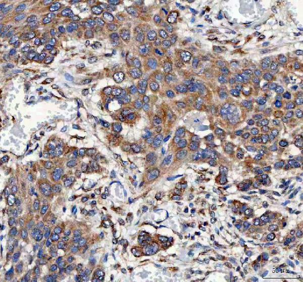
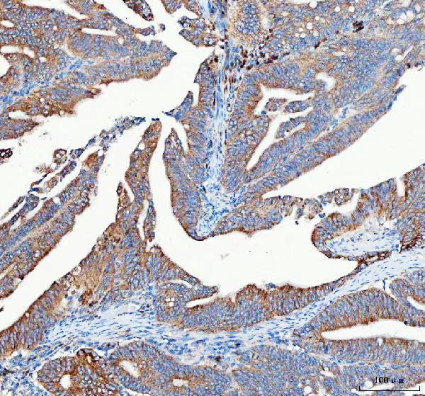
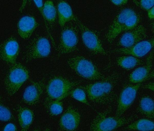
A04102-2 has IHC images from human paraffin sections and an IF/ICC image from HeLa cells; the catalog lists human, mouse, and rat reactivity (A04102-2 image captions; catalog reactivity).

Real IHC data IHC analysis of Mitofilin/IMMT using anti-Mitofilin/IMMT antibody (A04102-2). Mitofilin/IMMT was detected in a paraffin-embedded section of human esophageal squamous carcinoma tissue. Heat mediated antigen retrieval was performed in EDTA buffer (pH 8.0, epitope retrieval solution). The tissue section was blocked with 10% goat serum. The tissue section was then incubated with 2 μg/ml rabbit anti-Mitofilin/IMMT Antibody (A04102-2) overnight at 4°C. Peroxidase Conjugated Goat Anti-rabbit IgG was used as secondary antibody and incubated for 30 minutes at 37°C. The tissue section was developed using HRP Conjugated Rabbit IgG Super Vision Assay Kit (Catalog # SV0002) with DAB as the chromogen.
Anti-Mitofilin/IMMT Antibody ®
Cat # A04102-2

A04102-2 has IHC images from paraffin sections of human esophageal squamous carcinoma, rectum adenocarcinoma, lung cancer, and testicular germ cell tumors (A04102-2 IHC image captions). A04102-2 also has an IF/ICC image from HeLa cells (A04102-2 IF image caption).

Which to pick: For paraffin-section tissue IHC, choose A04102-2: its own IHC captions document human tissue staining with EDTA retrieval at pH 8.0 and 2 μg/ml primary antibody; the fixative is unreported (A04102-2 IHC image captions). For human IF/ICC, the same SKU has a HeLa cell image and catalog-listed IF/ICC use (A04102-2 IF image caption; catalog applications). For mouse or rat IHC, A04102-2 is catalog-listed as reactive with both species, although its supplied IHC images show human tissue only; clone status is unreported (catalog reactivity; A04102-2 IHC image captions; catalog clone field).

Each figure is that product's own IHC / IF validation image from its datasheet.

References

  1. UniProt Consortium. UniProt entry Q16891 (MIC60_HUMAN, MICOS complex subunit MIC60).
  2. Human Protein Atlas. IMMT tissue IHC expression (reliability: Supported).
  3. Human Protein Atlas. IMMT subcellular location (ICC-IF): Localized to the mitochondria..
  4. Human Protein Atlas. IMMT antibody validation summary (3 antibodies).
  5. Prognostic significance of IMMT expression in surgically-resected lung adenocarcinoma. Thoracic cancer 2019 — PMC6825906.
  6. Metabolic adaptation to IMMT deficiency through the ATF6-PPARγ axis is contingent on TP53 mutation status in breast cancer. Cell death & disease 2026 — PMC13261075.
  7. Suppressing mitochondrial inner membrane protein (IMMT) inhibits the proliferation of breast cancer cells through mitochondrial remodeling and metabolic regulation. Scientific reports 2024 — PMC11150385.
  8. Genetic ablation of Immt induces a lethal disruption of the MICOS complex. Life science alliance 2024 — PMC10927357.
  9. PubMed PMID:8039717 — UniProt-cited evidence.
  10. PubMed PMID:9168817 — UniProt-cited evidence.
  11. PubMed PMID:14702039 — UniProt-cited evidence.