IMPDH2 / Inosine-5'-monophosphate dehydrogenase 2 · IHC design guide

Design Immunohistochemistry for IMPDH2

Plan IMPDH2 staining in paraffin sections around the cytoplasmic and nuclear pattern reported across most tissues (HPA tissue IHC). This guide covers fixation consistency, antigen retrieval and controls for interpreting tissue staining.

Evidence assembled Oct 2026 · For research use; verify linked source records and product datasheet before use
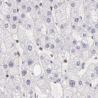
Immunohistochemistry protocol sheet for IMPDH2 (IHC for IMPDH2): expected localisation Cytoplasmic and nuclear staining (HPA tissue IHC), antibody A03021-2, validated IHC image, and IHC protocol steps
Printable IMPDH2 IHC protocol sheet — expected localisation Cytoplasmic and nuclear staining (HPA tissue IHC), antibody A03021-2, controls and protocol steps. Open the full IMPDH2 IHC guide →

IMPDH2 Immunohistochemistry Experimental Design Guide

Expected localisation, validated protocols, controls and antibodies — the at-a-glance facts below, then the full design guide.

Must know before staining
Expected localisation Cytoplasmic and nuclear staining (HPA tissue IHC)
Staining pattern Cytoplasmic and nuclear staining in most tissues (HPA tissue IHC)
Antigen retrieval EDTA pH 8.0 HIER, heat-mediated (datasheet A03021-2)
Positive control ⓘ Appendix+4 more · see all
Negative control ⓘ None in HPA (detected in all 45 tissues); use no-primary + isotype controls
Important caveats
Reasons your staining may differ from the expected pattern.
Fixation Keep fixation conditions consistent across sections. (standard IHC practice; not target-specific)
Caveat Staining may reflect proteins from more than one gene (HPA tissue IHC)
Regulation IMPDH2 predominates over IMPDH1 in tumors (UniProt)
Isoform / epitope No annotated isoforms or signal peptide (UniProt)
Section 1

Recommended IMPDH2 IHC & IF Protocols

The catalog antibody’s IHC-P protocol (datasheet A03021-2) is followed by four published IMPDH2 IHC protocols (PMC5429725; PMC6282329; PMC10197894; PMC8149691).

Recommended immunohistochemistry (IHC-P) protocol parameters
SampleParaffin-embedded human esophageal squamous carcinoma tissue; fixative not specified (datasheet A03021-2)
FixationImage fixative and duration unreported (datasheet A03021-2); verify before use.
Sectioning4–5 µm sections on charged slides (standard)
DeparaffinisationXylene, graded ethanol series to water (standard)
Antigen retrievalHeat retrieval: EDTA pH 8.0 (datasheet A03021-2); 20 min, 95–100 °C (standard)
Peroxidase block3% H2O2, 10 min, room temperature (standard)
Blocking10% goat serum (datasheet A03021-2)
Primary antibodyRabbit anti-IMPDH2, 2 μg/ml (datasheet A03021-2)
Primary incubationOvernight at 4 °C (datasheet A03021-2)
DetectionHRP-conjugated secondary, DAB chromogen (datasheet A03021-2)
CounterstainHematoxylin, blue, dehydrate and mount (standard)
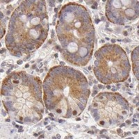
Expected resultIMPDH2-positive staining in glandular cells of appendix (HPA tissue IHC: High). HPA tissue profile: Cytoplasmic and nuclear expression in most tissues. No signal in the no-primary control.
💡Decision noteStart with heat-mediated EDTA retrieval at pH 8.0 for the catalog antibody (datasheet A03021-2). Use each paper’s stated retrieval condition when reproducing its protocol (PMC5429725; PMC6282329; PMC10197894).
Section 2

What Is the Expected IMPDH2 Staining Pattern?

IMPDH2 is a soluble protein found in the cytoplasm and nucleus (UniProt P12268: localization; no transmembrane segment). Expect staining in many tissues, including glandular, hematopoietic and immune cells (HPA: tissue IHC). HPA rates its tissue staining Approved, with medium agreement between staining and RNA data, and cautions that the antibody signal may include protein from more than one gene (HPA: reliability).

What am I looking at on my slide?
Cytoplasmic staining, with or without nuclear staining, in appendix or colon glandular cells.This matches the reported compartments and High staining in those cells (HPA: tissue IHC). Compare cells within the same section; a nuclear component alone does not invalidate an otherwise plausible pattern (UniProt P12268: localization).
Staining confined to a cell outline, lumen or extracellular deposit, with no intracellular signal.That distribution conflicts with the reported cytoplasmic and nuclear pattern and soluble topology (HPA: tissue IHC; UniProt P12268: topology). Check section morphology and detection background before assigning it to IMPDH2 (standard IHC practice).
Strong signal in cardiomyocytes, smooth muscle cells or fibroblasts, while nearby expected positive cells are unstained.HPA reports Low staining in those cell types, not absence (HPA: tissue IHC). The reversed contrast warrants checks for cross-reactivity or endogenous detection activity; it does not establish either cause by itself (standard IHC practice).
Uniform brown haze across cells and tissue spaces, obscuring cellular boundaries.Diffuse haze cannot be scored as the reported intracellular pattern (HPA: tissue IHC). Inspect a matched detection control and the counterstain; excess detection background can obscure localization (standard chromogenic IHC practice).
No cellular signal in a well-preserved appendix, colon or lymph node section.Glandular cells in appendix and colon, and germinal center cells in lymph node, are reported High (HPA: tissue IHC). A blank result prompts a check of the antibody and detection workflow; it does not prove biological absence.
💡Expected IMPDH2 appearanceCall a result positive when identifiable cells show intracellular cytoplasmic staining, sometimes with nuclear staining, with High signal possible in HPA-listed glandular or germinal center cells; outline-only or extracellular color is suspect (HPA: tissue IHC; UniProt P12268: localization).
How each factor affects the staining
Cell and tissue choiceAppendix and colon glandular cells, lung macrophages and lymph node germinal center cells are reported High; cardiomyocytes, smooth muscle cells and fibroblasts are Low (HPA: tissue IHC). Use these as relative expectations, not absolute positive and negative controls.
Compartment and topologyCytoplasm and nucleus are reported locations, and IMPDH2 has no transmembrane segment or signal peptide (UniProt P12268: localization and topology). Interpret intracellular staining against cell morphology; membrane-only color needs scrutiny.
Antibody specificityHPA rates tissue staining Approved but reports medium agreement with RNA and cautions that the signal may include protein from more than one gene (HPA: reliability). HPA001400 and CAB020717 each have Approved IHC status (HPA: antibody validation); status does not remove that caveat.
Processing and antigen retrievalNo signal peptide, propeptide or shedding is reported (UniProt P12268: processing). The supplied sources give no IMPDH2-specific fixation sensitivity or retrieval condition. Choose and document retrieval using the IHC-validated antibody's instructions and controls (standard IHC practice).
IF/ICC Q&AWhere should IF/ICC signal appear? Mainly in the cytosol, with additional rods and rings reported in cell imaging (HPA: subcellular ICC-IF); UniProt also notes cytoophidia after guanine-nucleotide depletion (UniProt P12268: localization). IF/ICC optimization belongs in its own guide.
Why is my staining missing, weak or wrong?
SituationLikely causeNext action
Expected high-staining cells show no chromogenic signal.An antibody, retrieval or detection step may have failed; the slide alone cannot identify which (standard IHC practice).Run a documented positive tissue control, then check the IHC-validated antibody's retrieval, dilution and detection instructions; HPA lists appendix or colon glandular cells as High (HPA: tissue IHC).
Only nuclei stain strongly.Nuclear IMPDH2 is reported, but HPA describes a cytoplasmic and nuclear tissue pattern (UniProt P12268: localization; HPA: tissue IHC).Inspect cytoplasmic signal in well-preserved positive cells and compare detection controls before calling nuclear-only staining specific (standard IHC practice).
Cell borders or luminal material stain more than cell interiors.That pattern does not fit the reported soluble intracellular locations (UniProt P12268: topology and localization).Review morphology and a matched detection control; score IMPDH2 only where a credible intracellular pattern is visible (standard IHC practice).
Low-staining reference cells appear stronger than expected.HPA labels cardiomyocytes, smooth muscle cells and fibroblasts Low, while warning that tissue antibody signal may include more than one gene (HPA: tissue IHC and reliability).Compare a high-staining cell population on the same run and check a detection control for endogenous activity; treat the apparent reversal as unresolved until controls agree (standard IHC practice).
Diffuse color obscures cell boundaries.Nonspecific antibody or detection background can prevent compartment scoring (standard chromogenic IHC practice).Check the matched control, blocking and detection conditions; adjust the documented IHC workflow, then reassess cytoplasmic and nuclear localization (standard IHC practice; HPA: tissue IHC).
A punctate or fiber-like pattern appears unexpectedly.UniProt reports cytoophidia under guanine-nucleotide depletion, and HPA reports rods and rings in ICC-IF; neither source establishes that every FFPE punctum is IMPDH2 (UniProt P12268: localization; HPA: subcellular ICC-IF).Document the pattern separately from routine diffuse staining and assess antibody and detection controls before assigning it to a specific IMPDH2 structure (standard IHC practice).

Sample controls for IMPDH2 IHC & IF

🧪Run esophagus first and score its squamous epithelial cells for IMPDH2 staining (HPA: High in esophageal squamous epithelial cells). HPA detects IMPDH2 in all 45 scored tissues, so there is no supported negative tissue; use no-primary and isotype controls, and treat any cells without specific staining on the positive slide as background rather than assuming a cell type is target-negative (HPA: no negative tissue rows; standard IHC practice).
Positive control tissue: Appendix (Glandular cells, HPA High)
Negative control tissue: None in HPA: IMPDH2 is detected in all 45 scored tissues. Use a no-primary (secondary-only) and an isotype control instead.
ICC-IF cell lines (HPA subcellular resource): HPA ICC-IF images show IMPDH2 in A-431, U-251MG, U2OS, ASC52telo, hTERT-RPE1 (serum starved), with annotated localisation: Cytosol (supported) (HPA subcellular).
Technical controls: Include a no-primary, secondary-only control; a rabbit IgG isotype control matched to the primary antibody’s clonality and concentration; and, if available, IMPDH2 knockout material or an immunogen peptide-block control (caption: rabbit anti-IMPDH2 primary; standard IHC practice). Block endogenous peroxidase before HRP/DAB detection and check the esophageal section for residual background (caption: HRP/DAB detection; standard IHC practice).
⚠️Feasibility: The selected A03021-2 paraffin-section caption does not report a fixative, and no target-specific fixation window or fixation effect is reported in the supplied evidence (selected-SKU tissue-IHC caption). Its staining used heat retrieval in EDTA at pH 8.0, but the caption does not establish that retrieval is required (selected-SKU tissue-IHC caption). Comparative ease of frozen-section IHC or IF and an esophagus-specific artefact are unreported; HPA ICC-IF images show mainly cytosolic staining with additional rods and rings in the listed cell lines (HPA: subcellular ICC-IF).

HPA tissue IHC evidence for IMPDH2

Comprehensive Human Protein Atlas IHC scoring per tissue (reliability: Approved — Medium consistency between antibody staining and RNA expression data. Caution, targets protein from more than one gene.). Rows are taken directly from the HPA tissue chart — click any row's HPA link to view the source.

Positive expression · recommended positive controls

TissueCell typeLevelEvidenceSource
Appendix Glandular cells High Protein (IHC) HPA →
Bone marrow Hematopoietic cells High Protein (IHC) HPA →
Colon Glandular cells High Protein (IHC) HPA →
Duodenum Glandular cells High Protein (IHC) HPA →
Esophagus Squamous epithelial cells High Protein (IHC) HPA →

Undetected expression · recommended negative controls

TissueCell typeLevelEvidenceSource
None in HPA: IMPDH2 is detected in all 45 scored tissues. Use a no-primary (secondary-only) and an isotype control instead.
Section 3

Advanced IMPDH2 IHC Tips

Use the catalog antibody’s paraffin-section IHC evidence as the starting point, then check staining against IMPDH2 localisation and tissue patterns.

Which retrieval conditions should I try first for IMPDH2 in paraffin sections?
Start with heat-mediated EDTA retrieval at pH 8.0 for paraffin sections (datasheet A03021-2). The selected tissue image used that buffer before overnight incubation at 4°C with 2 μg/ml primary antibody (datasheet A03021-2). Keep section thickness, heating, cooling, and detection conditions matched while comparing retrieval runs, so changes in staining remain interpretable (standard IHC practice). If staining is weak, test citrate buffer at pH 6.0 as a fallback on adjacent sections, with matched controls (standard IHC practice). Score signal alongside tissue preservation and background; stronger DAB alone does not establish specific IMPDH2 staining (standard IHC practice).
How should I assess whether fixation is affecting IMPDH2 IHC?
Target-specific fixation sensitivity is unknown because the selected paraffin-section caption does not report its fixative (datasheet A03021-2). For new material, document the fixative and duration, then process matched sections consistently before comparing IMPDH2 staining (standard IHC practice). A routine 10% neutral-buffered formalin process is a reasonable starting condition for paraffin IHC, but requires validation for this antibody (standard IHC practice). Compare morphology and staining in the same expected cell population rather than interpreting intensity across differently handled blocks in isolation (standard IHC practice). Repeat EDTA retrieval at pH 8.0 under matched conditions before attributing a difference to fixation (datasheet A03021-2; standard IHC practice).
Should IMPDH2 staining be cytoplasmic, nuclear, or arranged in rods?
Expect chiefly cytosolic staining, with possible nuclear expression in tissue sections (HPA subcellular: cytosol supported; HPA tissue IHC: cytoplasmic and nuclear expression). Rods and rings are an additional localisation, and guanine-nucleotide depletion can induce IMPDH2 cytoophidia (HPA subcellular: rods and rings approved; UniProt P12268). Record cytoplasmic and nuclear DAB separately for each cell population, using the counterstain to define nuclei (standard IHC practice). A thin DAB-positive structure may be difficult to distinguish from overlapping cells or precipitate in paraffin sections (standard IHC practice). If structures drive the conclusion, confirm their shape and cellular location with a spatially resolved method and appropriate controls (standard IHC practice).
How can I assess IMPDH2 specificity when a related enzyme is present?
The record lists 0 annotated IMPDH2 isoforms, but provides no antibody epitope map (UniProt P12268; supplied antibody evidence). IMPDH1 is the main species in normal leukocytes, whereas IMPDH2 predominates over IMPDH1 in tumors (UniProt P12268). The HPA tissue assessment warns that its staining may include protein from more than one gene, so its patterns alone cannot establish antibody specificity (HPA tissue IHC: Approved, medium consistency). Ask for epitope information and assess cross-reactivity with IMPDH1 before assigning every positive cell to IMPDH2 (standard IHC practice). IMPDH2 contains CBS domains at residues 114–173 and 179–237 and has reported modified residues; neither fact identifies this antibody’s binding site (UniProt P12268).
How should I follow up an IMPDH2 IHC pattern by multiplex IF?
Use IF as a separate validation experiment, starting from the expected cytosolic distribution and possible rods and rings (HPA subcellular: cytosol supported; rods and rings approved). Pair IMPDH2 with a marker for the cell type being evaluated; macrophages in lung are one documented high-staining population (HPA tissue IHC: High in lung macrophages). Choose fluorophores and acquisition channels after checking tissue autofluorescence, and include single-stain controls for multiplex interpretation (standard IF practice). IMPDH2 has no transmembrane segment, so permeabilisation should allow antibody access to intracellular epitopes, with its strength optimised against morphology (UniProt P12268 topology; standard IF practice). Do not transfer the paraffin IHC retrieval or incubation conditions directly to IF, which needs its own validation (datasheet A03021-2; standard IF practice).
What controls help identify nonspecific DAB staining in IMPDH2 sections?
The selected image used 10% goat serum blocking, 2 μg/ml primary antibody overnight at 4°C, and peroxidase-based DAB detection (datasheet A03021-2). Include a no-primary control and an endogenous peroxidase block to assess detection background in each tissue batch (standard IHC practice). Compare adjacent sections across a primary-antibody titration while keeping retrieval, DAB development, and imaging conditions matched (standard IHC practice). Inspect tissue edges, folds, necrotic regions, and pigment before scoring apparent cytoplasmic or nuclear positivity (standard IHC practice). Check whether the remaining cell distribution fits the reported broad cytoplasmic and nuclear profile, while treating that profile as supporting context rather than proof of specificity (HPA tissue IHC: profile; standard IHC practice).
How should I quantify IMPDH2 DAB staining across specimens? ⚠ ANSWER MARKED FOR VERIFICATION
Define the cell population and tissue region before scoring, because IMPDH2 tissue staining varies by cell type (HPA tissue IHC: positive and low-staining populations). For cellular expression, report the percentage of positive cells and an H-score from 0–300 using consistent intensity categories (standard IHC practice). Score cytoplasmic and nuclear compartments separately where both are visible, and exclude necrosis, folds, and poorly preserved regions (HPA tissue IHC: cytoplasmic and nuclear expression; standard IHC practice). If the question concerns infiltrating positive cells, report their density per mm² of viable tissue and the cell-type definition used (standard IHC practice). Normalise comparisons to the same eligible cell population or viable tissue area, with matched staining and imaging conditions (standard IHC practice).
When does an IMPDH2-positive DAB pattern warrant further validation?
A plausible result shows cellular cytoplasmic staining, sometimes with nuclear signal, in interpretable tissue rather than isolated marks (HPA tissue IHC: profile; standard IHC practice). High staining has been reported in lung macrophages and esophageal squamous epithelial cells, while cardiomyocytes are listed as low (HPA tissue IHC: positive and low-staining populations). Treat staining restricted to an unexpected compartment or cell population as a reason to check specificity and sample context (UniProt P12268 localisation; standard IHC practice). Edge-only signal, necrotic debris, and residual endogenous peroxidase activity can imitate DAB positivity and need control-section review (standard IHC practice). Even a matching tissue pattern is provisional because the HPA assessment cautions that staining may include protein from more than one gene (HPA tissue IHC: reliability description).
Boster reagents

Best IMPDH2 / Inosine-5'-monophosphate dehydrogenase 2 IHC Antibodies

The catalog includes IMPDH2 antibodies listed for IHC and IF in human, mouse, and rat (catalog applications and reactivity); image captions show human paraffin-section IHC and human IF/ICC (A03021-2 captions).

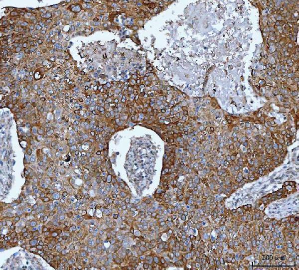
Real IHC data IHC analysis of IMPDH2 using anti-IMPDH2 antibody (A03021-2). IMPDH2 was detected in a paraffin-embedded section of human esophageal squamous carcinoma tissue. Heat mediated antigen retrieval was performed in EDTA buffer (pH 8.0, epitope retrieval solution). The tissue section was blocked with 10% goat serum. The tissue section was then incubated with 2 μg/ml rabbit anti-IMPDH2 Antibody (A03021-2) overnight at 4°C. Peroxidase Conjugated Goat Anti-rabbit IgG was used as secondary antibody and incubated for 30 minutes at 37°C. The tissue section was developed using HRP Conjugated Rabbit IgG Super Vision Assay Kit (Catalog # SV0002) with DAB as the chromogen.
Anti-IMPDH2 Antibody ®
Cat # A03021-2

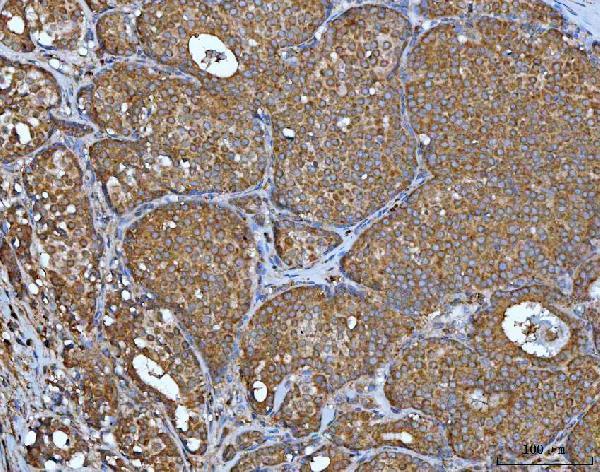
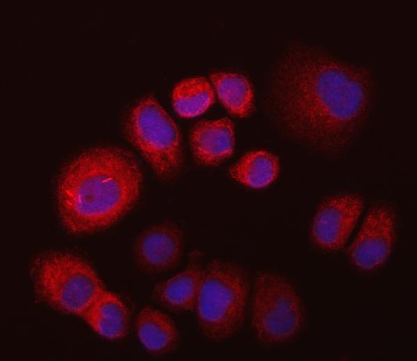
A03021-2 has IHC images from human paraffin sections of esophageal squamous carcinoma, breast cancer, cervical cancer, and ovarian cancer, plus IF images from T47D cells and a human breast cancer paraffin section (A03021-2 captions). M03021-1 is listed for IHC and IF/ICC in human, mouse, and rat, but has no IHC or IF image captions in the payload (M03021-1 catalog).

Which to pick: For tissue IHC, choose A03021-2 when a documented paraffin-section example matters; its IHC captions report EDTA retrieval at pH 8.0 and 2 μg/ml primary antibody, while the fixative is unreported (A03021-2 IHC captions). For IF/ICC, A03021-2 has human cell and tissue images; M03021-1 is a rabbit monoclonal listed for IF/ICC and IHC but has no corresponding image captions (A03021-2 IF captions; M03021-1 catalog). Both list human, mouse, and rat reactivity, but the supplied images document human samples only (catalog reactivity; A03021-2 captions).

Each figure is that product's own IHC / IF validation image from its datasheet.

References

  1. UniProt Consortium. UniProt entry P12268 (IMDH2_HUMAN, Inosine-5'-monophosphate dehydrogenase 2).
  2. Human Protein Atlas. IMPDH2 tissue IHC expression (reliability: Approved).
  3. Human Protein Atlas. IMPDH2 subcellular location (ICC-IF): Mainly localized to the cytosol. In addition localized to the rods & rings..
  4. Human Protein Atlas. IMPDH2 antibody validation summary (2 antibodies).
  5. High expression of IMPDH2 is associated with aggressive features and poor prognosis of primary nasopharyngeal carcinoma. Scientific reports 2017 — PMC5429725.
  6. IMPDH2 promotes colorectal cancer progression through activation of the PI3K/AKT/mTOR and PI3K/AKT/FOXO1 signaling pathways. Journal of experimental & clinical cancer research : CR 2018 — PMC6282329.
  7. LncRNA UCA1 Participates in De Novo Synthesis of Guanine Nucleotides in Bladder Cancer by Recruiting TWIST1 to Increase IMPDH1/2. International journal of biological sciences 2023 — PMC10197894.
  8. IMPDH2 and HPRT expression and a prognostic significance in preoperative and postoperative patients with osteosarcoma. Scientific reports 2021 — PMC8149691.
  9. PubMed PMID:2902093 — UniProt-cited evidence.
  10. PubMed PMID:1969416 — UniProt-cited evidence.
  11. PubMed PMID:7999076 — UniProt-cited evidence.