This website uses cookies to ensure you get the best experience on our website.


- Table of Contents
This guide uses cerebellar granular-layer processes as a positive reference for INA staining in paraffin sections (HPA tissue IHC). It covers chromogenic detection with an IHC-validated antibody used at 2 μg/mL in human brain sections (datasheet A03756-1).

Expected localisation, validated protocols, controls and antibodies — the at-a-glance facts below, then the full design guide.
| Expected localisation | Processes in cerebellar granular layer (HPA tissue IHC) | |
| Staining pattern | CNS neuronal cells and processes; some adrenal cells (HPA tissue IHC) | |
| Antigen retrieval | EDTA pH 8.0 HIER, heat-mediated (datasheet A03756-1) | |
| Positive control | Cerebellum+4 more · see all | |
| Negative control | Adipose tissue+4 more · see all |
| Fixation | Keep fixation consistent across sections. (standard IHC practice; not target-specific) | |
| Caveat | Leydig cells also show high staining (HPA tissue IHC) | |
| Regulation | Predominantly expressed in adult CNS (UniProt) | |
| Isoform / epitope | No annotated isoforms; one 1–499 chain (UniProt) |
The catalog antibody protocol (datasheet: A03756-1) is accompanied by two published chromogenic INA IHC protocols (PMC3617314; PMC5039014).
| Sample | Paraffin-embedded mouse brain tissue; fixative not specified (datasheet A03756-1) |
| Fixation | Image fixative and duration unreported (datasheet A03756-1); verify before use. |
| Sectioning | 4–5 µm sections on charged slides (standard) |
| Deparaffinisation | Xylene, graded ethanol series to water (standard) |
| Antigen retrieval | Heat retrieval: EDTA pH 8.0 (datasheet A03756-1); 20 min, 95–100 °C (standard) |
| Peroxidase block | 3% H2O2, 10 min, room temperature (standard) |
| Blocking | 10% goat serum (datasheet A03756-1) |
| Primary antibody | Rabbit anti-INA, 2-5μg/ml (datasheet A03756-1) |
| Primary incubation | Overnight at 4 °C (datasheet A03756-1) |
| Detection | Streptavidin-biotin complex (SABC), DAB chromogen (datasheet A03756-1) |
| Counterstain | Hematoxylin, blue, dehydrate and mount (standard) |
| Expected result | INA-positive staining in processes in granular layer of cerebellum (HPA tissue IHC: High). HPA tissue profile: Distinct expression in the CNS and in a subset of adrenal gland cells. No signal in the no-primary control. |
INA is a class-IV neuronal intermediate-filament protein found predominantly in adult CNS (UniProt Q16352). In paraffin-section IHC, expect staining in neuronal cells and processes, especially cerebellar granular-layer processes (HPA tissue IHC). Intermediate-filament localisation is supported by ICC-IF and should guide compartment checks without being treated as a tissue-specific IHC observation (HPA subcellular). INA has no transmembrane segment (UniProt Q16352 topology). The tissue IHC profile has Enhanced reliability (HPA tissue IHC).
| Strong staining in cerebellar granular-layer processes, with a filament-like pattern. | This matches the strongest CNS observation in the supplied tissue set: High staining in these processes (HPA tissue IHC). A filament-like appearance is consistent with INA’s intermediate-filament function (UniProt Q16352) and approved ICC-IF localisation (HPA subcellular). Score the processes separately from nearby cell bodies; the tissue observation identifies a compartment, not uniform staining of every cell in the layer. |
| Moderate staining in neuronal cells of caudate, cerebral cortex, or hippocampus. | Neuronal cells in each of these regions are reported at Medium intensity (HPA tissue IHC). Their signal provides a plausible positive result alongside the stronger cerebellar-process reference. Compare the labelled cells and their compartments with local tissue architecture; do not require every neuron or every CNS region to have the same intensity. |
| Predominantly nuclear or sharply cell-surface staining in a putative positive field. | Neither pattern fits the approved intermediate-filament location, with cytosol as an additional location (HPA subcellular), or INA’s lack of a transmembrane segment (UniProt Q16352 topology). Treat it as a localisation mismatch and investigate artefact or nonspecific staining. ICC-IF localisation guides this check, but does not prove the exact appearance of every paraffin-section tissue. |
| Strong staining in adipocytes or unrelated epithelial cells while the expected positive cells are unconvincing. | Adipocytes in adipose tissue and respiratory epithelial cells in bronchus are reported as Not detected (HPA tissue IHC). Check whether the colour follows the intended cell type; a discordant pattern raises cross-reactivity or endogenous detection activity as possibilities (general IHC practice). A tissue-level comparison alone cannot identify which mechanism caused it. |
| Widespread pale colour obscures neuronal processes, or no signal appears in a known-positive region. | Diffuse colour that fails to resolve cells or processes is background, not the distinct CNS pattern reported by HPA (HPA tissue IHC; general IHC practice). Conversely, an unstained cerebellar granular layer conflicts with its reported High process staining (HPA tissue IHC). Review the run controls before interpreting either result as an INA expression change. |
| Tissue and cell choice | Cerebellar granular-layer processes are High; caudate, cortical, and hippocampal neuronal cells are Medium (HPA tissue IHC). Adrenal glandular cells are Medium and testicular Leydig cells are High in the supplied HPA observations (HPA tissue IHC). Those findings mean a CNS-only scoring rule would miss reported positives, although each result still needs its stated cell context. |
| Compartment and protein organisation | INA has an intermediate-filament rod at residues 94–407, can self-assemble, and can associate with other neurofilament proteins (UniProt Q16352). HPA places it mainly on intermediate filaments and additionally in cytosol by ICC-IF (HPA subcellular). These facts support a filament-associated interpretation; they do not specify the appearance of every paraffin section. |
| Antibody evidence and limits | Two listed antibodies have Enhanced IHC status, while another has Approved ICC status without a listed IHC status (HPA antibodies). The tissue profile’s Enhanced reliability reflects consistency between staining and RNA expression (HPA tissue IHC). Use the application-specific validation when weighing an unexpected pattern; ICC approval alone does not establish paraffin-section IHC performance. |
| IF/ICC: should the same appearance be expected? | HPA ICC-IF reports mainly intermediate filaments and additional cytosol (HPA subcellular). That supports a compartment check in fluorescent images, while the paraffin-section tissue intensities come from IHC (HPA tissue IHC). Compare each result within its own application; no IF/ICC protocol or matching tissue-intensity claim is supplied here. |
| Processing and fixation evidence | UniProt lists one 1–499 chain, no signal peptide, no propeptide, and no transmembrane segment (UniProt Q16352). These features do not establish an epitope or antigen-retrieval setting. Target-specific fixation sensitivity is unreported in the supplied UniProt and HPA records; choose and assess retrieval using the antibody’s own IHC instructions and run controls (general IHC practice). |
| Situation | Likely cause | Next action |
|---|---|---|
| Cerebellar granular-layer processes show no detectable stain. | The expected High process signal is missing (HPA tissue IHC); section handling, retrieval, primary-antibody conditions, or detection may have failed (general IHC practice). | Confirm that the positive-control section and detection controls worked, then check the recorded retrieval, primary-antibody dilution, incubation, and detection steps against the antibody’s IHC instructions (general IHC practice). Do not infer INA loss from a failed run. |
| Signal is confined to nuclei or crisp cell borders. | This conflicts with the approved intermediate-filament and additional cytosolic locations (HPA subcellular) and with the absence of a transmembrane segment (UniProt Q16352 topology). | Re-examine the counterstain and cellular boundaries, then compare a known-positive region and appropriate negative controls under the same detection conditions (general IHC practice). Record the localisation mismatch before assigning a positive score. |
| Adipocytes or bronchial epithelial cells stain strongly. | Those cell types are Not detected in the supplied tissue observations (HPA tissue IHC); nonspecific primary binding or endogenous detection activity could produce discordant colour (general IHC practice). | Run a no-primary control and the detection-system controls appropriate to the chromogen, then compare staining with an expected positive compartment (general IHC practice; HPA tissue IHC). A control result can narrow the cause; the unexpected tissue alone cannot. |
| A diffuse wash of colour masks neuronal cells and processes. | Broad colour without discernible structures prevents comparison with the distinct CNS staining pattern (HPA tissue IHC); incomplete blocking, washing, or excessive detection signal are possible general causes (general IHC practice). | Inspect the no-primary control, wash and blocking records, and chromogen development; adjust the general IHC workflow only after identifying the step responsible (general IHC practice). Rescore once the processes can be distinguished. |
| Cerebellar processes stain, but cortical neurons look weaker. | That difference can match High cerebellar-process versus Medium cortical-neuronal observations (HPA tissue IHC). | Score intensity by the specified tissue and cell type, with the same run controls and scoring threshold (HPA tissue IHC; general IHC practice). Do not call the cortical result negative solely because it is weaker than the cerebellar reference. |
| An ICC-IF image looks filamentous, but the paraffin-section result is discordant. | The approved ICC-IF compartment and the IHC tissue observations describe different applications (HPA subcellular; HPA tissue IHC); the listed ICC-approved antibody lacks a stated IHC status (HPA antibodies). | Review the IHC antibody’s application-specific validation and the paraffin-section positive and negative controls before transferring an ICC-IF interpretation to IHC (HPA antibodies; general IHC practice). |
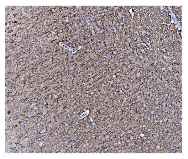
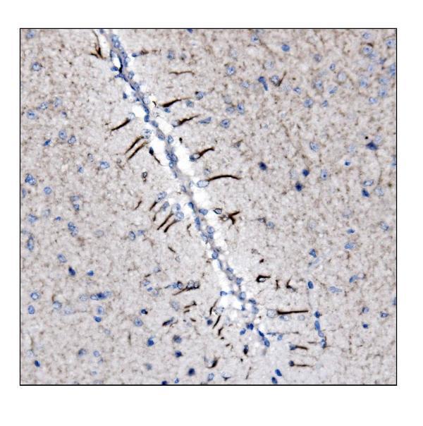
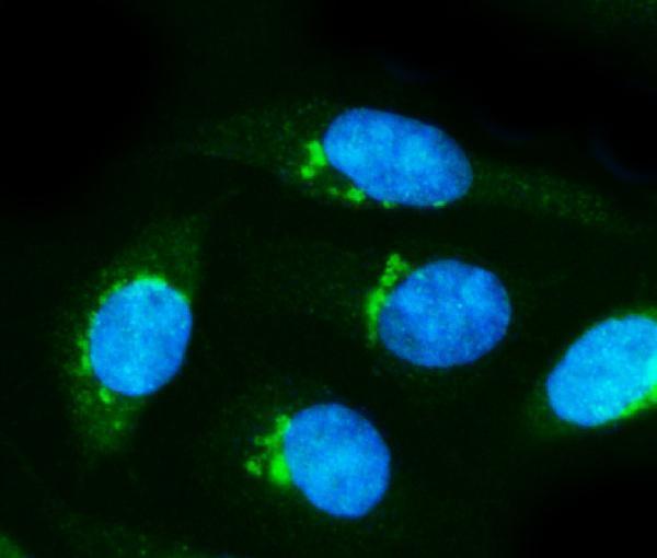


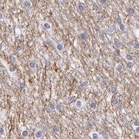
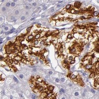
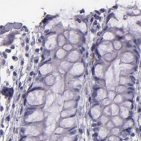


Comprehensive Human Protein Atlas IHC scoring per tissue (reliability: Enhanced — High consistency between antibody staining and RNA expression data.). Rows are taken directly from the HPA tissue chart — click any row's HPA link to view the source.
| Tissue | Cell type | Level | Evidence | Source |
|---|---|---|---|---|
| Cerebellum | Processes in granular layer | High | Protein (IHC) | HPA → |
| Testis | Leydig cells | High | Protein (IHC) | HPA → |
| Adrenal gland | Glandular cells | Medium | Protein (IHC) | HPA → |
| Caudate | Neuronal cells | Medium | Protein (IHC) | HPA → |
| Cerebral cortex | Neuronal cells | Medium | Protein (IHC) | HPA → |
| Tissue | Cell type | Level | Evidence | Source |
|---|---|---|---|---|
| Adipose tissue | Adipocytes | Not detected | Protein (IHC) | HPA → |
| Appendix | Glandular cells | Not detected | Protein (IHC) | HPA → |
| Bone marrow | Hematopoietic cells | Not detected | Protein (IHC) | HPA → |
| Breast | Adipocytes | Not detected | Protein (IHC) | HPA → |
| Bronchus | Respiratory epithelial cells | Not detected | Protein (IHC) | HPA → |
Use neuronal process staining, matched controls, and the catalog antibody’s documented paraffin section conditions to troubleshoot INA chromogenic IHC (HPA tissue IHC; datasheet A03756-1).
Anti-INA antibodies have IHC images from paraffin sections of human, mouse and rat tissues, plus IF images from human cells and rodent brain sections (catalog image captions).
A03756-1 has IHC images from human, mouse and rat brain paraffin sections and IF images from SiHa cells and rat brain sections (A03756-1 image captions). PB10003 has IHC images from human glioma and mouse and rat brain paraffin sections and IF images from A549 cells and mouse and rat brain sections (PB10003 image captions).
Which to pick: For tissue IHC, choose A03756-1 for brain sections or PB10003 for the documented human glioma example; each IHC caption describes a paraffin section with EDTA pH 8.0 retrieval, and neither reports the fixative (A03756-1 and PB10003 IHC image captions). For IF/ICC, choose by the documented sample: A03756-1 for SiHa cells or PB10003 for A549 cells (A03756-1 and PB10003 IF image captions). Both list IHC and IF/ICC applications and human, mouse and rat reactivity, with IHC images across all three species; neither catalog entry specifies a clone, so clonality does not distinguish them (catalog applications, reactivity and clone fields; IHC image captions).