This website uses cookies to ensure you get the best experience on our website.


- Table of Contents
Plan INF2 IHC in paraffin sections around cytoplasmic staining in most tissues (HPA tissue IHC). Start with the catalog antibody’s 2–5 μg/mL IHC range (datasheet A02710-2) and compare cell-specific staining with controls.

Expected localisation, validated protocols, controls and antibodies — the at-a-glance facts below, then the full design guide.
| Expected localisation | Cytoplasmic staining in most tissues (HPA tissue IHC) | |
| Staining pattern | Cytoplasmic staining across most tissues (HPA tissue IHC) | |
| Antigen retrieval | EDTA pH 8.0 HIER, heat-mediated (datasheet A02710-2) | |
| Positive control | Appendix+4 more · see all | |
| Negative control | Adipose tissue+4 more · see all |
| Fixation | Keep fixation consistent across paraffin sections. (standard IHC practice; not target-specific) | |
| Caveat | Staining has medium consistency with RNA expression (HPA tissue IHC) | |
| Regulation | Low tissue specificity (HPA tissue RNA) | |
| Isoform / epitope | 3 isoforms; check whether the epitope is shared (UniProt) |
The catalog antibody uses EDTA pH 8.0 retrieval (datasheet A02710-2). The published IHC protocols below cover glioblastoma, INF2 morphants, and human colorectal tissue (PMC7391617; PMC4457406; PMC12128233).
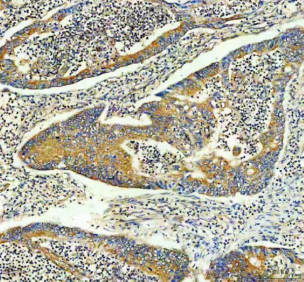
| Sample | Paraffin-embedded human colon cancer tissue; fixative not specified (datasheet A02710-2) |
| Fixation | Image fixative and duration unreported (datasheet A02710-2); verify before use. |
| Sectioning | 4–5 µm sections on charged slides (standard) |
| Deparaffinisation | Xylene, graded ethanol series to water (standard) |
| Antigen retrieval | Heat retrieval: EDTA pH 8.0 (datasheet A02710-2); 20 min, 95–100 °C (standard) |
| Peroxidase block | 3% H2O2, 10 min, room temperature (standard) |
| Blocking | 10% goat serum (datasheet A02710-2) |
| Primary antibody | Rabbit anti-INF2, 2-5μg/ml (datasheet A02710-2) |
| Primary incubation | Overnight at 4 °C (datasheet A02710-2) |
| Detection | HRP-conjugated secondary, DAB chromogen (datasheet A02710-2) |
| Counterstain | Hematoxylin, blue, dehydrate and mount (standard) |
| Expected result | INF2-positive staining in glandular cells of appendix (HPA tissue IHC: High). HPA tissue profile: Cytoplasmic expression in most tissues. No signal in the no-primary control. |
INF2 should appear mainly in the cytoplasm of many tissue cell types, with a perinuclear distribution also consistent with its annotated location (HPA tissue IHC: cytoplasmic expression in most tissues; UniProt Q27J81: cytoplasm, perinuclear region). Glandular cells in appendix, gallbladder, rectum and stomach, and placental trophoblastic cells, show high staining (HPA tissue IHC: High). INF2 has no transmembrane segment (UniProt Q27J81 topology). HPA rates tissue IHC reliability Approved, with medium consistency against RNA expression (HPA tissue IHC).
| Cytoplasmic, possibly perinuclear, staining in appendix glandular cells or placental trophoblastic cells. | This fits INF2 localization and the reported high staining in those cells (UniProt Q27J81: cytoplasm, perinuclear region; HPA tissue IHC: High). Compare signal within the named cell population; staining elsewhere in the section alone is a less direct check of the reported pattern. |
| Predominantly nuclear-only or crisp membrane-only staining in an otherwise positive tissue. | This departs from the reported tissue IHC cytoplasmic pattern and UniProt location (HPA tissue IHC; UniProt Q27J81). Check the control section and detection conditions before assigning the signal to INF2. HPA reports nuclear bodies in ICC-IF, so isolated nuclear puncta need separate interpretation (HPA subcellular ICC-IF). |
| Strong staining of adipocytes, cardiomyocytes or skeletal myocytes, especially without the expected positive cells. | HPA reports these cell types as not detected (HPA tissue IHC). Consider cross-reactivity or endogenous detection activity and compare with an appropriate reagent control (general IHC practice). The HPA observation is a reference pattern, not proof that every positive cell is false. |
| Uniform brown haze across cells and extracellular areas, obscuring cell boundaries. | Diffuse background limits a compartment-level call. Review blocking, washing and detection controls for nonspecific signal or endogenous activity (general chromogenic IHC practice). Do not score the haze as cytoplasmic INF2 solely because HPA describes broad tissue expression (HPA tissue IHC). |
| No staining in appendix glandular cells or placental trophoblastic cells on an otherwise interpretable section. | That conflicts with HPA's high-staining examples (HPA tissue IHC). Check whether the IHC-validated antibody and detection run yield a signal in a positive control, then review retrieval and reagent performance (general IHC practice). Absence in one section does not establish absent INF2 protein. |
| Tissue and cell selection | Appendix, gallbladder, rectum and stomach glandular cells and placental trophoblastic cells are high-staining examples; adipocytes and cardiomyocytes are reported not detected (HPA tissue IHC). Score the specified cells, since a whole-section average can conceal their pattern. |
| Compartment and topology | UniProt places INF2 in cytoplasm and the perinuclear region and lists no transmembrane segment (UniProt Q27J81). HPA tissue IHC describes cytoplasmic expression in most tissues (HPA tissue IHC). These annotations support a cellular pattern check, not a precise organelle assignment by chromogenic IHC. |
| Antibody evidence | HPA000724 is Approved for IHC, while the tissue profile has medium consistency with RNA data (HPA antibodies; HPA tissue IHC). Interpret an unexpected cell-specific result alongside controls; Approved does not make every staining event target-specific. |
| Isoforms and epitope | UniProt lists three INF2 isoforms and domains at residues 2–330, 554–946 and 974–989 (UniProt Q27J81). No antibody epitope or isoform coverage is supplied, so a difference in staining cannot be assigned to an isoform from this record. |
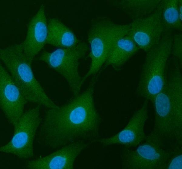
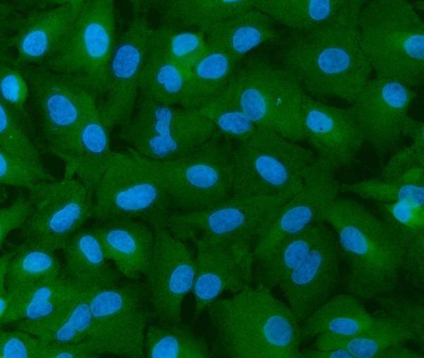
| IF/ICC Q: What pattern is reported? | A: HPA reports mainly endoplasmic reticulum localization, with additional nuclear bodies, in ICC-IF (HPA subcellular ICC-IF). HPA000724 has Uncertain ICC validation (HPA antibodies). This IF observation can inform interpretation but is not an IHC organelle-level claim. |
| Situation | Likely cause | Next action |
|---|---|---|
| A high-staining reference cell population is blank. | Possible run-level loss of detectable signal; HPA reports high staining in appendix glandular cells and placental trophoblastic cells (HPA tissue IHC). | Inspect the positive control and detection run, then review the antibody's IHC retrieval conditions and reagent performance (general IHC practice). Record the result as unresolved until the control works. |
| Most cells show similar brown haze. | Background may obscure the HPA-reported cytoplasmic pattern (HPA tissue IHC); nonspecific binding or endogenous detection activity are general IHC possibilities. | Compare reagent controls, blocking and wash conditions, and the distribution of signal outside cells (general chromogenic IHC practice). Score defined cells only after background permits that distinction. |
| Signal is sharply membrane-bound. | A membrane-dominant result conflicts with cytoplasmic tissue staining and the absence of a transmembrane segment (HPA tissue IHC; UniProt Q27J81 topology). | Check localization in a HPA high-staining reference population and review detection controls (HPA tissue IHC; general IHC practice). Treat persistent membrane-only staining as unconfirmed. |
| Adipocytes or muscle cells stain more strongly than nearby reference cells. | HPA reports adipocytes, cardiomyocytes and skeletal myocytes as not detected (HPA tissue IHC); cross-reactivity or endogenous activity is possible (general IHC practice). | Compare the named cell types with a positive reference section and reagent controls before calling the unexpected staining INF2 (HPA tissue IHC; general IHC practice). |
| Scoring varies greatly between regions of one section. | INF2 staining is cell-type dependent in the supplied tissue examples: some glandular cells are High, while other listed populations are Low or Not detected (HPA tissue IHC). | Identify and score the relevant cell population in each region rather than averaging all tissue elements; document the observed compartment and intensity (HPA tissue IHC; general IHC practice). |
| ICC-IF shows nuclear puncta, but tissue IHC is mainly cytoplasmic. | HPA lists nuclear bodies as an additional ICC-IF location and cytoplasmic expression as the tissue IHC profile (HPA subcellular ICC-IF; HPA tissue IHC). | Interpret each assay against its own HPA observation. HPA000724 has Uncertain ICC validation, so verify an ICC-IF punctate result independently before relying on it (HPA antibodies). |


Comprehensive Human Protein Atlas IHC scoring per tissue (reliability: Approved — Medium consistency between antibody staining and RNA expression data.). Rows are taken directly from the HPA tissue chart — click any row's HPA link to view the source.
| Tissue | Cell type | Level | Evidence | Source |
|---|---|---|---|---|
| Appendix | Glandular cells | High | Protein (IHC) | HPA → |
| Gallbladder | Glandular cells | High | Protein (IHC) | HPA → |
| Placenta | Trophoblastic cells | High | Protein (IHC) | HPA → |
| Rectum | Glandular cells | High | Protein (IHC) | HPA → |
| Stomach | Glandular cells | High | Protein (IHC) | HPA → |
| Tissue | Cell type | Level | Evidence | Source |
|---|---|---|---|---|
| Adipose tissue | Adipocytes | Not detected | Protein (IHC) | HPA → |
| Heart muscle | Cardiomyocytes | Not detected | Protein (IHC) | HPA → |
| Ovary | Ovarian stroma cells | Not detected | Protein (IHC) | HPA → |
| Skeletal muscle | Myocytes | Not detected | Protein (IHC) | HPA → |
| Smooth muscle | Smooth muscle cells | Not detected | Protein (IHC) | HPA → |
Troubleshoot INF2 staining in paraffin sections by checking retrieval, cellular localisation and controls before comparing staining intensity across samples.
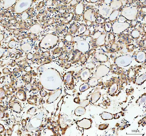
Anti-INF2 antibodies have IHC images from human colon and thyroid cancer and mouse and rat brain paraffin sections (A02710-2 captions), plus A549 cell IF images (A02710-1 and A02710-2 captions).
A02710-2 has paraffin-section IHC images from human colon and thyroid cancer and mouse and rat brain, plus an A549 cell IF image (A02710-2 captions). A02710-1 lists IHC and IF/ICC applications and has an A549 cell IF image; its IHC image is unreported (A02710-1 catalog and caption).
Which to pick: Choose A02710-2 for tissue IHC because its own captions document paraffin sections; the fixative is unreported (A02710-2 IHC captions). For IF/ICC, either SKU has an A549 cell image, and both list IF/ICC; A02710-2 is listed as polyclonal, while A02710-1 clonality is unreported (catalog and IF captions). For work across species, both list human, mouse and rat reactivity, while A02710-2 additionally has IHC images in all three species (catalog and A02710-2 IHC captions).