This website uses cookies to ensure you get the best experience on our website.


- Table of Contents
Plan paraffin-section INPP5D IHC around the expected cytoplasmic staining in lymphoid tissue (HPA tissue IHC). Use tonsil as a positive control (HPA tissue IHC; datasheet A03358), then assess staining by cell population (standard IHC practice).

Expected localisation, validated protocols, controls and antibodies — the at-a-glance facts below, then the full design guide.
| Expected localisation | Cytoplasmic in lymphoid tissues (HPA tissue IHC) | |
| Staining pattern | Strong cytoplasmic staining in germinal center cells (HPA tissue IHC) | |
| Antigen retrieval | Citrate pH 6 HIER, heat-mediated (datasheet A03358) | |
| Positive control | Appendix+4 more · see all | |
| Negative control | Adipose tissue+4 more · see all |
| Fixation | Keep fixation consistent across sections (standard IHC practice; not target-specific) | |
| Caveat | Staining varies among hematopoietic cell populations (UniProt) | |
| Regulation | Varies with hematopoietic maturation (UniProt) | |
| Isoform / epitope | Three isoforms; epitope differences are unspecified (UniProt) |
The catalog antibody has a datasheet IHC-P protocol (datasheet: A03358). One published chromogenic SHIP1 protocol uses paraffin-embedded kidney sections (PMC12992027).
| Sample | Paraffin-embedded human tonsil tissue; fixative not specified (datasheet A03358) |
| Fixation | Image fixative and duration unreported (datasheet A03358); verify before use. |
| Sectioning | 4–5 µm sections on charged slides (standard) |
| Deparaffinisation | Xylene, graded ethanol series to water (standard) |
| Antigen retrieval | Heat retrieval: Citrate pH 6, 20 min (datasheet A03358) |
| Peroxidase block | 3% H2O2, 10 min, room temperature (standard) |
| Blocking | 10% goat serum (datasheet A03358) |
| Primary antibody | Rabbit anti-INPP5D, 2μg/ml (datasheet A03358) |
| Primary incubation | Overnight at 4 °C (datasheet A03358) |
| Detection | Streptavidin-biotin complex (SABC), DAB chromogen (datasheet A03358) |
| Counterstain | Hematoxylin, blue, dehydrate and mount (standard) |
| Expected result | INPP5D-positive staining in germinal center cells of appendix (HPA tissue IHC: High). HPA tissue profile: High cytoplasmic expression in lymphoid tissues. No signal in the no-primary control. |
INPP5D should mainly give cytoplasmic staining in lymphoid and hematopoietic cells (HPA tissue IHC: high cytoplasmic expression in lymphoid tissues; UniProt Q92835: immune and hematopoietic expression). Membrane-associated signal can occur because INPP5D moves to the plasma membrane upon activation, although it has no transmembrane segment (UniProt Q92835: localization and topology). HPA rates the tissue IHC pattern Enhanced, citing agreement between staining and RNA expression (HPA tissue IHC: reliability).
| Strong cytoplasmic staining in lymph node or tonsil germinal center cells, with identifiable hematoxylin-stained nuclei. | This matches HPA's high staining in those cells and its cytoplasmic lymphoid profile (HPA tissue IHC: lymph node, tonsil, profile). Score the cells that stain, not the entire tissue as uniformly positive; UniProt reports substantial variation within immune populations (UniProt Q92835: tissue specificity). |
| Predominantly nuclear staining, with little corresponding cytoplasmic signal, in an otherwise positive lymphoid section. | Treat a nuclear-only pattern as suspect: HPA describes cytoplasmic tissue staining, and UniProt lists cytoplasmic and membrane-associated locations, not a nuclear location (HPA tissue IHC: profile; UniProt Q92835: subcellular location). Review the negative control and detection steps before scoring it as INPP5D (general IHC practice). |
| Strong staining in adipocytes or bronchial respiratory epithelial cells while expected immune cells are weak. | HPA reports INPP5D as not detected in those cell types (HPA tissue IHC: adipose tissue, bronchus). Suspect nonspecific antibody binding or endogenous detection activity; check staining against a matched control and inspect whether positive immune cells retain the expected pattern (general IHC practice). |
| Diffuse chromogen across stroma, lumens, and many unrelated cell types, obscuring cellular boundaries. | A broad haze cannot be assigned to INPP5D from these sources; HPA describes a cell-specific, mainly cytoplasmic pattern (HPA tissue IHC: profile and tissue entries). Investigate background from antibody concentration, incomplete blocking, or detection chemistry using the relevant controls (general IHC practice). |
| No convincing staining in bone marrow hematopoietic cells or lymph node germinal center cells. | Both are reported as high-staining populations (HPA tissue IHC: bone marrow, lymph node). A blank result in a known-positive control raises a technical concern, though expression varies among immune cells (UniProt Q92835: tissue specificity); verify section quality, antibody performance, and detection controls before interpreting the study tissue (general IHC practice). |
| Cell identity and tissue context | HPA reports high staining in germinal center cells, marrow hematopoietic cells, splenic red pulp cells, and some nonimmune populations, including kidney proximal-tubule microvilli (HPA tissue IHC: tissue entries). Interpret each compartment against its own annotated cell type; UniProt's immune-cell specificity does not make every nonimmune HPA signal an artefact (UniProt Q92835: tissue specificity). |
| Cytoplasm versus membrane | UniProt places INPP5D in cytoplasm, cell membrane, membrane rafts, and cytoskeleton, and describes stimulus-dependent movement to membrane ruffles (UniProt Q92835: subcellular location). Membrane-edge staining can therefore be plausible when accompanied by an appropriate cellular pattern; it is not evidence that INPP5D is an integral membrane protein (UniProt Q92835: topology). |
| Variation within immune populations | UniProt reports INPP5D in at least 74% of immature CD34+ cells but only 10% of more mature CD33+ cells, and in most T cells but a minority of B cells (UniProt Q92835: tissue specificity). Do not require equal intensity across every immune cell in a section. |
| Antibody evidence and isoforms | HPA lists two antibodies with Enhanced IHC validation, HPA070455 and CAB016300 (HPA antibodies: IHC validation). UniProt lists three isoforms (UniProt Q92835: isoforms). These records do not identify the recognized epitope or establish whether either antibody detects all isoforms, so avoid assigning a weak region to an isoform. |
| IF/ICC question: where should signal appear? | HPA's ICC-IF summary supports a cytosolic location and lists Hep-G2 images (HPA subcellular ICC-IF: summary, image list). UniProt also allows activation-associated membrane recruitment (UniProt Q92835: localization). These observations inform image interpretation; they do not establish an IF/ICC protocol or make the cell line an IHC tissue control. |
| Situation | Likely cause | Next action |
|---|---|---|
| Known-positive lymphoid or marrow control is blank. | The result conflicts with HPA's high staining in germinal center or marrow hematopoietic cells (HPA tissue IHC: lymph node, bone marrow); the failed step is undetermined. | Confirm that the expected cells are present, then review deparaffinization, retrieval, primary antibody, and detection controls one step at a time (general IHC practice). No INPP5D-specific retrieval condition or fixation sensitivity is supplied by HPA or UniProt. |
| The whole section stains, including cell types HPA lists as not detected. | Nonspecific binding or endogenous detection activity is possible (general IHC practice); adipocytes and bronchial respiratory epithelium are HPA-negative reference cells (HPA tissue IHC: adipose tissue, bronchus). | Compare a no-primary control and an appropriate detection control; adjust blocking or antibody concentration only after locating the source of background (general IHC practice). |
| Most apparent signal sits over nuclei. | A nuclear-dominant result does not fit HPA's cytoplasmic tissue profile or UniProt's listed locations (HPA tissue IHC: profile; UniProt Q92835: subcellular location). | Check counterstain and chromogen boundaries at higher magnification, compare the no-primary control, and repeat with an IHC-validated antibody if the nuclear pattern persists (general IHC practice; HPA antibodies: IHC validation). |
| Only some immune cells in a positive region stain. | Expression can vary by immune population; UniProt reports different frequencies in CD34+ versus CD33+ cells and T versus B cells (UniProt Q92835: tissue specificity). | Identify the stained population morphologically and score its distribution and intensity separately. Use an independent cell marker if lineage assignment matters (general IHC practice). |
| A sharp peripheral signal appears beside cytoplasmic staining. | Activated INPP5D can move to the plasma membrane and membrane ruffles (UniProt Q92835: subcellular location); section-edge artefact is also possible (general IHC practice). | Check whether the peripheral pattern follows intact cells and recurs in the expected population; compare section edges and the no-primary control before accepting it as membrane-associated signal (general IHC practice). |
| Kidney proximal-tubule microvilli stain while nearby cells do not. | HPA records high staining in proximal-tubule microvilli (HPA tissue IHC: kidney), even though UniProt emphasizes immune and hematopoietic expression (UniProt Q92835: tissue specificity). | Assess the signal in the annotated microvillar location rather than rejecting it solely for being outside lymphoid tissue. Compare appropriate controls and avoid extending that observation to all renal cells (general IHC practice; HPA tissue IHC: kidney). |
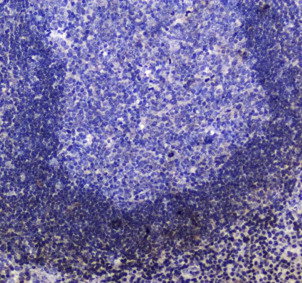
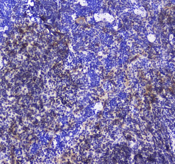
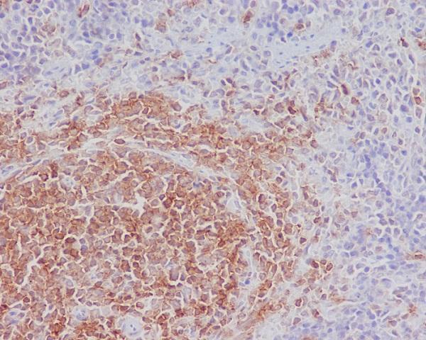


Comprehensive Human Protein Atlas IHC scoring per tissue (reliability: Enhanced — High consistency between antibody staining and RNA expression data.). Rows are taken directly from the HPA tissue chart — click any row's HPA link to view the source.
| Tissue | Cell type | Level | Evidence | Source |
|---|---|---|---|---|
| Appendix | Germinal center cells | High | Protein (IHC) | HPA → |
| Bone marrow | Hematopoietic cells | High | Protein (IHC) | HPA → |
| Cerebellum | Granular cells - cytoplasm/membrane | High | Protein (IHC) | HPA → |
| Kidney | Proximal tubules (microvilli) | High | Protein (IHC) | HPA → |
| Lymph node | Germinal center cells | High | Protein (IHC) | HPA → |
| Tissue | Cell type | Level | Evidence | Source |
|---|---|---|---|---|
| Adipose tissue | Adipocytes | Not detected | Protein (IHC) | HPA → |
| Bronchus | Respiratory epithelial cells | Not detected | Protein (IHC) | HPA → |
| Cervix | Glandular cells | Not detected | Protein (IHC) | HPA → |
| Duodenum | Endocrine cells | Not detected | Protein (IHC) | HPA → |
| Epididymis | Glandular cells | Not detected | Protein (IHC) | HPA → |
Use the catalog antibody’s paraffin-section evidence and tissue-specific expression patterns to troubleshoot INPP5D chromogenic IHC (datasheet A03358; HPA tissue IHC).
IHC images show INPP5D staining in paraffin sections of human tonsil and mouse and rat spleen (A03358 image captions). An ICC/IF-listed antibody has no IF figure (catalog: P03358-1).
A03358 lists IHC and Human, Mouse and Rat reactivity, with paraffin-section images from human tonsil and mouse and rat spleen (catalog: A03358 applications/reactivity; A03358 image captions). M03358 lists IHC and Human reactivity, while its paraffin-section image depicts rat spleen (catalog: M03358 applications/reactivity; M03358 image caption).
Which to pick: For cross-species tissue IHC, choose rabbit polyclonal A03358: its listed reactivity covers Human, Mouse and Rat, and its own captions show paraffin-section IHC in all three species; the fixative is unreported (catalog: A03358 host/clonality/reactivity; A03358 image captions). Rabbit monoclonal M03358 is another IHC-listed option, but its rat spleen caption differs from its Human-only reactivity listing; the caption does not report a fixative (catalog: M03358 clone/applications/reactivity; M03358 image caption). For IF/ICC, choose P03358-1 based on its IF/ICC application listing and Human reactivity; no IF figure is provided (catalog: P03358-1 applications/reactivity/IF images).