INPPL1 / Phosphatidylinositol 3,4,5-trisphosphate 5-phosphatase 2 · IHC design guide

Design Immunohistochemistry for INPPL1

Plan chromogenic INPPL1 IHC-P using cytoplasmic tissue staining as the expected pattern (HPA tissue IHC). Start with the catalog antibody at 0.5–1 μg/mL (datasheet A01790), and use placenta trophoblasts as a high-staining reference (HPA tissue IHC).

Evidence assembled Oct 2026 · For research use; verify linked source records and product datasheet before use
Immunohistochemistry protocol sheet for INPPL1 (IHC for INPPL1): expected localisation Cytoplasmic staining across tissues (HPA tissue IHC), antibody A01790, validated IHC image, and IHC protocol steps
Printable INPPL1 IHC protocol sheet — expected localisation Cytoplasmic staining across tissues (HPA tissue IHC), antibody A01790, controls and protocol steps. Open the full INPPL1 IHC guide →

INPPL1 Immunohistochemistry Experimental Design Guide

Expected localisation, validated protocols, controls and antibodies — the at-a-glance facts below, then the full design guide.

Must know before staining
Expected localisation Cytoplasmic staining across tissues (HPA tissue IHC)
Staining pattern Cytoplasmic staining across many tissue cell types (HPA tissue IHC)
Antigen retrieval Citrate pH 6 HIER, heat-mediated (datasheet A01790)
Positive control ⓘ Bronchus+4 more · see all
Negative control ⓘ Adipose tissue
Important caveats
Reasons your staining may differ from the expected pattern.
Fixation Keep fixation consistent across sections (standard IHC practice; not target-specific); Matched tissue-IHC evidence does not establish the fixation claim. Validate the specimen-specific method before use. (selected-SKU IHC image A01790)
Caveat Activation can shift SHIP2 toward the membrane (UniProt)
Regulation Macrophages express it; blood monocytes do not (UniProt)
Isoform / epitope 2 isoforms; epitope coverage is unspecified (UniProt)
Section 1

Recommended INPPL1 IHC & IF Protocols

The catalog antibody protocol uses citrate pH 6 retrieval (datasheet A01790). Two published IHC protocols add brain and colorectal tissue examples (PMC11150309; PMC5341996).

Recommended immunohistochemistry (IHC-P) protocol parameters
SampleParaffin-embedded human gastric cancer tissue; fixative not specified (datasheet A01790)
FixationImage fixative and duration unreported (datasheet A01790); verify before use.
Sectioning4–5 µm sections on charged slides (standard)
DeparaffinisationXylene, graded ethanol series to water (standard)
Antigen retrievalHeat retrieval: Citrate pH 6, 20 min (datasheet A01790)
Peroxidase block3% H2O2, 10 min, room temperature (standard)
Blocking10% goat serum (datasheet A01790)
Primary antibodyRabbit anti-INPPL1, 0.5-1μg/ml (datasheet A01790)
Primary incubationOvernight at 4 °C (datasheet A01790)
DetectionStreptavidin-biotin complex (SABC), DAB chromogen (datasheet A01790)
CounterstainHematoxylin, blue, dehydrate and mount (standard)
Expected resultINPPL1-positive staining in respiratory epithelial cells of bronchus (HPA tissue IHC: High). HPA tissue profile: Ubiquitous cytoplasmic expression. No signal in the no-primary control.
💡Decision noteStart with heat-mediated citrate pH 6 retrieval (datasheet A01790); both published protocols also use citrate retrieval (PMC11150309; PMC5341996).
Section 2

What Is the Expected INPPL1 Staining Pattern?

INPPL1 (SHIP2) is mainly cytosolic, with reported cytoskeletal, membrane and nuclear locations; it has no transmembrane segment (UniProt O15357). In tissue IHC, expect predominantly cytoplasmic staining, including strong staining in selected epithelial and glandular cells and Purkinje cells (HPA tissue IHC). HPA describes the tissue pattern as ubiquitous cytoplasmic expression, but rates its IHC evidence Approved, with medium consistency between staining and RNA data (HPA tissue IHC).

What am I looking at on my slide?
Clear cytoplasmic staining in bronchial respiratory epithelium, colonic or duodenal glands, or cerebellar Purkinje cells.These are reported High cell-level examples (HPA tissue IHC). Judge the named cells, rather than the whole section, as the positive reference. A cytoplasmic pattern agrees with HPA tissue IHC and the principal cytosolic location (UniProt O15357).
A sharp nuclear-only, uniformly surface-only, or extracellular deposit replaces cytoplasmic staining.Treat a dominant pattern in an unexpected compartment as suspect, especially if the expected cytoplasmic signal is absent (HPA tissue IHC). UniProt also lists nuclear and membrane-associated locations, so a minor signal there is not automatically artefactual (UniProt O15357). Check the control and morphology before assigning localisation.
Adipocytes stain as strongly as adjacent expected-positive cells.HPA reports adipocytes as Not detected (HPA tissue IHC). Strong adipocyte staining warrants a cross-reactivity or detection-artifact check; the HPA rating is Approved with medium staining–RNA consistency, so this comparison is a warning flag rather than an absolute specificity test (HPA tissue IHC).
Color coats stroma, lumina or most cells without clear cellular boundaries.Diffuse deposit obscures whether signal occupies the reported cytoplasmic compartment (HPA tissue IHC). Uneven reagent coverage, nonspecific detection or endogenous enzyme activity are general chromogenic IHC possibilities; compare a no-primary control and inspect tissue morphology (general IHC practice).
A reported high-staining cell population shows no signal.First verify that the expected cells are present and preserved: HPA's High calls are cell-specific, including respiratory epithelial, glandular and Purkinje cells (HPA tissue IHC). A blank positive reference calls for a run-level check before concluding that the test sample lacks INPPL1 (general IHC practice).
💡Expected INPPL1 appearanceCall an IHC result positive when identifiable cells show predominantly cytoplasmic signal, with High staining in an HPA-listed positive cell population; widespread extracellular deposit or equally strong adipocyte staining is suspect (HPA tissue IHC).
How each factor affects the staining
Cell choice for interpretationHPA reports High staining in specified cell populations across bronchus, cerebellum, colon, duodenum, esophagus, fallopian tube, oral mucosa and placenta, while adipocytes are Not detected (HPA tissue IHC). Score the listed cells within their tissue context; low staining is also reported in parathyroid glandular cells, ovarian follicle cells and splenic red-pulp cells (HPA tissue IHC).
Compartment and cell stateThe cytosol is a principal location, while UniProt also reports cytoskeleton, membrane, projections, nucleus and spindle pole (UniProt O15357). Redistribution to membrane ruffles can follow activation and depends on stimulus and cell type (UniProt O15357). These locations support cautious interpretation of focal peripheral signal, not an expectation that every paraffin section shows it.
Antibody evidenceThe listed antibody, HPA037601, is IHC Approved; HPA describes medium consistency between antibody staining and RNA expression (HPA antibodies; HPA tissue IHC). Use the reported pattern as a practical comparator, while treating an unexpected compartment or cell type as a reason to review controls rather than proof of INPPL1.
Isoforms and topologyUniProt lists 2 isoforms and no transmembrane segment or signal peptide (UniProt O15357). The supplied evidence gives no epitope mapping, so it cannot establish isoform-selective staining. A surface-only interpretation also needs scrutiny against the reported cytoplasmic tissue pattern (HPA tissue IHC).
Why is my staining missing, weak or wrong?
SituationLikely causeNext action
The positive reference section is blank.The expected cell population may be absent from the examined area, or the staining run may have failed (HPA tissue IHC; general IHC practice).Locate an HPA-listed High cell population on the section, then check reagent coverage, retrieval, primary-antibody step and detection controls (HPA tissue IHC; general IHC practice). No INPPL1-specific retrieval condition is established by these sources.
All cells stain, including adipocytes.A broad deposit may reflect nonspecific detection or endogenous activity; adipocytes are reported Not detected (general IHC practice; HPA tissue IHC).Compare a no-primary control and review blocking and detection steps; for enzyme-based detection, check the relevant endogenous-activity control (general IHC practice). Reassess against identifiable HPA-positive cells (HPA tissue IHC).
Only nuclei or extracellular spaces carry strong color.The dominant pattern conflicts with HPA's ubiquitous cytoplasmic IHC description, although UniProt includes the nucleus among reported locations (HPA tissue IHC; UniProt O15357).Inspect morphology and background controls, then repeat interpretation in an HPA-listed positive cell population before calling the compartment specific (HPA tissue IHC; general IHC practice).
Peripheral staining appears in a subset of otherwise cytoplasmic cells.Stimulus- and cell-dependent membrane redistribution is reported, but its occurrence in this section is unknown (UniProt O15357).Record peripheral and cytoplasmic signal separately; require convincing cell boundaries and a clean control before assigning membrane-associated staining (UniProt O15357; general IHC practice).
Weakly stained cells are mistaken for a failed section.HPA reports Low staining in parathyroid glandular cells, ovarian follicle cells and splenic red-pulp cells (HPA tissue IHC).Interpret intensity by the named cell type and compare an HPA-listed High population processed in the same run (HPA tissue IHC; general IHC practice).
Can ICC/IF images define the expected paraffin IHC pattern?HPA ICC/IF supports a mainly cytosolic location and approves an additional Golgi location; its antibody status is ICC Supported (HPA subcellular; HPA antibodies).Use ICC/IF as a localisation cross-check only; use HPA tissue IHC for the paraffin-section cell pattern, without requiring visible Golgi staining in IHC (HPA subcellular; HPA tissue IHC).

Sample controls for INPPL1 IHC & IF

🧪Run bronchus first: respiratory epithelial cells should stain (HPA: High in bronchus respiratory epithelial cells). Use adipose tissue as the negative tissue, where adipocytes should lack specific staining (HPA: Not detected in adipocytes); on the bronchus slide, HPA-unlisted cells should show only background DAB signal, but they are not validated target-negative cells.
Positive control tissue: Bronchus (Respiratory epithelial cells, HPA High)
Negative control tissue: Adipose tissue (HPA Not detected)
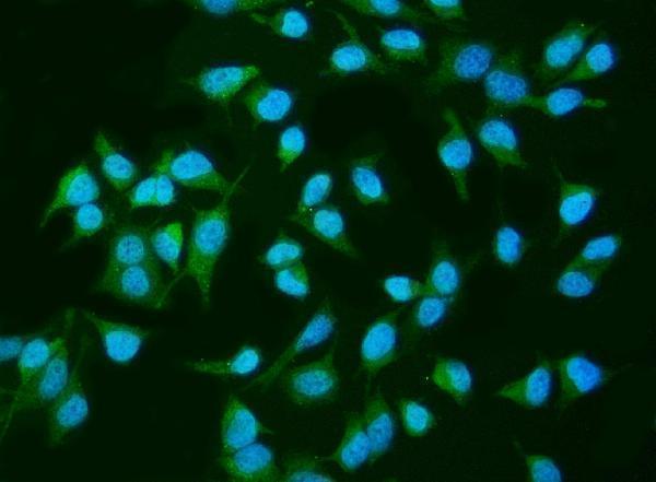
ICC-IF cell lines (HPA subcellular resource): HPA ICC-IF images show INPPL1 in A-431, U-251MG, U2OS, with annotated localisation: Cytosol (supported) (HPA subcellular).
Technical controls: Include no-primary (secondary-only) and concentration-matched nonimmune rabbit IgG controls, plus INPPL1 knockout material processed in parallel as a biological negative (caption: rabbit anti-INPPL1 antibody; standard IHC practice). Quench endogenous peroxidase and check for endogenous biotin signal because the caption uses biotinylated secondary antibody, streptavidin–biotin detection and DAB (caption: biotinylated secondary, SABC and DAB; standard IHC practice).
⚠️Feasibility: The selected-SKU paraffin-section caption reports citrate retrieval at pH 6 for 20 minutes, but does not establish that retrieval is required; its fixative is unreported (caption: citrate retrieval; fixative not stated). A target-specific fixation window or fixation effect is unreported, and the supplied evidence does not establish whether frozen sections or IF are easier (supplied caption: paraffin-section IHC; HPA: ICC-IF images). In bronchus, respiratory epithelial staining should be distinguished from any signal in inflammatory cells by morphology and the negative controls, since endogenous peroxidase can produce chromogenic background (HPA: High in bronchus respiratory epithelial cells; standard IHC practice).

HPA tissue IHC evidence for INPPL1

Comprehensive Human Protein Atlas IHC scoring per tissue (reliability: Approved — Medium consistency between antibody staining and RNA expression data.). Rows are taken directly from the HPA tissue chart — click any row's HPA link to view the source.

Positive expression · recommended positive controls

TissueCell typeLevelEvidenceSource
Bronchus Respiratory epithelial cells High Protein (IHC) HPA →
Cerebellum Purkinje cells High Protein (IHC) HPA →
Colon Glandular cells High Protein (IHC) HPA →
Duodenum Glandular cells High Protein (IHC) HPA →
Esophagus Squamous epithelial cells High Protein (IHC) HPA →

Undetected expression · recommended negative controls

TissueCell typeLevelEvidenceSource
Adipose tissue Adipocytes Not detected Protein (IHC) HPA →
Section 3

Advanced INPPL1 IHC Tips

Troubleshoot INPPL1 staining in paraffin sections by checking retrieval, cellular distribution, controls and cell type before interpreting signal intensity.

How should I retrieve INPPL1 in paraffin sections when staining is weak?
Use heat-mediated citrate pH 6 antigen retrieval for paraffin sections (datasheet A01790). The selected gastric cancer tissue image used 20 minutes of retrieval in that buffer before chromogenic detection (caption A01790). If staining is weak, compare a fresh section processed for the same duration with a shorter retrieval time, keeping antibody concentration and detection constant (standard IHC practice). Check whether signal improves in intact cells without a parallel rise in diffuse background or tissue damage (standard IHC practice). Record the heating method and cooling conditions so the comparison can be repeated (standard IHC practice).
Could fixation explain weak or uneven INPPL1 staining?
Target-specific sensitivity of INPPL1 to fixation is unknown from the supplied evidence; the selected paraffin-section caption does not state a fixative (caption A01790). Compare sections with documented fixation and processing histories while holding citrate pH 6 retrieval and detection constant (datasheet A01790; standard IHC practice). Inspect section edges, folds and damaged areas before attributing uneven staining to the antibody (standard IHC practice). Use a section with expected epithelial staining as a processing comparison, such as colon glandular cells (HPA: High in colon glandular cells). Report the fixative as unknown when the only source is this caption (caption A01790).
Should INPPL1 stain cytoplasm, membrane, or nuclei in tissue?
Expect a predominantly cytoplasmic pattern in tissue IHC (HPA: ubiquitous cytoplasmic expression). INPPL1 is mainly cytosolic, while membrane, cytoskeletal, nuclear and projection-associated locations are also recorded (UniProt O15357 subcellular location). Its membrane recruitment can vary with stimulation and cell type, so a peripheral accent should be assessed in intact cells and alongside the dominant cytoplasmic signal (UniProt O15357 subcellular location). Compare matched regions at the same chromogen exposure and examine whether apparent nuclear staining follows damaged or crowded cells (standard IHC practice). An isolated compartment pattern warrants independent antibody or biological validation before it is assigned to INPPL1 (standard IHC practice).
How can isoforms or epitope placement affect INPPL1 IHC results?
INPPL1 has 2 annotated isoforms, and the supplied caption does not identify the antibody epitope (UniProt O15357 isoforms; caption A01790). Its SH2 domain spans residues 21–117, the 5-phosphatase domain 419–732, and the SAM domain 1196–1258 (UniProt O15357 domains). Check the antibody's documented immunogen before claiming that staining represents both isoforms or a particular domain (standard IHC practice). Documented phosphorylation sites include serine 132 and tyrosine 886, but the supplied evidence does not establish their effect on this antibody's staining (UniProt O15357 modified residues; caption A01790). Use an independent epitope or orthogonal assay to investigate a discordant pattern (standard IHC practice).
How should I investigate INPPL1 localisation by IF after tissue IHC?
Treat IF as a separate assay: the selected product evidence describes paraffin-section chromogenic IHC, while IF images are reported for A-431, U-251MG and U2OS cells (caption A01790; HPA subcellular). For tissue multiplexing, pair INPPL1 with an epithelial cell marker when examining respiratory epithelial cells, which show high tissue staining (HPA: High in bronchus respiratory epithelial cells; standard IF practice). Choose a fluorophore channel after checking tissue autofluorescence and include single-label controls (standard IF practice). INPPL1 has no transmembrane segment and is mainly cytosolic, so use controlled permeabilisation to access intracellular epitopes (UniProt O15357 topology; HPA subcellular). Compare cytosolic and peripheral signal without treating the IHC caption as IF validation (standard IF practice; caption A01790).
What should I check when INPPL1 DAB staining is diffuse?
First compare a no-primary control with the stained section to identify secondary-reagent or detection background (standard IHC practice). The selected workflow used 10% goat serum blocking, 1 μg/ml primary antibody overnight at 4°C, and a biotin-based DAB system (caption A01790). For a peroxidase DAB workflow, verify the peroxidase block and inspect whether pigment or endogenous enzyme activity persists in controls (standard IHC practice). If background increases across all cells, adjust blocking, washing and primary concentration systematically while keeping retrieval constant (standard IHC practice). Preserve a reference section because widespread cytoplasmic expression is expected for INPPL1 (HPA: ubiquitous cytoplasmic expression).
How can I quantify INPPL1 staining without confusing cell composition with expression? ⚠ ANSWER MARKED FOR VERIFICATION
Score chromogenic staining in defined, intact cell populations using percent positive cells and an intensity-based H-score (standard IHC practice). Record the scoring thresholds and analyze matched tissue compartments under the same retrieval, antibody and imaging conditions (standard IHC practice). Normalize positive-cell counts to the number of eligible cells, or report positive-cell density per mm² of viable tissue when cell counting is impractical (standard IHC practice). Keep respiratory epithelial cells separate from surrounding cells because HPA reports high staining in that population (HPA: High in bronchus respiratory epithelial cells). Exclude folds, necrosis and section edges from the measured area, and report those exclusions (standard IHC practice).
When is an unexpected INPPL1 signal more likely to be artefact?
Check whether staining occupies intact cytoplasm, the dominant tissue pattern reported for INPPL1 (HPA: ubiquitous cytoplasmic expression). High signal in an expected population, such as colon glandular cells, supports interpretation only when control sections are clean (HPA: High in colon glandular cells; standard IHC practice). Treat isolated staining at section edges, folds or necrotic areas as suspect and compare it with adjacent intact tissue (standard IHC practice). An exclusively nuclear or extracellular DAB deposit warrants review because INPPL1 is mainly cytosolic, despite additional nuclear and membrane annotations (HPA subcellular; UniProt O15357 subcellular location). Exclude endogenous peroxidase-related signal with a no-primary control before assigning unexpected staining to INPPL1 (standard IHC practice).
Boster reagents

Best INPPL1 / Phosphatidylinositol 3,4,5-trisphosphate 5-phosphatase 2 IHC Antibodies

Two anti-INPPL1 antibodies have IHC images from human tissues and mouse and rat brain, plus IF/ICC cell images (catalog image captions).

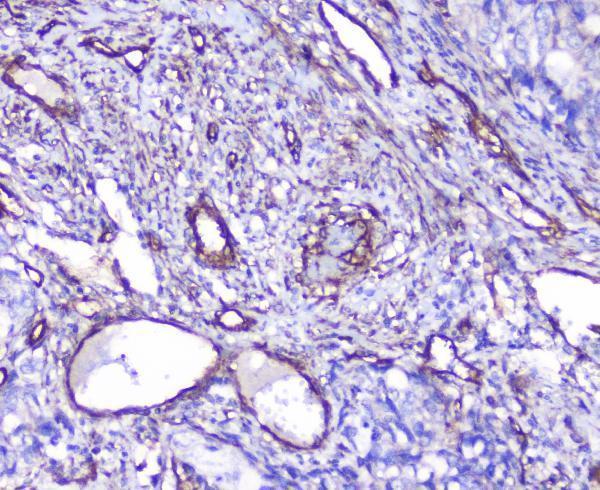
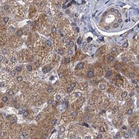
Real IHC data IHC analysis of INPPL1 using anti-INPPL1 antibody (A01790). INPPL1 was detected in paraffin-embedded section of human gastric cancer tissue. Heat mediated antigen retrieval was performed in citrate buffer (pH6, epitope retrieval solution) for 20 mins. The tissue section was blocked with 10% goat serum. The tissue section was then incubated with 1μg/ml rabbit anti-INPPL1 Antibody (A01790) overnight at 4°C. Biotinylated goat anti-rabbit IgG was used as secondary antibody and incubated for 30 minutes at 37°C. The tissue section was developed using Strepavidin-Biotin-Complex (SABC)(Catalog # SA1022) with DAB as the chromogen.
Anti-INPPL1 Antibody ®
Cat # A01790
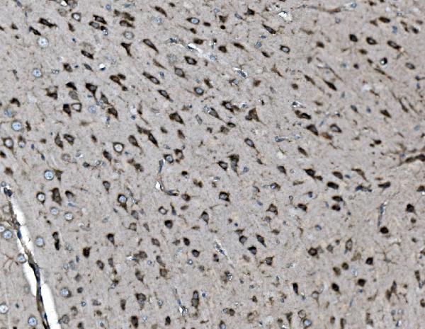
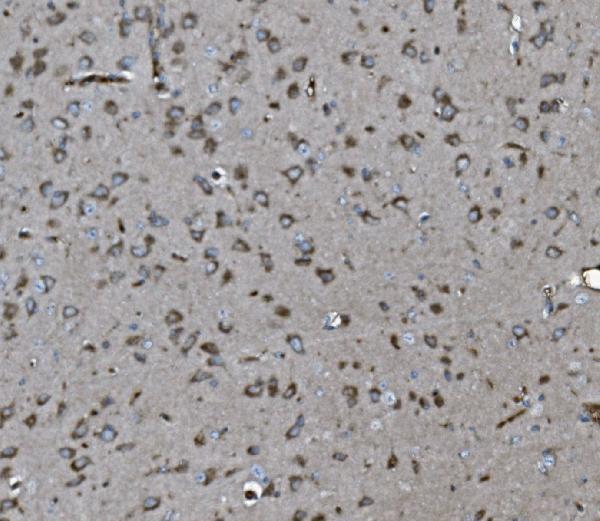
Real IHC data IHC analysis of INPPL1 using anti-INPPL1 antibody (M01790). INPPL1 was detected in paraffin-embedded section of mouse brain tissue. Heat mediated antigen retrieval was performed in EDTA buffer (pH8.0, epitope retrieval solution). The tissue section was blocked with 10% goat serum. The tissue section was then incubated with 2μg/ml mouse anti-INPPL1 Antibody (M01790) overnight at 4°C. Biotinylated goat anti-mouse IgG was used as secondary antibody and incubated for 30 minutes at 37°C. The tissue section was developed using Strepavidin-Biotin-Complex (SABC) (Catalog # SA1021) with DAB as the chromogen.
Anti-INPPL1 Antibody ® (monoclonal, 8C13)
Cat # M01790

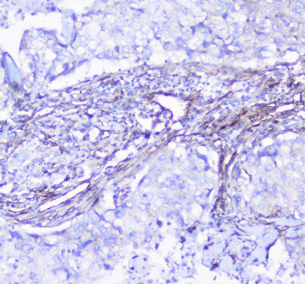
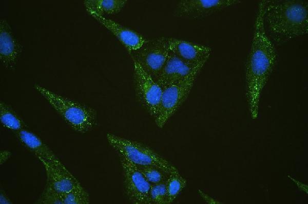
A01790 has IHC images from paraffin-embedded human gastric, lung, ovarian, and placental tissue, and an IF/ICC image from U20S cells (A01790 image captions). M01790 has IHC images from paraffin-embedded mouse and rat brain and human ovarian and renal tumors, and an IF/ICC image from HELA cells (M01790 image captions).

Which to pick: For human tissue IHC, choose A01790 when its citrate pH 6 retrieval matches your plan (A01790 IHC captions); choose M01790 for mouse or rat brain examples using EDTA pH 8 retrieval (M01790 IHC captions). For IF/ICC, both have cell images; A01790 is rabbit-hosted, while M01790 is monoclonal clone 8C13 and mouse-hosted (catalog entries; IF image captions). For cross-species tissue IHC, M01790 has human, mouse, and rat paraffin-section examples (M01790 IHC captions). The fixative is unreported for both antibodies' IHC examples (A01790 and M01790 IHC captions).

Each figure is that product's own IHC / IF validation image from its datasheet.

References

  1. UniProt Consortium. UniProt entry O15357 (SHIP2_HUMAN, Phosphatidylinositol 3,4,5-trisphosphate 5-phosphatase 2).
  2. Human Protein Atlas. INPPL1 tissue IHC expression (reliability: Approved).
  3. Human Protein Atlas. INPPL1 subcellular location (ICC-IF): Mainly localized to the cytosol. In addition localized to the Golgi apparatus..
  4. Human Protein Atlas. INPPL1 antibody validation summary (1 antibodies).
  5. Alteration of gene expression and protein solubility of the PI 5-phosphatase SHIP2 are correlated with Alzheimer's disease pathology progression. Acta neuropathologica 2024 — PMC11150309.
  6. Lipid phosphatase SHIP2 functions as oncogene in colorectal cancer by regulating PKB activation. Oncotarget 2016 — PMC5341996.
  7. SHIP2 deletion in cartilage does not modulate osteoarthritis in male mice. Osteoarthritis and cartilage open 2026 — PMC13572538.
  8. PubMed PMID:8530088 — UniProt-cited evidence.
  9. PubMed PMID:9367831 — UniProt-cited evidence.
  10. PubMed PMID:16554811 — UniProt-cited evidence.