This website uses cookies to ensure you get the best experience on our website.
- Table of Contents
Facts about Interleukin-1 receptor-associated kinase 3.

| Human | |
|---|---|
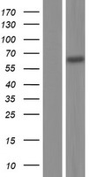
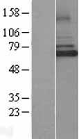
| Gene Name: | IRAK3 |
| Uniprot: | Q9Y616 |
| Entrez: | 11213 |

| Belongs to: |
|---|
| protein kinase superfamily |

ASRT5; EC 2.7.11.1; FLJ13601; IL-1 receptor-associated kinase M; interleukin-1 receptor-associated kinase 3; IRAK3; IRAK-3; IRAKM; IRAK-M
Mass (kDA):
67.767 kDA

| Human | |
|---|---|
| Location: | 12q14.3 |
| Sequence: | 12; NC_000012.12 (66189214..66254622) |
Expressed predominantly in peripheral blood lymphocytes.


PMID: 10383454 by Wesche H., et al. IRAK-M is a novel member of the Pelle/interleukin-1 receptor- associated kinase (IRAK) family.
PMID: 17503328 by Balaci L., et al. IRAK-M is involved in the pathogenesis of early-onset persistent asthma.