IRF5 / Interferon regulatory factor 5 · IHC design guide

Design Immunohistochemistry for IRF5

Plan paraffin-section IRF5 IHC around cytoplasmic staining in selected tissues (HPA tissue IHC). Start with PB9646 at 0.5–1 μg/mL (datasheet: PB9646), and score nuclear staining separately because activated IRF5 translocates to the nucleus (UniProt).

Evidence assembled Oct 2026 · For research use; verify linked source records and product datasheet before use
Immunohistochemistry protocol sheet for IRF5 (IHC for IRF5): expected localisation Cytoplasmic staining in selected tissues (HPA tissue IHC), antibody PB9646, validated IHC image, and IHC protocol steps
Printable IRF5 IHC protocol sheet — expected localisation Cytoplasmic staining in selected tissues (HPA tissue IHC), antibody PB9646, controls and protocol steps. Open the full IRF5 IHC guide →

IRF5 Immunohistochemistry Experimental Design Guide

Expected localisation, validated protocols, controls and antibodies — the at-a-glance facts below, then the full design guide.

Must know before staining
Expected localisation Cytoplasmic staining in selected tissues (HPA tissue IHC)
Staining pattern Cytoplasmic signal in selected tissues, including lymphoid areas (HPA tissue IHC)
Antigen retrieval EDTA pH 8.0 HIER, heat-mediated (datasheet PB9646)
Positive control ⓘ Appendix+4 more · see all
Negative control ⓘ Adipose tissue+4 more · see all
Important caveats
Reasons your staining may differ from the expected pattern.
Fixation Keep fixation consistent across sections (standard IHC practice; not target-specific). Selected-image fixative and duration unreported (datasheet PB9646); verify before use.
Caveat Activation can shift IRF5 into nuclei (UniProt)
Regulation Expression regulation is unreported (UniProt)
Isoform / epitope 6 isoforms; epitope coverage is unknown (UniProt)
Section 1

Recommended IRF5 IHC & IF Protocols

The catalog antibody protocol is complemented by 3 published IRF5 IHC protocols (PMC10412693; PMC12467263; PMC4969615).

Recommended immunohistochemistry (IHC-P) protocol parameters
SampleParaffin-embedded mouse spleen tissue; fixative not specified (datasheet PB9646)
FixationImage fixative and duration unreported (datasheet PB9646); verify before use.
Sectioning4–5 µm sections on charged slides (standard)
DeparaffinisationXylene, graded ethanol series to water (standard)
Antigen retrievalHeat retrieval: EDTA pH 8.0 (datasheet PB9646); 20 min, 95–100 °C (standard)
Peroxidase block3% H2O2, 10 min, room temperature (standard)
Blocking10% goat serum (datasheet PB9646)
Primary antibodyRabbit anti-IRF5, 0.5-1μg/ml (datasheet PB9646)
Primary incubationOvernight at 4 °C (datasheet PB9646)
DetectionStreptavidin-biotin complex (SABC), DAB chromogen (datasheet PB9646)
CounterstainHematoxylin, blue, dehydrate and mount (standard)
Expected resultIRF5-positive staining in lymphoid tissue of appendix (HPA tissue IHC: High). HPA tissue profile: Cytoplasmic expression in selected tissues. No signal in the no-primary control.
💡Decision noteStart with EDTA pH 8.0 heat retrieval for the catalog antibody (datasheet: PB9646); the published protocols use other retrieval methods where stated (PMC12467263; PMC4969615).
Section 2

What Is the Expected IRF5 Staining Pattern?

IRF5 is a cytoplasmic and nuclear protein that can move into the nucleus after activation (UniProt Q13568: subcellular location). In paraffin sections, expect mainly cytoplasmic staining in selected tissues, including hematopoietic and lymphoid cell populations (HPA: tissue IHC, Enhanced reliability). IRF5 has no transmembrane segment, so a membrane-only pattern is unexpected (UniProt Q13568: topology).

What am I looking at on my slide?
Cytoplasmic staining in bone marrow hematopoietic cells or appendix lymphoid tissue, with nearby cells less intensely stained.This fits the reported high staining in those cell populations and the overall cytoplasmic tissue profile (HPA: bone marrow and appendix, High; tissue IHC profile). Judge the named cells, since a whole-section average can hide a cell-specific pattern (general IHC practice).
Nuclear staining accompanies cytoplasmic staining in an otherwise plausible positive cell population.Nuclear localisation is biologically possible: IRF5 shuttles from cytoplasm to nucleus following MYD88-linked activation and phosphorylation (UniProt Q13568: subcellular location). Nuclear colour alone does not establish pathway activation; compare localisation within identified cells and the study’s controls (general IHC practice).
Signal appears only as a sharp cell-border or membrane outline.That compartment does not match the reported cytoplasmic tissue profile or cytoplasm-to-nucleus shuttling (HPA: tissue IHC profile; UniProt Q13568: subcellular location). IRF5 has no transmembrane segment (UniProt Q13568: topology). Review morphology and detection controls before assigning the outline to IRF5 (general IHC practice).
Strong staining appears in a cell population reported as not detected, such as adipocytes in adipose tissue.The discrepancy raises possible nonspecific binding, endogenous detection activity, or cell misidentification; it does not by itself prove cross-reactivity (HPA: adipose adipocytes, Not detected; general IHC practice). Check the stained cell’s identity and an appropriate detection control. HPA also notes staining in some unannotated cells or structures (HPA: reliability description).
Colour spreads through extracellular areas or broadly across unlike cell populations, obscuring cell boundaries.That distribution is difficult to reconcile with HPA’s selected-cell cytoplasmic pattern (HPA: tissue IHC profile). Diffuse colour can reflect background from antibody or detection reagents; compare a no-primary control and assess whether identifiable positive cells remain distinct (general IHC practice).
💡Expected IRF5 appearanceCall an IHC result positive when identifiable cells show distinct cytoplasmic staining, especially high staining in bone marrow hematopoietic cells or appendix lymphoid tissue (HPA: tissue IHC, High); nuclear staining can be plausible in activated cells (UniProt Q13568: subcellular location), whereas a membrane-only outline or diffuse cell-independent colour warrants investigation (UniProt Q13568: topology; general IHC practice).
How each factor affects the staining
Choice of comparison tissueBone marrow hematopoietic cells and appendix lymphoid tissue are reported High; adipose adipocytes are Not detected (HPA: tissue IHC). These provide contrasting cell-level expectations, but the absence of colour in one population cannot validate every staining condition (general IHC practice).
Activation-dependent localisationIRF5 can move into the nucleus after MYD88-linked activation and phosphorylation (UniProt Q13568: subcellular location). Score nuclear and cytoplasmic compartments separately; tissue IHC describes predominantly cytoplasmic expression and does not establish the activation state of an individual stained cell (HPA: tissue IHC profile).
Antibody-level IHC evidenceHPA rates IHC staining Enhanced for HPA046700 and Approved for HPA076024; neither has an ICC validation status in the supplied antibody record (HPA: antibody validation). Treat the tissue pattern as a guide and verify the actual antibody used in the experiment (general IHC practice).
Q: What should IF/ICC show?A: Cytoplasmic and potentially nuclear IRF5 are biologically plausible (UniProt Q13568: subcellular location). The supplied HPA subcellular record provides no main location or ICC-IF images, and the listed antibodies have no ICC validation status (HPA: subcellular and antibody records); it cannot confirm an IF image pattern.
Why is my staining missing, weak or wrong?
SituationLikely causeNext action
No staining in bone marrow hematopoietic cells or appendix lymphoid tissue.These are reported High, so a blank result conflicts with the expected cell-level pattern (HPA: tissue IHC). The record does not identify which workflow step failed.Confirm the intended cells are present, then check primary-antibody use, retrieval, detection reagents and counterstain against the assay controls (general IHC practice). Do not infer an IRF5-specific fixation effect from this result.
Adipose adipocytes stain as strongly as the proposed positive cells.Adipocytes are reported Not detected in adipose tissue (HPA: tissue IHC); apparent colour could arise from misidentified cells or nonspecific or endogenous detection signal (general IHC practice).Recheck cell morphology and compare a no-primary detection control; interpret staining in neighbouring non-adipocytes separately (general IHC practice).
Only a crisp membrane outline is visible.The reported tissue pattern is cytoplasmic, and IRF5 lacks a transmembrane segment (HPA: tissue IHC profile; UniProt Q13568: topology).Review the same cells for cytoplasmic colour, then compare detection controls and antibody validation before scoring the outline as IRF5 (general IHC practice; HPA: antibody validation).
Nuclei stain, but cytoplasmic staining is weak or absent.IRF5 can translocate to the nucleus after activation, but HPA’s tissue IHC profile is cytoplasmic in selected tissues (UniProt Q13568: subcellular location; HPA: tissue IHC profile).Score the compartments separately in identified cells and compare a known-positive tissue and detection control; avoid calling activation from nuclear colour alone (general IHC practice).
Diffuse colour obscures the expected positive cells.Broad cell-independent colour does not resemble the selected-cell pattern (HPA: tissue IHC profile) and may reflect background from staining reagents (general IHC practice).Compare a no-primary control, inspect blocking and washing, and adjust detection conditions under the assay’s standard validation process (general IHC practice).
A high-staining HPA tissue shows uneven results across cells or fields.HPA reports particular cell populations as High, not uniform staining of an entire organ (HPA: tissue IHC). Its Enhanced tissue-level reliability still notes medium agreement with RNA and staining in unannotated structures (HPA: reliability description).Identify the scored cell population and document compartment and intensity by field; use morphology and controls when assessing unexpected structures (general IHC practice).

Sample controls for IRF5 IHC & IF

🧪Run spleen first: cells in the red pulp should stain for IRF5 (HPA: High in spleen red-pulp cells). Use adipose tissue as the negative tissue: adipocytes should lack detectable staining (HPA: Not detected in adipocytes); cells without specific signal on the spleen slide should show only counterstain.
Positive control tissue: Appendix (Lymphoid tissue, HPA High)
Negative control tissue: Adipose tissue (HPA Not detected)
ICC-IF cell lines (HPA subcellular resource): HPA carries no ICC-IF cell line for IRF5; derive a cell-line control from the positive tissue's cell type (Lymphoid tissue) and confirm it by RNA or western blot first.
Technical controls: Include a no-primary, secondary-only slide, a concentration-matched nonimmune rabbit IgG control for the caption’s rabbit primary, and IRF5-knockout tissue as a biological negative (PB9646 caption: rabbit primary; standard IHC practice). In spleen red pulp, quench endogenous peroxidase and check endogenous biotin because the caption uses biotin-based detection with DAB (PB9646 caption: SABC/DAB; standard IHC practice).
⚠️Feasibility: A target-specific fixation window or fixation effect is unreported, and the selected paraffin-section caption does not state the fixative (PB9646 caption). The reported mouse-spleen IHC condition uses heat-mediated EDTA retrieval at pH 8.0; whether retrieval is essential is unreported (PB9646 caption). The supplied evidence does not establish that frozen sections or IF/ICC are easier, and spleen red pulp can give endogenous peroxidase or biotin background with the reported SABC/DAB detection (HPA subcellular: no ICC-IF images; PB9646 caption: SABC/DAB; standard IHC practice).

HPA tissue IHC evidence for IRF5

Comprehensive Human Protein Atlas IHC scoring per tissue (reliability: Enhanced — Medium consistency between antibody staining and RNA expression data. Antibody staining in cells/structures not annotated, view images.). Rows are taken directly from the HPA tissue chart — click any row's HPA link to view the source.

Positive expression · recommended positive controls

TissueCell typeLevelEvidenceSource
Appendix Lymphoid tissue High Protein (IHC) HPA →
Bone marrow Hematopoietic cells High Protein (IHC) HPA →
Fallopian tube Ciliated cells (tip of cilia) High Protein (IHC) HPA →
Lymph node Non-germinal center cells High Protein (IHC) HPA →
Spleen Cells in red pulp High Protein (IHC) HPA →

Undetected expression · recommended negative controls

TissueCell typeLevelEvidenceSource
Adipose tissue Adipocytes Not detected Protein (IHC) HPA →
Adrenal gland Glandular cells Not detected Protein (IHC) HPA →
Breast Adipocytes Not detected Protein (IHC) HPA →
Caudate Glial cells Not detected Protein (IHC) HPA →
Cerebellum Cells in granular layer Not detected Protein (IHC) HPA →
Section 3

Advanced IRF5 IHC Tips

Troubleshoot IRF5 chromogenic IHC in paraffin sections by checking retrieval, cellular compartment, controls, and scoring before interpreting differences between samples.

Which antigen retrieval should I use for IRF5 paraffin sections?
Start with heat-mediated EDTA retrieval at pH 8.0 for IRF5 paraffin-section IHC (datasheet PB9646). The mouse spleen example paired this retrieval with a 10% goat serum block and 1 μg/ml primary antibody overnight at 4°C (caption PB9646). The fixative and heating duration were unreported, so record both locally and compare serial sections before attributing weak staining to low IRF5 abundance (caption PB9646; standard IHC practice). If staining remains weak, test a secondary retrieval condition on matched sections while holding primary concentration and detection constant (standard IHC practice). Compare cell morphology and compartment staining with spleen red-pulp cells as a reference (HPA: High in spleen red pulp; UniProt Q13568 localisation).
How should fixation differences be handled when comparing IRF5 IHC sections?
Target-specific fixation sensitivity is unknown from the supplied IHC evidence; the PB9646 paraffin-section caption does not report a fixative (caption PB9646). Document fixative, fixation duration, tissue thickness, and processing history for each case before comparing staining intensity (standard IHC practice). Apply the same heat-mediated EDTA retrieval at pH 8.0 to matched sections so processing differences can be assessed consistently (datasheet PB9646; standard IHC practice). Check cellular morphology and exclude folds, damaged edges, and poorly preserved regions from scoring (standard IHC practice). HPA staining patterns and IRF5 phosphorylation annotations do not establish how fixation affects this antibody's staining (HPA tissue IHC; UniProt Q13568 modified residues).
How should I interpret cytoplasmic versus nuclear IRF5 staining?
Score cytoplasmic and nuclear compartments separately: IRF5 shuttles between them and translocates to nuclei after MYD88-dependent activation and phosphorylation (UniProt Q13568 localisation). Predominantly cytoplasmic staining agrees with the selected-tissue IHC profile, which includes high staining in spleen red-pulp cells (HPA tissue IHC). Use a counterstain to define nuclei, then report the fraction of positive nuclei within the cell population being assessed (standard IHC practice). Cytoplasmic staining alone does not establish pathway inactivity, and nuclear staining alone does not prove activation without supporting evidence (UniProt Q13568 localisation; standard IHC interpretation). Exclude pigment or precipitate outside intact cells before reporting a compartment shift (standard IHC practice).
Can this IHC antibody distinguish IRF5 isoforms or phosphorylation states?
IRF5 has 6 annotated isoforms, but the supplied PB9646 tissue caption provides neither epitope sequence nor isoform coverage (UniProt Q13568 isoforms; caption PB9646). Assign isoform specificity only when immunogen mapping and independent validation support it (standard IHC practice). Phosphorylation is annotated at multiple residues, including TBK1-linked sites 158 and 293, but those annotations do not establish phospho-selective binding by the catalog antibody (UniProt Q13568 modified residues). When cases stain differently, compare controls and consider an independently validated antibody with a mapped distinct epitope before proposing splice-variant differences (standard IHC practice). Report compartment and scoring criteria without assigning an isoform from DAB intensity alone (standard IHC practice).
How should I assess IRF5 in multiplex immunofluorescence?
Validate IF separately from the supplied PB9646 chromogenic paraffin-section IHC example, whose fixative is unreported (caption PB9646; standard IF practice). Pair IRF5 with a marker for the expected hematopoietic cell population and verify each channel separately before interpreting merged images (HPA: High in bone-marrow hematopoietic cells; standard IF practice). Choose fluorophores with emission separated from tissue autofluorescence, and inspect unstained and single-color controls (standard IF practice). IRF5 occupies cytoplasm and nucleus and has no transmembrane segment, so permeabilise for intracellular antibody access while preserving nuclear morphology (UniProt Q13568 topology and localisation; standard IF practice). Quantify IRF5 within segmented cells, reporting nuclear and cytoplasmic signals separately (standard IF practice).
How can I distinguish IRF5 staining from DAB background?
The PB9646 example uses a biotinylated secondary antibody, avidin-biotin amplification, and DAB; include no-primary and secondary-only controls (caption PB9646; standard IHC practice). Block endogenous peroxidase before DAB development and assess endogenous biotin when using avidin-biotin detection (standard IHC practice). Compare pigment, edge deposits, and damaged regions with staining confined to intact cells (standard IHC practice). Cell identity helps interpretation: HPA reports high staining in selected hematopoietic compartments and no detected staining in adipocytes (HPA tissue IHC). If background persists, titrate reagents around the reported 1 μg/ml primary concentration on adjacent sections while retaining EDTA retrieval at pH 8.0 (caption PB9646; standard IHC practice).
How should I quantify IRF5 IHC across heterogeneous sections? ⚠ ANSWER MARKED FOR VERIFICATION
Define the cell population before scoring because HPA reports high staining in spleen red-pulp and bone-marrow hematopoietic cells, but no detected staining in adipocytes (HPA tissue IHC). Use an H-score, percentage of positive cells, or positive-cell density per mm², with one prespecified threshold across the batch (standard IHC practice). Normalize positive-cell counts to viable cells in the target population or to assessed tissue area, and document excluded necrotic regions (standard IHC practice). Score nuclear and cytoplasmic signals separately because IRF5 can shuttle between those compartments (UniProt Q13568 localisation). Keep retrieval, DAB development, counterstain, and image acquisition consistent, and report controls with each score (standard IHC practice).
What makes an IRF5-positive cell convincing rather than artefactual?
Look for staining within intact cytoplasm or nuclei in an identified cell population; IRF5 occupies both compartments, and HPA reports staining in selected hematopoietic cells (UniProt Q13568 localisation; HPA tissue IHC). Question signal confined to section edges, necrosis, folds, or extracellular deposits, especially when no-primary controls look similar (standard IHC practice). Assess endogenous peroxidase and biotin before attributing diffuse brown staining to IRF5 in the PB9646 avidin-biotin DAB workflow (caption PB9646; standard IHC practice). Compare reference compartments: HPA reports high staining in spleen red pulp and no detected staining in adipocytes (HPA tissue IHC). Nuclear enrichment may support relocalisation but cannot by itself prove MYD88 activation or phosphorylation (UniProt Q13568 localisation; standard IHC interpretation).
Boster reagents

Best IRF5 / Interferon regulatory factor 5 IHC Antibodies

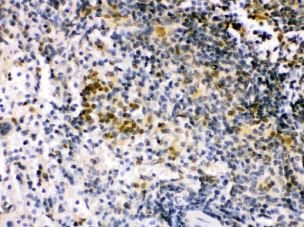
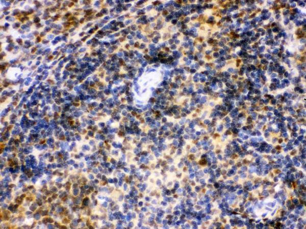
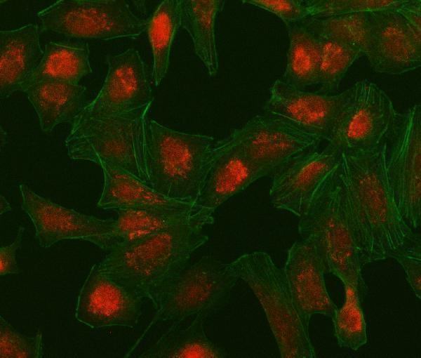
Anti-IRF5 antibodies have IHC images from paraffin sections of mouse and rat spleen and human mammary cancer, plus IF/ICC imaging in human A549 cells (PB9646 and A00958-1 image captions).

Real IHC data IHC analysis of IRF5 using anti-IRF5 antibody (PB9646). IRF5 was detected in a paraffin-embedded section of mouse spleen tissue. Heat mediated antigen retrieval was performed in EDTA buffer (pH 8.0, epitope retrieval solution). The tissue section was blocked with 10% goat serum. The tissue section was then incubated with 1 μg/ml rabbit anti-IRF5 Antibody (PB9646) overnight at 4°C. Biotinylated goat anti-rabbit IgG was used as secondary antibody and incubated for 30 minutes at 37°C. The tissue section was developed using Strepavidin-Biotin-Complex (SABC) (Catalog # SA1022) with DAB as the chromogen.
Anti-IRF5 Antibody ®
Cat # PB9646
Real IF data IF analysis of IRF5 using anti-IRF5 antibody (A00958-1). IRF5 was detected in an immunocytochemical section of A549 cells. Enzyme antigen retrieval was performed using IHC enzyme antigen retrieval reagent (AR0022) for 15 mins. The cells were blocked with 10% goat serum. And then incubated with 5 μg/mL rabbit anti-IRF5 Antibody (A00958-1) overnight at 4°C. Cy3 Conjugated Goat Anti-Rabbit IgG (BA1032) was used as secondary antibody at 1:100 dilution and incubated for 30 minutes at 37°C. The tissue section was developed using Phalloidin-iFluor 488 Conjugated. Visualize using a fluorescence microscope and filter sets appropriate for the label used.
Anti-IRF5 Antibody ®
Cat # A00958-1

PB9646 has IHC images from paraffin sections of mouse and rat spleen and human mammary cancer (PB9646 image captions). A00958-1 has an IF/ICC image from human A549 cells (A00958-1 image caption).

Which to pick: For paraffin-section IHC, pick PB9646: its mouse spleen image used EDTA pH 8 retrieval and 1 μg/ml primary antibody overnight; the fixative is unreported (PB9646 image caption). For IF/ICC, pick A00958-1, imaged in A549 cells at 5 μg/ml (A00958-1 IF image caption). For tissue IHC across human, mouse and rat, PB9646 has listed reactivity and paraffin-section images in all three species (PB9646 catalog reactivity and image captions).

Each figure is that product's own IHC / IF validation image from its datasheet.

References

  1. UniProt Consortium. UniProt entry Q13568 (IRF5_HUMAN, Interferon regulatory factor 5).
  2. Human Protein Atlas. IRF5 tissue IHC expression (reliability: Enhanced).
  3. Human Protein Atlas. IRF5 subcellular location (ICC-IF): Highest expression in HDLM-2: 112.9 nTPM.
  4. Human Protein Atlas. IRF5 antibody validation summary (2 antibodies).
  5. Dysregulation of PLOD2 Promotes Tumor Metastasis and Invasion in Hepatocellular Carcinoma. Journal of clinical and translational hepatology 2023 — PMC10412693.
  6. Interferon Regulator Factor 5: A Novel Inflammatory Marker and Promising Therapeutic Target in Ulcerative Colitis. Biomedicines 2025 — PMC12467263.
  7. Specific detection of interferon regulatory factor 5 (IRF5): A case of antibody inequality. Scientific reports 2016 — PMC4969615.
  8. Securidaca inappendiculata-Derived Xanthones Protected Joints from Degradation in Male Rats with Collagen-Induced Arthritis by Regulating PPAR-γ Signaling. Journal of inflammation research 2021 — PMC7896782.
  9. PubMed PMID:11303025 — UniProt-cited evidence.
  10. PubMed PMID:15695821 — UniProt-cited evidence.
  11. PubMed PMID:15805103 — UniProt-cited evidence.