This website uses cookies to ensure you get the best experience on our website.


- Table of Contents
Plan paraffin IHC around nuclear IRF9 staining in most tissues (HPA tissue IHC). Compare high-staining respiratory or glandular cells with reported unstained cells, and disregard membrane staining as off target (HPA tissue IHC).

Expected localisation, validated protocols, controls and antibodies — the at-a-glance facts below, then the full design guide.
| Expected localisation | Nuclear staining in most tissues (HPA tissue IHC) | |
| Staining pattern | Nuclear signal in respiratory and glandular cells (HPA tissue IHC) | |
| Antigen retrieval | EDTA pH 8.0 HIER, heat-mediated (datasheet A04485) | |
| Positive control | Bronchus+4 more · see all | |
| Negative control | Adipose tissue+4 more · see all |
| Fixation | Keep fixation consistent across sections (standard IHC practice; not target-specific) | |
| Caveat | Disregard cell membrane staining as off target (HPA tissue IHC) | |
| Regulation | Type I IFN promotes nuclear translocation (UniProt) | |
| Isoform / epitope | One 1–393 chain; no isoforms annotated (UniProt) |
The catalog antibody uses EDTA pH 8.0 retrieval (datasheet: A04485). The published IHC protocols below cover pulmonary artery, lung tumor, prostate, and xenograft sections (PMC8672195; PMC7827113; PMC3753051; PMC13426216).
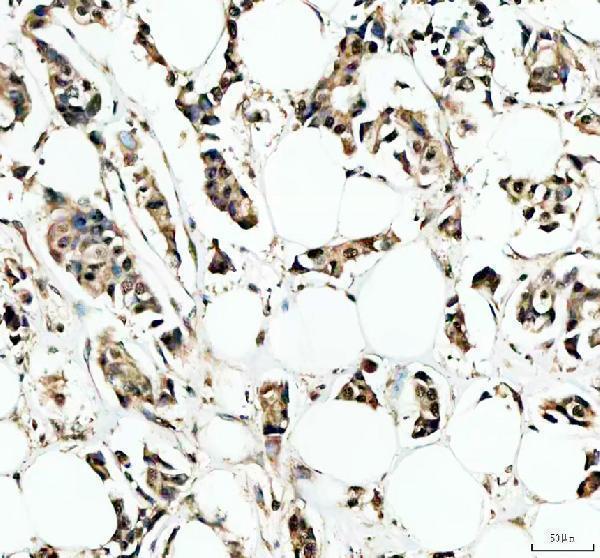
| Sample | Paraffin-embedded human breast cancer tissue; fixative not specified (datasheet A04485) |
| Fixation | Image fixative and duration unreported (datasheet A04485); verify before use. |
| Sectioning | 4–5 µm sections on charged slides (standard) |
| Deparaffinisation | Xylene, graded ethanol series to water (standard) |
| Antigen retrieval | Heat retrieval: EDTA pH 8.0 (datasheet A04485); 20 min, 95–100 °C (standard) |
| Peroxidase block | 3% H2O2, 10 min, room temperature (standard) |
| Blocking | 10% goat serum (datasheet A04485) |
| Primary antibody | Rabbit anti-IRF9, 2-5μg/ml (datasheet A04485) |
| Primary incubation | Overnight at 4 °C (datasheet A04485) |
| Detection | HRP-conjugated secondary, DAB chromogen (datasheet A04485) |
| Counterstain | Hematoxylin, blue, dehydrate and mount (standard) |
| Expected result | IRF9-positive staining in respiratory epithelial cells of bronchus (HPA tissue IHC: High). HPA tissue profile: Nuclear expression in most tissues. No signal in the no-primary control. |
For paraffin-section IHC, expect predominantly nuclear IRF9 staining in many tissues, including respiratory, glandular and urothelial cells (HPA: nuclear expression in most tissues; High in the listed cell types). IRF9 can also reside in the cytoplasm and moves into the nucleus after type I interferon activation (UniProt Q00978: subcellular location). It has no transmembrane segment (UniProt Q00978: topology). HPA rates its tissue IHC pattern Approved but reports low agreement with RNA data (HPA: reliability description).
| Distinct nuclear staining in bronchial or nasopharyngeal respiratory epithelium, or endometrial glandular cells (HPA: High in these cells). | This fits the reported tissue IHC pattern (HPA: nuclear expression in most tissues). Compare staining with nearby cell types and a negative control; HPA's Approved rating carries a low RNA-agreement caveat (HPA: reliability description). |
| Strong staining outlines cell borders instead of nuclei. | Treat a membrane pattern as suspect: HPA viewed membrane staining as off target and disregarded it (HPA: reliability description). IRF9 lacks a transmembrane segment (UniProt Q00978: topology). Review morphology and detection controls before scoring these cells as positive. |
| An apparently strong positive result occurs mainly in adipocytes or cardiomyocytes. | Those cells are reported as not detected in the sampled tissues (HPA: adipocytes in adipose tissue; cardiomyocytes in heart muscle). Check cell identification and controls. Cross-reactivity or endogenous detection activity is possible, but the slide alone cannot establish either cause. |
| Colour spreads across nuclei, cytoplasm and empty spaces without clear cell boundaries. | Diffuse background prevents reliable compartment scoring. Check the no-primary control and compare with a known-positive section; review blocking, washes and detection conditions as general IHC troubleshooting. HPA's tissue pattern alone cannot identify the source of background. |
| No convincing signal appears in a bronchial section's respiratory epithelial cells. | This differs from HPA's High staining report for those cells (HPA: bronchus). Confirm that the expected cells are present, then review the antibody and detection workflow with controls. A negative section does not by itself prove that IRF9 is absent. |
| Cell context and activation | UniProt places IRF9 in cytoplasm and nucleus and reports nuclear translocation after IFN-alpha/beta activation (UniProt Q00978: subcellular location). Interpret a nuclear IHC result in tissue context; compartment alone does not establish whether a specimen received an interferon stimulus. |
| Strength of the tissue reference | HPA reports High staining in bronchial respiratory epithelium and endometrial glands, Medium in breast glands, and no detection in adipocytes (HPA: tissue IHC). These are useful comparisons, but its Approved pattern has low agreement with RNA data (HPA: reliability description). |
| Antibody validation | The listed antibody HPA001862 is Approved for IHC, while its ICC status is Supported (HPA: antibody validation). Do not describe its IHC result as Enhanced: the supplied validation record does not assign that status (HPA: antibody validation). |
| IF/ICC Q: Should cytosolic staining match paraffin-section IHC? | A: HPA reports supported cytosolic localisation in ICC-IF, while its tissue IHC profile is mainly nuclear (HPA: subcellular and tissue IHC). UniProt lists both compartments and activation-dependent nuclear translocation (UniProt Q00978: subcellular location). Assess each application against its own evidence. |
| Situation | Likely cause | Next action |
|---|---|---|
| Expected epithelial nuclei are faint or unstained. | The section may lack the expected cells, or the IHC workflow may have produced insufficient detectable signal; neither cause is established by HPA. | Verify the cells on the counterstained section. Run a reported High tissue alongside it and check primary-antibody and chromogenic-detection steps (HPA: High in bronchial respiratory epithelium). |
| Cell borders stain more strongly than nuclei. | HPA specifically regarded membrane staining as off target (HPA: reliability description). | Exclude border-only staining from positive nuclear scoring. Compare a no-primary control and inspect the reported nuclear pattern in a positive tissue (HPA: nuclear tissue profile). |
| A reported negative cell type appears strongly positive. | Cell misidentification, cross-reactivity or endogenous detection activity may explain the result; the cause remains unproven (HPA: no detection in adipocytes and cardiomyocytes). | Recheck morphology and the no-primary control, then compare with a reported High cell type on a separately assessed section (HPA: bronchial respiratory epithelial cells). |
| Most of the section has diffuse chromogenic colour. | General IHC background can obscure cell boundaries and nuclear localisation; the HPA tissue record does not identify its source. | Check the no-primary control, blocking and washes, then reassess whether nuclei can be distinguished from surrounding tissue before scoring. |
| Nuclear and cytoplasmic staining coexist. | Both locations are compatible with UniProt's IRF9 annotation; nuclear translocation follows IFN-alpha/beta activation (UniProt Q00978: subcellular location). | Score nuclear and cytoplasmic compartments separately. Compare the nuclear component with HPA tissue IHC, without inferring activation state from staining alone (HPA: nuclear tissue profile). |
| A change in antigen retrieval seems to alter staining. | The supplied HPA and UniProt records provide no target-specific retrieval or fixation sensitivity. | Record the retrieval conditions and compare controlled sections under the same scoring criteria. Treat any change as an assay observation until independently verified; do not assign it an IRF9-specific mechanism. |


Comprehensive Human Protein Atlas IHC scoring per tissue (reliability: Approved — Low consistency between antibody staining and RNA expression data. Cell membrane staining viewed as off target and disregarded.). Rows are taken directly from the HPA tissue chart — click any row's HPA link to view the source.
| Tissue | Cell type | Level | Evidence | Source |
|---|---|---|---|---|
| Bronchus | Respiratory epithelial cells | High | Protein (IHC) | HPA → |
| Endometrium | Glandular cells | High | Protein (IHC) | HPA → |
| Epididymis | Glandular cells | High | Protein (IHC) | HPA → |
| Fallopian tube | Glandular cells | High | Protein (IHC) | HPA → |
| Nasopharynx | Respiratory epithelial cells | High | Protein (IHC) | HPA → |
| Tissue | Cell type | Level | Evidence | Source |
|---|---|---|---|---|
| Adipose tissue | Adipocytes | Not detected | Protein (IHC) | HPA → |
| Adrenal gland | Glandular cells | Not detected | Protein (IHC) | HPA → |
| Heart muscle | Cardiomyocytes | Not detected | Protein (IHC) | HPA → |
| Hippocampus | Glial cells | Not detected | Protein (IHC) | HPA → |
| Parathyroid gland | Glandular cells | Not detected | Protein (IHC) | HPA → |
Troubleshoot IRF9 staining by checking retrieval, compartment, controls and scoring against the tested paraffin section workflow and reported tissue patterns.
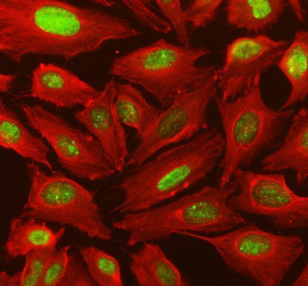
A04485 has human paraffin-section IHC and U2OS-cell IF images (A04485 IHC/IF captions).
A04485 will render with an IHC image from a human breast cancer paraffin section (A04485 IHC caption). Its IF image shows U2OS cells (A04485 IF caption).
Which to pick: Choose A04485 for human paraffin-section IHC; its own image caption reports EDTA pH 8 retrieval and 2 μg/mL primary antibody, but does not report the fixative (A04485 IHC caption). For IF/ICC, A04485 has an IF image at 5 μg/mL, while M04485 is a rabbit monoclonal, clone 21I34, listed for ICC/IF without a supplied image (A04485 IF caption; catalog: M04485 clone, applications and image fields). Neither SKU has documented reactivity beyond human (catalog: A04485 and M04485 reactivity).