This website uses cookies to ensure you get the best experience on our website.


- Table of Contents
Plan ISL1 chromogenic IHC in paraffin sections around its nuclear staining pattern and the cell populations reported as positive (HPA tissue IHC). The guide covers fixation, staining controls and interpretation of sparse positive cells (HPA tissue IHC).

Expected localisation, validated protocols, controls and antibodies — the at-a-glance facts below, then the full design guide.
| Expected localisation | Nuclear staining in a few tissues (HPA tissue IHC) | |
| Staining pattern | Nuclear staining in selected cell populations (HPA tissue IHC) | |
| Antigen retrieval | EDTA pH 8.0 HIER, heat-mediated (datasheet A02969-3) | |
| Positive control | Adrenal gland+4 more · see all | |
| Negative control | Adipose tissue+4 more · see all |
| Fixation | Keep fixation consistent across sections (standard IHC practice; not target-specific). Selected-image fixative and duration unreported (datasheet A02969-3); verify before use. | |
| Caveat | Sparse positive cells may be missed in sections (HPA tissue IHC) | |
| Regulation | Expression varies by cell type (HPA tissue IHC) | |
| Isoform / epitope | No isoforms or cleavage products annotated (UniProt) |
The catalog antibody’s IHC-P protocol is accompanied by published ISL1 IHC methods for neuroendocrine tumors and neural tissue (PMC7665972; PMC4898681).
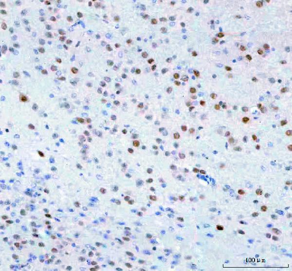
| Sample | Paraffin-embedded mouse brain tissue; fixative not specified (datasheet A02969-3) |
| Fixation | Image fixative and duration unreported (datasheet A02969-3); verify before use. |
| Sectioning | 4–5 µm sections on charged slides (standard) |
| Deparaffinisation | Xylene, graded ethanol series to water (standard) |
| Antigen retrieval | Heat retrieval: EDTA pH 8.0 (datasheet A02969-3); 20 min, 95–100 °C (standard) |
| Peroxidase block | 3% H2O2, 10 min, room temperature (standard) |
| Blocking | 10% goat serum (datasheet A02969-3) |
| Primary antibody | Rabbit anti-ISL1, 2-5 μg/ml (datasheet A02969-3) |
| Primary incubation | Overnight at 4 °C (datasheet A02969-3) |
| Detection | HRP-conjugated secondary, DAB chromogen (datasheet A02969-3) |
| Counterstain | Hematoxylin, blue, dehydrate and mount (standard) |
| Expected result | ISL1-positive staining in glandular cells of adrenal gland (HPA tissue IHC: Medium). HPA tissue profile: Nuclear expression in few tissues. No signal in the no-primary control. |
ISL1 is a nuclear transcription factor with no transmembrane segment (UniProt P61371). In paraffin-section IHC, expect staining in nuclei of selected cells, including adrenal glandular cells, kidney tubular cells and testicular Leydig cells; HPA reports medium staining in each (HPA tissue IHC). HPA describes expression in few tissues, but rates its tissue IHC evidence Approved, pending external verification (HPA tissue IHC).
| Distinct nuclear stain in a subset of adrenal glandular cells, kidney tubular cells or testicular Leydig cells. | This fits the reported compartment and cell distribution; medium staining is reported for these cells (HPA tissue IHC; UniProt P61371). Score the named cell population and its nuclear signal. HPA's Approved rating is pending external verification, so confirm the pattern with controls (HPA tissue IHC; standard IHC practice). |
| Predominantly cytoplasmic, membranous or extracellular chromogen, with little nuclear staining. | That distribution conflicts with the expected nuclear IHC profile and the absence of a transmembrane segment (HPA tissue IHC; UniProt P61371). Treat it as suspect staining; review morphology, the no-primary control and detection conditions before assigning ISL1 positivity (standard IHC practice). |
| Stain appears in cells expected to be negative, such as adipocytes in adipose tissue or hematopoietic cells in bone marrow. | Both named populations were not detected in HPA tissue IHC (HPA tissue IHC). Check whether signal follows the cell nuclei; unexpected staining can reflect antibody cross-reactivity or endogenous detection activity (standard IHC practice). A negative reference is a comparison, not proof that every specimen must be unstained. |
| Diffuse chromogen obscures nuclei across multiple cell types or tissue regions. | A widespread haze cannot establish the selective nuclear pattern reported for ISL1 (HPA tissue IHC). Compare with a no-primary control, inspect reagent background and adjust blocking, washing or detection conditions as indicated (standard IHC practice). Do not score obscured nuclei as positive. |
| No nuclear signal in an adrenal gland section containing identifiable glandular cells. | HPA reports medium staining in adrenal glandular cells, so this is a useful positive-reference failure to investigate (HPA tissue IHC). Verify that the intended cells are present and review antibody concentration, retrieval and detection with the catalog IHC-P protocol (standard IHC practice); HPA does not establish ISL1-specific fixation sensitivity. |
| Compartment and cell distribution (UniProt P61371; HPA tissue IHC) | ISL1 is nuclear, while HPA reports expression in few tissues and medium staining in selected cell populations. Judge nuclear signal within the named cells; a tissue-wide positive/negative label can conceal a small positive population (HPA tissue IHC). |
| Evidence strength and antibody validation (HPA tissue IHC; HPA antibodies) | HPA's tissue IHC rating is Approved with external verification pending. Its IHC-Approved antibody is HPA057416; HPA048613 is ICC-Approved with no IHC status supplied (HPA antibodies). Validation of one antibody or application does not establish the other's IHC performance. |
| Antigen retrieval and detection setup (standard IHC practice) | Follow the catalog antibody's IHC-P instructions and assess changes against positive and negative controls (standard IHC practice). The supplied HPA and UniProt records give no ISL1-specific retrieval condition or fixation-effect evidence; neither source supports predicting a rescue from altered retrieval. |
| Situation | Likely cause | Next action |
|---|---|---|
| Adrenal glandular nuclei are blank (HPA tissue IHC). | A missed target cell population or an unsuccessful staining run are possibilities (standard IHC practice). | Confirm cell identity and section quality, then check the catalog antibody's IHC-P instructions, retrieval, dilution and detection controls (standard IHC practice). |
| Only cytoplasmic or edge staining appears. | The location conflicts with nuclear ISL1 and HPA's nuclear IHC profile (UniProt P61371; HPA tissue IHC). | Compare with a no-primary control and examine section edges and morphology; score only convincing nuclear staining in the relevant cells (standard IHC practice). |
| Adipocytes or bone-marrow hematopoietic cells stain strongly. | HPA reports these cells as not detected; cross-reactivity or endogenous detection activity may account for unexpected signal (HPA tissue IHC; standard IHC practice). | Check nuclear localisation, run a no-primary control and review blocking or endogenous-activity controls appropriate to the detection chemistry (standard IHC practice). |
| Weak haze makes nuclear scoring uncertain. | Background or insufficient separation between signal and counterstain may obscure nuclei (standard IHC practice). | Compare control sections, review washing and blocking, and adjust counterstain or detection conditions using the IHC-P workflow (standard IHC practice). |
| A tissue called negative contains a few stained nuclei. | HPA's profile is selective, and its listed cell-level observations do not make every cell in a tissue equivalent (HPA tissue IHC). | Identify the stained cell type and compare its nuclear pattern with HPA's cell-level entries before interpreting the result (HPA tissue IHC; standard IHC practice). |
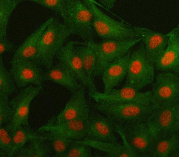
| IF/ICC question: should extranuclear fluorescence be expected? | HPA reports mainly nucleoplasmic signal, with additional nuclear-speckle and mitochondrial localisation in ICC-IF (HPA subcellular). | Interpret that observation within the separate IF/ICC application; it does not override the nuclear expectation for paraffin-section IHC, and HPA's ICC approval is antibody-specific (HPA tissue IHC; HPA antibodies). |


Comprehensive Human Protein Atlas IHC scoring per tissue (reliability: Approved — Pending external verification.). Rows are taken directly from the HPA tissue chart — click any row's HPA link to view the source.
| Tissue | Cell type | Level | Evidence | Source |
|---|---|---|---|---|
| Adrenal gland | Glandular cells | Medium | Protein (IHC) | HPA → |
| Bronchus | Respiratory epithelial cells | Medium | Protein (IHC) | HPA → |
| Caudate | Glial cells | Medium | Protein (IHC) | HPA → |
| Cerebellum | Cells in molecular layer | Medium | Protein (IHC) | HPA → |
| Fallopian tube | Glandular cells | Medium | Protein (IHC) | HPA → |
| Tissue | Cell type | Level | Evidence | Source |
|---|---|---|---|---|
| Adipose tissue | Adipocytes | Not detected | Protein (IHC) | HPA → |
| Bone marrow | Hematopoietic cells | Not detected | Protein (IHC) | HPA → |
| Breast | Adipocytes | Not detected | Protein (IHC) | HPA → |
| Cervix | Glandular cells | Not detected | Protein (IHC) | HPA → |
| Colon | Endothelial cells | Not detected | Protein (IHC) | HPA → |
Troubleshoot ISL1 staining by checking retrieval, nuclear localisation and cell identity before interpreting chromogenic signal (UniProt P61371; HPA tissue IHC).
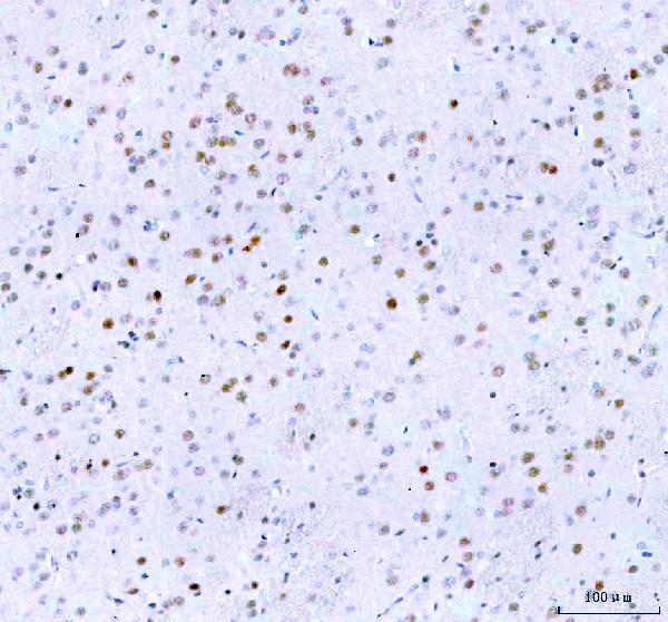
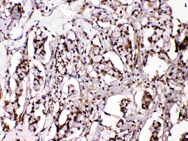
A02969-3 has IHC images in mouse and rat brain and IF images in A549 cells and mouse and rat brain (catalog image captions); A02969-1 has human mammary cancer IHC (catalog image caption).
A02969-3 will render with its mouse brain paraffin-section IHC figure; its additional captions show rat brain IHC and IF in A549 cells and mouse and rat brain (A02969-3 image captions). A02969-1 will render with its human mammary cancer paraffin-section IHC figure (A02969-1 IHC caption).
Which to pick: For human mammary cancer tissue IHC, choose A02969-1; its figure used citrate retrieval at pH 6 for 20 minutes and 1 μg/ml primary antibody (A02969-1 IHC caption). For IF/ICC or mouse and rat brain IHC, choose A02969-3: it lists IF and ICC, reports Human, Mouse and Rat reactivity, and its brain IHC captions used EDTA retrieval at pH 8 and 2 μg/ml primary antibody (A02969-3 applications, reactivity and IHC captions). Both IHC figures show paraffin sections, and neither caption reports the fixative (A02969-1 and A02969-3 IHC captions).