This website uses cookies to ensure you get the best experience on our website.
- Table of Contents
1 Citations
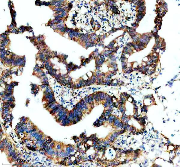
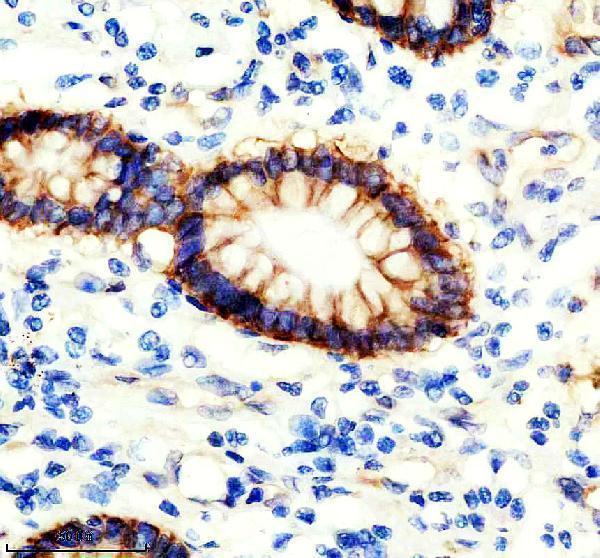
Facts about Integrin alpha-2.

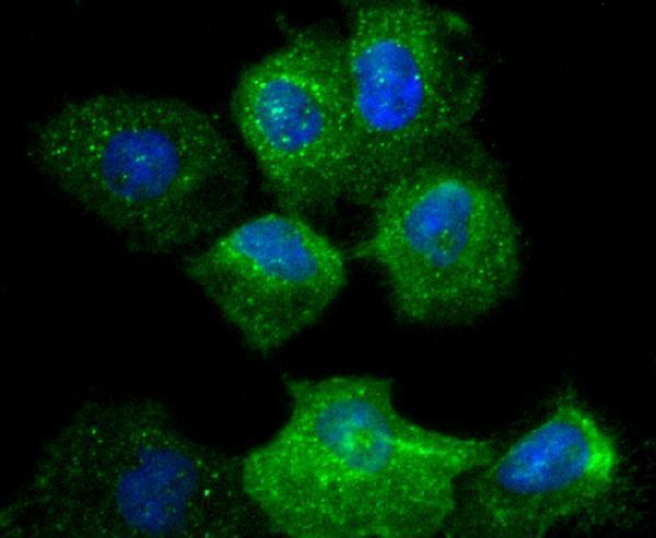
It's responsible for adhesion of platelets and other cells to collagens, modulation of collagen and collagenase gene expression, force generation and organization of newly synthesized extracellular matrix. .
| Human | |
|---|---|
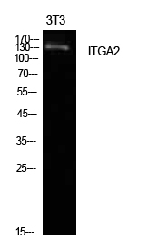
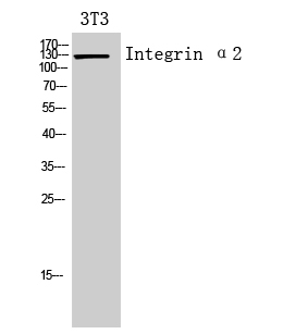
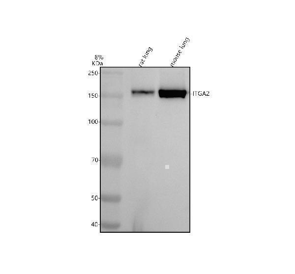
| Gene Name: | ITGA2 |
| Uniprot: | P17301 |
| Entrez: | 3673 |

| Belongs to: |
|---|
| integrin alpha chain family |

alpha-2 subunit; CD49b antigen; CD49b; Integrin alpha 2; integrin, alpha 2 (CD49B, alpha 2 subunit of VLA-2 receptor); ITGA2; VLA 2; VLA-2 alpha
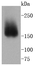
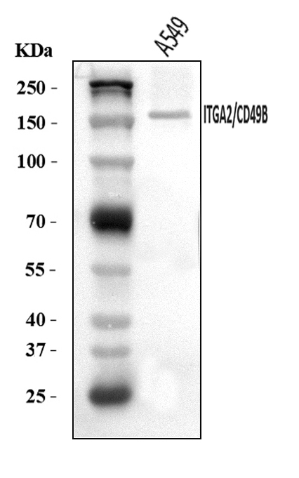
Mass (kDA):
129.295 kDA

| Human | |
|---|---|
| Location: | 5q11.2 |
| Sequence: | 5; NC_000005.10 (52989352..53094779) |
Membrane; Single-pass type I membrane protein.


PMID: 2545729 by Takada Y., et al. The primary structure of the VLA-2/collagen receptor alpha 2 subunit (platelet GPIa): homology to other integrins and the presence of a possible collagen-binding domain.
PMID: 8276836 by Zutter M.M., et al. The human alpha 2 integrin gene promoter. Identification of positive and negative regulatory elements important for cell-type and developmentally restricted gene expression.