This website uses cookies to ensure you get the best experience on our website.


- Table of Contents
Plan ITPA staining in paraffin sections around the reported cytoplasmic tissue pattern (HPA tissue IHC). This guide pairs the catalog antibody’s 2–5 μg/ml IHC range (datasheet A02304) with positive and negative tissue controls drawn from the staining profile (HPA tissue IHC).

Expected localisation, validated protocols, controls and antibodies — the at-a-glance facts below, then the full design guide.
| Expected localisation | General cytoplasmic tissue staining (HPA tissue IHC) | |
| Staining pattern | Cytoplasmic staining in multiple cell types (HPA tissue IHC) | |
| Antigen retrieval | EDTA pH 8.0 HIER, heat-mediated (datasheet A02304) | |
| Positive control | Adipose tissue+4 more · see all | |
| Negative control | Esophagus+4 more · see all |
| Fixation | Keep paraffin-section fixation consistent (standard IHC practice; not target-specific); Matched tissue-IHC evidence does not establish the fixation claim. Validate the specimen-specific method before use. (selected-SKU IHC image A02304) | |
| Caveat | Skin keratinocytes may be unstained despite broad expression (HPA tissue IHC) | |
| Regulation | No stimulus regulator specified (UniProt) | |
| Isoform / epitope | 3 isoforms; check whether the epitope is shared (UniProt) |
The catalog antibody’s IHC-P protocol (datasheet: A02304) is accompanied by published ITPA staining methods for mouse paraffin sections (PMC7710303) and renal carcinoma tissue microarrays (PMC5347726).
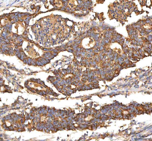
| Sample | Paraffin-embedded human colon adenocarcinoma tissue; fixative not specified (datasheet A02304) |
| Fixation | Image fixative and duration unreported (datasheet A02304); verify before use. |
| Sectioning | 4–5 µm sections on charged slides (standard) |
| Deparaffinisation | Xylene, graded ethanol series to water (standard) |
| Antigen retrieval | Heat retrieval: EDTA pH 8.0 (datasheet A02304); 20 min, 95–100 °C (standard) |
| Peroxidase block | 3% H2O2, 10 min, room temperature (standard) |
| Blocking | 10% goat serum (datasheet A02304) |
| Primary antibody | Rabbit anti-ITPA, 2-5 μg/ml (datasheet A02304) |
| Primary incubation | Overnight at 4 °C (datasheet A02304) |
| Detection | HRP-conjugated secondary, DAB chromogen (datasheet A02304) |
| Counterstain | Hematoxylin, blue, dehydrate and mount (standard) |
| Expected result | ITPA-positive staining in adipocytes of adipose tissue (HPA tissue IHC: Medium). HPA tissue profile: General cytoplasmic expression. No signal in the no-primary control. |
ITPA is cytoplasmic and has no transmembrane segment (UniProt Q9BY32). In paraffin sections, expect general cytoplasmic staining across cell types, with medium staining documented in selected adipocytes, glandular cells, respiratory epithelial cells and neuronal cells (HPA tissue IHC). HPA rates its tissue IHC profile Approved, with medium consistency between antibody staining and RNA expression data (HPA tissue IHC).
| Cytoplasmic staining in adrenal glandular cells or bronchial respiratory epithelial cells. | This fits the reported general cytoplasmic pattern; both cell populations have medium staining (HPA tissue IHC). Score the identified cells and their cytoplasm, because nearby unstained structures do not change the recorded result for those cells (general IHC practice). |
| Predominantly membrane, extracellular or sharply nuclear staining in an IHC section. | Membrane or extracellular emphasis conflicts with cytoplasmic localisation and the absence of a transmembrane segment or signal peptide (UniProt Q9BY32; HPA tissue IHC). A nuclear IHC pattern also warrants review, but nuclear signal alone cannot be dismissed: HPA reports supported nucleoplasmic staining in ICC-IF (HPA subcellular). |
| Strong staining in skin keratinocytes or esophageal squamous epithelial cells. | Both populations are listed as not detected by tissue IHC (HPA tissue IHC). Review cell identification, the no-primary control and detection chemistry for cross-reactivity or endogenous activity (general IHC practice). A discordant result calls for investigation rather than an automatic biological conclusion. |
| Broad, even colour over cells and surrounding tissue, without clear cell boundaries. | This is difficult to score as cytoplasmic ITPA, whose tissue IHC profile is generally cytoplasmic (HPA tissue IHC). Compare a no-primary control and inspect washing, blocking and chromogen development; excess background can obscure a real cell-associated signal (general IHC practice). |
| No staining in a section expected to contain adrenal glandular cells. | Those cells have medium staining in the HPA tissue IHC record (HPA tissue IHC). Confirm their presence and assess section quality, retrieval, antibody dilution and detection controls before calling the sample negative (general IHC practice). HPA's Approved rating still carries only medium staining-to-RNA consistency (HPA tissue IHC). |
| Tissue and cell selection | HPA reports medium staining in adipocytes, adrenal glandular cells, bronchial respiratory epithelial cells and selected neuronal cells; skin keratinocytes and esophageal squamous epithelial cells are not detected (HPA tissue IHC). Match the scored cell population to the record. |
| Compartment and topology | UniProt places ITPA in the cytoplasm and records no transmembrane segment or signal peptide (UniProt Q9BY32). These annotations support a cytoplasmic IHC expectation, without establishing how fixation or antigen retrieval affects staining. |
| Antibody evidence | HPA022824 is Approved for IHC; HPA073963 has no supplied IHC rating (HPA antibodies). HPA's tissue profile is Approved but has medium consistency with RNA expression data (HPA tissue IHC). Interpret unexpected patterns with that limit in view. |
| IF/ICC Q&A: Is nuclear signal expected? | Yes: HPA reports supported nucleoplasmic and cytosolic localisation in ICC-IF (HPA subcellular). That observation informs compartment interpretation; it does not establish an IHC staining intensity or an IF/ICC protocol. |
| Isoforms and epitope coverage | UniProt lists three ITPA isoforms (UniProt Q9BY32). The supplied records do not map the catalog antibody's epitope to those isoforms, so they cannot establish which variants its IHC staining detects. |
| Situation | Likely cause | Next action |
|---|---|---|
| Expected positive tissue has no signal. | The expected adrenal glandular-cell staining is medium, not guaranteed in every section (HPA tissue IHC); a failed staining step is also possible (general IHC practice). | Verify the relevant cells are present, then check a documented positive section, retrieval, antibody dilution and detection reagents together (general IHC practice). |
| The whole section has diffuse brown colour. | Nonspecific background or endogenous detection activity can obscure cell boundaries (general IHC practice); this does not match HPA's general cytoplasmic profile (HPA tissue IHC). | Compare no-primary and detection controls, then review blocking, washing and chromogen development before scoring cells (general IHC practice). |
| Skin keratinocytes stain strongly. | That cell population is listed as not detected (HPA tissue IHC); cross-reactivity or endogenous detection activity is possible (general IHC practice). | Confirm the cells and compare control sections and a no-primary control. Record the discrepancy instead of treating the signal as established ITPA expression (general IHC practice; HPA tissue IHC). |
| Signal outlines membranes or lies outside cells. | This conflicts with cytoplasmic localisation and absent membrane-spanning or secretory features (UniProt Q9BY32; HPA tissue IHC). | Inspect section morphology and control staining, then reassess whether the apparent outline is background, an adjacent cell or a true intracellular signal (general IHC practice). |
| IHC shows prominent nuclear staining. | The tissue IHC profile is generally cytoplasmic, while ICC-IF supports nucleoplasmic as well as cytosolic localisation (HPA tissue IHC; HPA subcellular). | Document the compartment and compare controls and the cytoplasmic signal. Treat a nuclear-only IHC result as unresolved rather than automatically specific or artifactual (general IHC practice; HPA subcellular). |
| Staining differs across areas of one section. | Cell populations can have different recorded staining levels, including low or not-detected entries (HPA tissue IHC); uneven staining is also a technical possibility (general IHC practice). | Score each identified cell population separately and compare morphology and control staining across the section before assigning a tissue-wide result (general IHC practice). |


Comprehensive Human Protein Atlas IHC scoring per tissue (reliability: Approved — Medium consistency between antibody staining and RNA expression data.). Rows are taken directly from the HPA tissue chart — click any row's HPA link to view the source.
| Tissue | Cell type | Level | Evidence | Source |
|---|---|---|---|---|
| Adipose tissue | Adipocytes | Medium | Protein (IHC) | HPA → |
| Adrenal gland | Glandular cells | Medium | Protein (IHC) | HPA → |
| Appendix | Glandular cells | Medium | Protein (IHC) | HPA → |
| Breast | Glandular cells | Medium | Protein (IHC) | HPA → |
| Bronchus | Respiratory epithelial cells | Medium | Protein (IHC) | HPA → |
| Tissue | Cell type | Level | Evidence | Source |
|---|---|---|---|---|
| Esophagus | Squamous epithelial cells | Not detected | Protein (IHC) | HPA → |
| Salivary gland | Glandular cells | Not detected | Protein (IHC) | HPA → |
| Skin | Keratinocytes | Not detected | Protein (IHC) | HPA → |
| Tonsil | Germinal center cells | Not detected | Protein (IHC) | HPA → |
| Vagina | Squamous epithelial cells | Not detected | Protein (IHC) | HPA → |
Troubleshoot ITPA staining in paraffin sections using the catalog antibody’s tissue image and independent localisation evidence (A02304 caption; HPA tissue IHC; HPA subcellular).
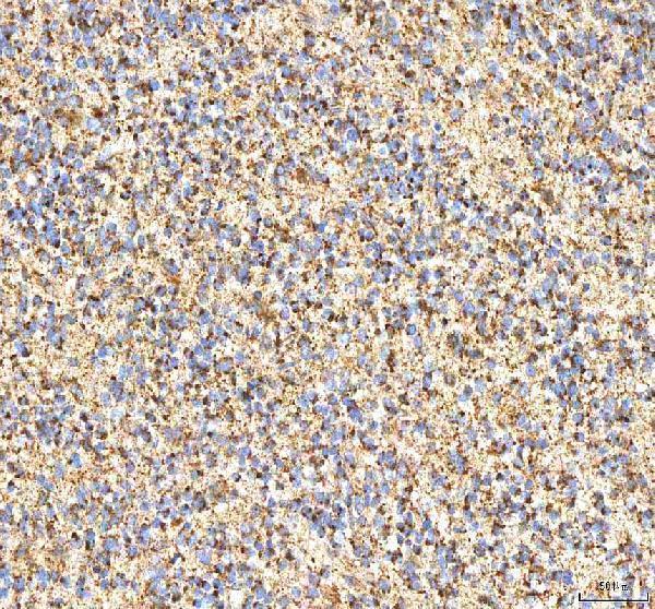
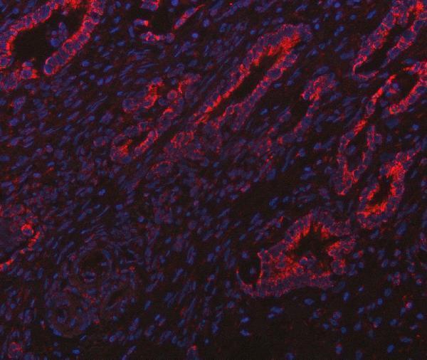
A02304 has documented paraffin-section IHC in human cancer tissues and IF in a human intestinal cancer section (catalog image captions); its listed reactivity includes human, mouse and rat (catalog applications/reactivity).
A02304 is the SKU with a rendered IHC figure, showing a human colon adenocarcinoma paraffin section; its other captions document human glioblastoma, liver cancer and lung cancer IHC, plus intestinal cancer IF (catalog image captions). M02304 lists human IHC, but has no IHC or IF image caption in the payload (catalog applications/reactivity; image captions).
Which to pick: Choose A02304 for paraffin-section IHC because its own caption documents that preparation and 2 μg/ml primary antibody (A02304 IHC caption); the fixative is unreported (A02304 IHC caption). For IF, choose A02304 based on its human intestinal cancer section image at 5 μg/ml; ICC evidence is unreported (A02304 IF caption). A02304 lists human, mouse and rat reactivity, while rabbit monoclonal M02304 lists human reactivity and IHC without a figure caption (catalog applications/reactivity; M02304 clone listing and image captions).